


26 August 2026: Articles 
Perioperative Airway Management in Treacher Collins Syndrome With Obstructive Sleep Apnea: A Case Report
Challenging differential diagnosis, Unusual setting of medical care, Congenital defects / diseases, Educational Purpose (only if useful for a systematic review or synthesis)
Yaqiong Jiang ABCEF 1, Zhikeng Deng E 1, Bo Yao F 1, Xiaohua Liang BC 1, Zhaojun Qin ACF 2*DOI: 10.12659/AJCR.952912
Am J Case Rep 2026; 27:e952912


Abstract
BACKGROUND: Treacher Collins syndrome (TCS) is a congenital disorder resulting from maldevelopment of the first and second pharyngeal arches. A major high-risk comorbidity is obstructive sleep apnea (OSA), characterized by recurrent upper airway collapse during sleep, leading to intermittent hypoxia and sleep fragmentation. This condition requires a tailored anesthetic strategy due to an anticipated difficult airway during intubation and increased vulnerability to postoperative respiratory failure.
CASE REPORT: A 28-year-old man with TCS and polysomnography-confirmed mild OSA underwent orthognathic surgery. Preoperative evaluation included cone-beam computed tomography (CBCT), which objectively quantified retrolingual airway stenosis. After a multidisciplinary briefing, anesthesia was induced with intravenous remimazolam, propofol, sufentanil, and cisatracurium; the airway was secured through videolaryngoscopy-guided nasotracheal intubation. This technique optimized glottic visualization, and controlled induction was feasible because mask ventilation difficulty was not anticipated. The surgical procedure (Le Fort I osteotomy, bilateral sagittal split osteotomy, and genioplasty) was performed to expand the upper airway and correct skeletal deformities. Comparisons of preoperative and postoperative CBCT images and lateral cephalometric radiographs demonstrated clear upper airway expansion and improvement in airway obstruction. Postoperative anesthetic management prioritized a strict opioid-sparing analgesic protocol and monitored extubation, resulting in an uneventful recovery.
CONCLUSIONS: Management of patients with TCS and OSA during orthognathic surgery relies on a proactive, imaging-informed approach. Key elements include the use of advanced imaging for precise anatomic risk stratification, selection of an intubation technique that provides definitive airway visualization, and implementation of a structured multidisciplinary plan with vigilant postoperative monitoring to mitigate delayed respiratory complications.
Keywords: airway management, Anesthesiology, obstructive sleep apnea, orthognathic surgery, Treacher Collins syndrome, Case Reports
Introduction
Treacher Collins syndrome (TCS) is a rare congenital craniofacial disorder affecting 1 in 50 000 births. It results from abnormal development of the first and second branchial arches, leading to characteristic craniofacial anomalies and conductive hearing loss [1]. Facial bone hypoplasia frequently contributes to upper airway compromise and predisposes affected individuals to obstructive sleep apnea (OSA) [2]. OSA is characterized by recurrent upper airway collapse during sleep, resulting in intermittent hypoxia and sleep fragmentation; it occurs in approximately 46% of patients with TCS, a substantially higher prevalence relative to the general population [3]. OSA also increases risks associated with airway management, including difficulties with intubation and ventilation; it thus requires careful perioperative evaluation.
This report describes a patient with TCS and OSA who underwent orthognathic surgery; it focuses on personalized anesthetic strategies for difficult airway management in this specific craniofacial phenotype.
Case Report
HISTORY AND PHYSICAL EXAMINATION:
A 28-year-old man presented with a 20-year history of facial asymmetry and mandibular retrognathia complicated by TCS and mild OSA. He weighed 51.6 kg and had a body mass index (BMI) of 16.7 kg/m2. His medical history included cleft palate repair at 7 months of age, septoplasty in 2018, bilateral zygomatic reconstruction in 2022, and ongoing orthodontic treatment. He had no significant systemic illnesses. Polysomnography confirmed mild OSA, with an overall apnea-hypopnea index of 13.4 events/hour. Vital signs on admission were blood pressure 103/66 mm Hg, heart rate 79 bpm, oxygen saturation 100%, respiratory rate 20 breaths/min, and temperature 36.5 °C. Physical examination revealed typical features of TCS, including bilateral zygomatic hypoplasia, upper eyelid ptosis, down-slanting palpebral fissures, retrognathia, and micrognathia (Figure 1). Airway assessment demonstrated a Modified Mallampati Class II airway, an interincisor distance limited to 25 mm, and partial uvular obstruction (Figure 2). Cervical spine mobility was normal; however, mandibular retrognathia limited airway improvement during neck extension.
Imaging studies, including cone-beam computed tomography (CBCT) and lateral cephalometry, showed mandibular retrognathia and an inferiorly and posteriorly displaced hyoid bone, resulting in tongue base collapse and clinically significant pharyngeal airway narrowing (Figure 3). Preoperative chest radiography and laboratory tests, including a complete blood count, biochemical profile, and coagulation studies, all exhibited results within normal limits. Electrocardiography demonstrated sinus arrhythmia with intermittent incomplete right bundle branch block.
After comprehensive anesthetic evaluation, the patient was deemed suitable for the planned orthognathic procedures: Le Fort I osteotomy, bilateral sagittal split ramus osteotomy, and genioplasty, aimed at improving both airway dimensions and facial aesthetics.
INTRAOPERATIVE MANAGEMENT:
Prior to anesthesia induction, vital signs were blood pressure 115/75 mm Hg, heart rate 64 bpm, oxygen saturation 100%, respiratory rate 17 breaths/min, and temperature 36.5 °C. Peripheral intravenous access was established, and the patient was reassured. After supine positioning and preoxygenation, a urinary catheter was inserted for intraoperative fluid monitoring and bladder management. Anesthesia was induced with intravenous remimazolam (5 mg), propofol (100 mg), and sufentanil (30 μg) for sedation and analgesia, followed by cisatracurium (10 mg) for neuromuscular blockade. After confirming an adequate depth of anesthesia and muscle relaxation, nasotracheal intubation was performed using a 6.0-mm endotracheal tube under videolaryngoscopic guidance, with clear visualization of the vocal cords. The tube was secured with a nasal suture to prevent intraoperative dislodgement (Figure 4).
Anesthesia was maintained using a balanced intravenous-inhalational technique comprising continuous infusions of propofol (2.5 mg/kg/h) and remifentanil (0.15 μg/kg/min), supplemented with 1% inhaled sevoflurane. Invasive arterial monitoring was established to enable continuous blood pressure measurement and arterial blood gas analysis; anesthetic doses were titrated according to surgical stimulation and the patient’s physiological responses. After an 8-hour procedure characterized by stable hemodynamics and no episodes of airway obstruction or hypoxemia, the patient met extubation criteria and was successfully extubated. For postoperative pain management, patient-controlled analgesia consisted of a low-dose opioid (sufentanil 70 μg), a nonsteroidal anti-inflammatory drug (flurbiprofen axetil 100 mg), and an antiemetic (ondansetron 8 mg). Additionally, a temporary tongue suture was placed intraoperatively and removed the following day when airway stability had been confirmed. Postoperative imaging performed before discharge (Figure 3) demonstrated increased airway width and volume. Recovery was uneventful, and the patient was discharged on postoperative day 5.
Discussion
In the present case, management of a patient with TCS and comorbid OSA required a highly individualized approach. Unlike general difficult-airway management principles, this case highlights an imaging-informed, obstruction-targeted workflow for adult, low-BMI patients with TCS and OSA undergoing orthognathic surgery. Although general principles for difficult airway management remain applicable, this case demonstrates how specific anatomic and clinical features should guide perioperative planning. The following discussion is organized around the key management decisions made in this case and their educational implications.
The present case involved a complex and potentially high-risk airway scenario—improper management could result in severe complications, including hypoxic brain injury or death [4]. Perioperative planning was guided by the 2023 expert consensus recommending a multidimensional airway assessment that encompassed 5 domains: medical history, physical examination, comprehensive scoring systems, imaging, and adjunctive tests [5]. Key findings included the underlying diagnosis of TCS, maxillomandibular hypoplasia, a restricted interincisal distance (25 mm), and comorbid OSA. Preoperative imaging confirmed clinically significant airway narrowing. Taken together, these findings established a clear multilevel obstructive risk profile. Notably, this young, non-obese patient (BMI 16.7 kg/m2) lacked conventional OSA risk factors such as obesity hypoventilation syndrome, making craniofacial anatomy the primary etiologic factor [6,7]. This presentation differs from many OSA cases in which obesity is the predominant contributor.
Preoperative CBCT and cephalometric analysis served as essential tools for surgical planning and perioperative decision-making. These studies objectively demonstrated posterior-inferior displacement of the hyoid bone and considerable narrowing of the oropharyngeal airway at its narrowest point (Figure 3A, 3C), allowing accurate localization of the anticipated site of airway collapse. Accordingly, our management strategy included: (a) nasotracheal intubation to secure a patent airway proximal to the area of constriction, and (b) hyoid suspension to reduce the risk of postoperative tongue-base obstruction. This imaging-guided approach to anticipating airway compromise represents an important clinical lesson. Furthermore, comparative postoperative imaging (Figure 3) objectively demonstrated substantial increases in airway diameter and volume, providing definitive evidence of the surgical outcome.
In terms of airway management, both videolaryngoscopy (VL) and awake flexible bronchoscopy are effective techniques for managing difficult airways. Although evidence suggests comparable efficacy between the 2 approaches [8], VL was selected in the present case because: (a) controlled induction was feasible given the patient’s mild OSA and the absence of anticipated mask ventilation difficulty; (b) nasal patency was adequate, and VL provides superior glottic visualization for nasotracheal tube placement; and (c) VL is generally faster and better tolerated by patients, with less coughing during the procedure. The successful first-attempt intubation demonstrates that VL may be particularly effective in patients with TCS whose primary airway challenge arises from fixed craniofacial skeletal abnormalities, rather than dynamic soft-tissue collapse or severe trismus. In a related case, VL-assisted intubation combined with a laryngeal mask airway was successfully utilized; stable vital signs were maintained throughout the procedure [9].
The postoperative period presents additional challenges for patients with TCS and OSA, primarily due to the elevated risk of respiratory complications and substantial soft-tissue edema following orthognathic surgery, particularly mandibular procedures such as sagittal split osteotomy [10]. In low-BMI patients with OSA, postoperative edema may further increase the risk of airway obstruction after extubation [11]. To proactively address the risk of tongue-base collapse, a temporary tongue suture was placed intraoperatively and removed the following day when airway stability had been confirmed. This technique can temporarily enlarge the retroglossal space and may help avoid tracheostomy or prolonged intubation. Its use in patients undergoing mandibular advancement surgery warrants further investigation.
Due to the patient’s OSA and the associated risk of opioid-induced respiratory depression, an opioid-sparing analgesic strategy was prioritized. Compared with opioid-based analgesia, this approach may reduce postoperative nausea, vomiting, and pain while minimizing respiratory complications [12]. In the present case, patient-controlled analgesia consisted of a low-dose opioid (sufentanil 70 μg), a nonsteroidal anti-inflammatory drug (flurbiprofen axetil 100 mg), and an antiemetic (ondansetron 8 mg), which provided effective pain control while limiting the risk of opioid-related respiratory depression. This outcome supports consideration of opioid-sparing analgesia as the preferred analgesic strategy for this patient population to promote postoperative respiratory stability.
Maxillomandibular advancement with genioplasty effectively improves OSA by expanding the upper airway [2,13]. In the present case, postoperative imaging (Figure 3) demonstrated increased airway width and volume, findings consistent with clinical improvement and indicative of successful multilevel airway expansion. Additionally, the procedure improved the patient’s facial profile and occlusion, supporting a role for orthognathic surgery as an effective treatment for OSA in patients with TCS through its enhancements of function and aesthetics. A notable limitation of this case was the absence of postoperative polysomnography, the gold standard for objectively assessing the resolution of sleep-disordered breathing.
Ultimately, the successful outcome resulted from coordinated multidisciplinary collaboration among anesthesiologists, surgeons, and nursing staff. Within this framework, the surgeon developed and executed the surgical plan while preparing for potential emergencies; the anesthesiologist performed the preoperative assessment, planned airway management, and maintained intraoperative physiologic stability; and the nursing staff provided comprehensive perioperative care. This structured briefing process and clear delineation of responsibilities were critical to patient safety and effective perioperative management.
This case offers several key clinical insights: (a) imaging, particularly mid-sagittal and 3-dimensional CBCT, facilitates the diagnosis and planning of airway stenosis management; (b) OSA is a heterogeneous condition, and non-obese patients with TCS require treatment strategies directed at anatomic airway obstruction; (c) VL can serve as a primary airway management strategy in selected patients with TCS; (d) temporary tongue sutures can help prevent postoperative airway obstruction; and (e) multidisciplinary briefings enhance patient safety through clear role assignment and perioperative planning.
Conclusions
The successful anesthetic management of this patient with TCS and mild OSA was not incidental but resulted from a stepwise “assess-decide-execute” strategy tailored to syndromic craniofacial patients with OSA. This approach highlights 3 key elements: comprehensive risk assessment based on imaging and clinical findings, appropriate technical interventions, and effective multidisciplinary collaboration. This practical framework may be broadly applicable to other patients with syndromic craniofacial anomalies and comorbid OSA.
Figures
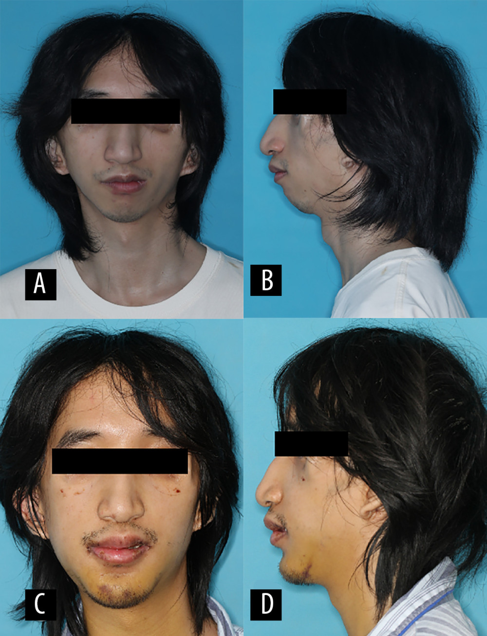 Figure 1. Facial photographs. (A, B) Preoperative frontal and lateral views. (C, D) Postoperative frontal and lateral views after orthognathic surgery.
 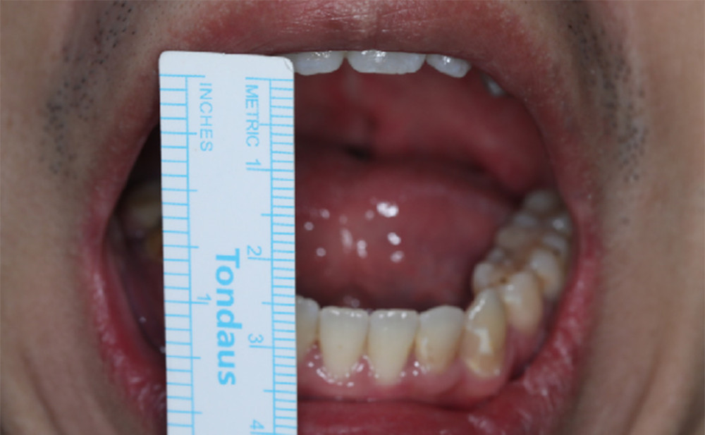 Figure 2. Clinical assessment of mouth opening.
 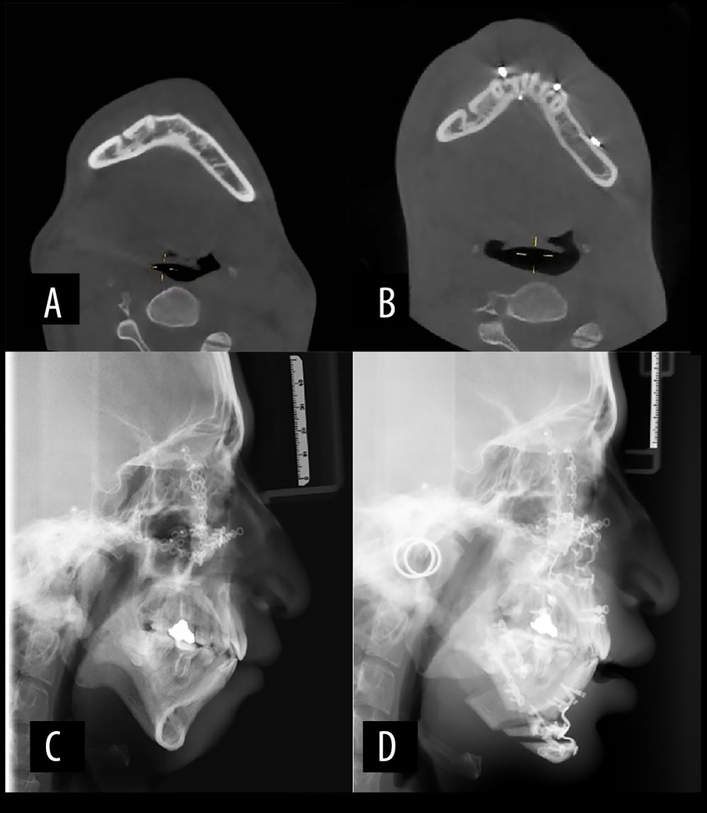 Figure 3. Imaging comparison of skeletal and airway changes after orthognathic surgery. (A, B) Preoperative and postoperative axial cone beam computed tomography images demonstrating the minimum airway width. (C, D) Preoperative and postoperative lateral cephalograms.
 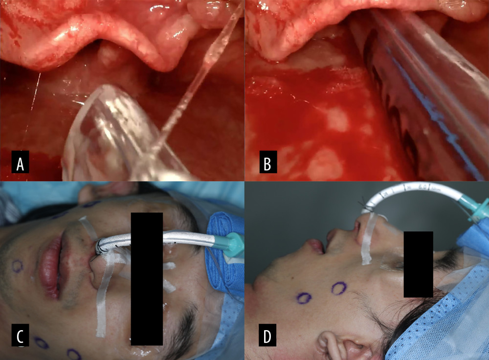 Figure 4. Videolaryngoscopy-guided tracheal intubation. (A) Glottic view before tube insertion. (B) Endotracheal tube positioned within the trachea. (C, D) Frontal and lateral views of the patient after successful intubation.
 References
1. Beaumont CA, Dunaway DJ, Padwa BL, Extracraniofacial anomalies in Treacher Collins syndrome: A multicentre study of 248 patients: Int J Oral Maxillofac Surg, 2021; 50; 1471-76
2. Paradowska-Stolarz A, Sluzalec-Wieckiewicz K, Mikulewicz M, Obstructive sleep apnoea in patients with Treacher Collins syndrome: A narrative review: J Clin Med, 2025; 14; 4741
3. Plomp RG, Bredero-Boelhouwer HH, Joosten KF, Obstructive sleep apnoea in Treacher Collins syndrome: Prevalence, severity and cause: Int J Oral Maxillofac Surg, 2012; 41; 696-701
4. Apfelbaum JL, Hagberg CA, Connis RT, 2022 American Society of Anesthesiologists practice guidelines for management of the difficult airway: Anesthesiology, 2022; 136; 31-81
5. Xia M, Ma W, Zuo M, Expert consensus on difficult airway assessment: Hepatobiliary Surg Nutr, 2023; 12; 545-66
6. Zhang W, Zhang H, Xi X, Association of craniofacial skeletal and soft tissue characteristics with severe obstructive sleep apnea in age-specific and BMI-specific patient groups: J Craniofac Surg, 2025; 36; 1967-71
7. Jordan AS, McSharry DG, Malhotra A, Adult obstructive sleep apnoea: Lancet, 2014; 383; 736-47
8. Alhomary M, Ramadan E, Curran E, Videolaryngoscopy vs. fibreoptic bronchoscopy for awake tracheal intubation: A systematic review and meta-analysis: Anaesthesia, 2018; 73; 1151-61
9. Mohan S, Del Rosario TJ, Pruett BE, Anesthetic management of Treacher Collins syndrome in an outpatient surgical center: Am J Case Rep, 2021; 22; e931974
10. Roesslein M, Chung F, Obstructive sleep apnoea in adults: Peri-operative considerations: A narrative review: Eur J Anaesthesiol, 2018; 35; 245-55
11. Woo HJ, Lim JH, Ahn JC, Characteristics of obstructive sleep apnea patients with a low body mass index: Emphasis on the obstruction site determined by drug-induced sleep endoscopy: Clin Exp Otorhinolaryngol, 2020; 13; 415-21
12. La Via L, Iannella G, Pace A, Anesthesiologic management of adult and pediatric patients with obstructive sleep apnea: Healthcare (Basel), 2025; 13; 2183
13. Zaghi S, Holty JE, Certal V, Maxillomandibular advancement for treatment of obstructive sleep apnea: A meta-analysis: JAMA Otolaryngol Head Neck Surg, 2016; 142; 58-66
Figures
Figure 1. Facial photographs. (A, B) Preoperative frontal and lateral views. (C, D) Postoperative frontal and lateral views after orthognathic surgery.Figure 2. Clinical assessment of mouth opening.Figure 3. Imaging comparison of skeletal and airway changes after orthognathic surgery. (A, B) Preoperative and postoperative axial cone beam computed tomography images demonstrating the minimum airway width. (C, D) Preoperative and postoperative lateral cephalograms.Figure 4. Videolaryngoscopy-guided tracheal intubation. (A) Glottic view before tube insertion. (B) Endotracheal tube positioned within the trachea. (C, D) Frontal and lateral views of the patient after successful intubation. In Press
Case report 
Am J Case Rep In Press; DOI: 10.12659/AJCR.953248
Case report
Am J Case Rep In Press; DOI: 10.12659/AJCR.953317
Case report 
Am J Case Rep In Press; DOI: 10.12659/AJCR.954643
Case report 
Am J Case Rep In Press; DOI: 10.12659/AJCR.954074
Most Viewed Current Articles
07 Dec 2021 : Case report  28,712,323
DOI :10.12659/AJCR.934347
Am J Case Rep 2021; 22:e934347
06 Dec 2021 : Case report  180,503
DOI :10.12659/AJCR.934406
Am J Case Rep 2021; 22:e934406
21 Jun 2024 : Case report
124,239
DOI :10.12659/AJCR.944371
Am J Case Rep 2024; 25:e944371
08 May 2026 : Case report
94,936
DOI :10.12659/AJCR.952158
Am J Case Rep 2026; 27:e952158