Logo American Journal of Case Reports

Call: 1.631.629.4328
Mon-Fri 10 am - 2 pm EST

Contact Us

Logo American Journal of Case Reports Logo American Journal of Case Reports Logo American Journal of Case Reports

25 August 2026: Articles  USA

Canine-Associated Capnocytophaga canimorsus Bacteremia in an Older Adult Without Classical Immunosuppressive Risk Factors

Rare disease

Ingrid L. Hirte ORCID logo ABEF 1*, Jack W. McHugh ORCID logo BEF 2, Nathaniel E. Miller ORCID logo EF 1

DOI: 10.12659/AJCR.952980

Am J Case Rep 2026; 27:e952980

0 Comments

Abstract

0:00

BACKGROUND: Several Capnocytophaga species exist, some predominantly colonizing the human oral cavity and others residing in feline or canine oral flora. Capnocytophaga canimorsus is a gram-negative facultative anaerobe commonly found in the oral cavity of dogs and, less commonly, cats. C. canimorsus infections are rare in humans. Although beta-lactamase production has been described among Capnocytophaga species, it is less common in C. canimorsus; therefore, penicillin may remain appropriate when susceptibility is confirmed.

CASE REPORT: An older man was admitted to the Family Medicine Inpatient Service with erythema and edema of the right hand after sustaining a hand laceration several days prior to admission; the wound had been licked multiple times by his dog. The patient developed worsening erythema and tenderness of the dorsal right hand, accompanied by progressive leukocytosis. He was transitioned to intravenous ampicillin-sulbactam. Blood cultures grew C. canimorsus. Because beta-lactamase testing results were pending at discharge, the patient was prescribed oral amoxicillin-clavulanate.

CONCLUSIONS: C. canimorsus is a rare zoonotic pathogen but can cause severe disease, particularly in patients with cirrhosis, asplenia, or alcohol use disorder; in the present case, advanced age may have contributed to vulnerability through immunosenescence. Growth of gram-negative bacilli in aerobic or anaerobic blood cultures from patients with zoonotic exposure should raise clinical suspicion for this organism. When Capnocytophaga infection is suspected in a hospitalized patient, empiric treatment with ampicillin-sulbactam should be initiated while awaiting culture confirmation and susceptibility results.

Keywords: Bacteremia, Capnocytophaga, Dog Diseases, Zoonoses

Introduction

Capnocytophaga species are facultative anaerobic, gram-negative bacilli that can cause various infections in humans. There are 9 recognized Capnocytophaga species, which can be categorized into 2 main groups: human oral-associated and zoonotic-associated. Human oral species include C. gingivalis, C. granulosa, C. haemolytica, C. leadbetteri, C. ochracea, and C. sputigena; zoonotic species include C. canimorsus, C. canis, and C. cynodegmi, which are most commonly found in the oral cavities of dogs and cats [1,2].

C. canimorsus is the species most commonly implicated in human infection, although such infections remain rare [3]. Capnocytophaga infections are estimated to occur in fewer than 1 case per million people [4]. Infections typically result from dog or cat bites, scratches, or close animal contact, such as licking of human wounds [3]. Early manifestations may include localized cellulitis, purulent discharge, lymphangitis, and regional lymphadenopathy. This case illustrates canine-associated C. canimorsus bacteremia after wound exposure to dog saliva and highlights advanced age as a potential contributor to host vulnerability outside the classically recognized risk groups.

Case Report

A 95-year-old man with a medical history notable for diastolic heart failure, dementia, hypertension, stage 3a chronic kidney disease, and thrombocytopenia presented with fever, erythema, and swelling of the dorsum of his right hand. He had sustained a laceration to his right hand 5 days before presentation and reported that his dog had repeatedly licked the wound in the interim.

Vital signs in the emergency department were notable for a blood pressure of 137/63 mm Hg, heart rate of 95 beats/min, respiratory rate of 18 breaths/min, oxygen saturation of 94% on room air, and temperature of 38.5 °C. Initial laboratory studies were remarkable for normocytic anemia with a hemoglobin level of 7.1 g/dL (reference range, 11.6–15.0 g/dL), leukocyte count of 4.2 × 10⁹/L (reference range, 3.4–9.6 × 10⁹/L), lymphopenia of 0.06 × 10⁹/L (reference range, 0.95–3.07 × 10⁹/L), erythrocyte sedimentation rate and C-reactive protein (CRP) within their normal ranges, creatinine of 1.60 mg/dL (reference range, 0.74–1.35 mg/dL), blood urea nitrogen of 33 mg/dL (reference range, 8–24 mg/dL), lactate below 2.2 mmol/L, and an elevated N-terminal pro-B-type natriuretic peptide (NT-proBNP) level of 1264 pg/mL (reference range, ≤ 540 pg/mL). Urinalysis revealed proteinuria without evidence of nitrites or leukocytes. Tests for influenza, respiratory syncytial virus, and severe acute respiratory syndrome coronavirus 2 (SARS-CoV-2) demonstrated negative findings. Blood cultures were obtained. Chest radiography showed a small chronic right-sided pleural effusion. Radiography of the right hand demonstrated only advanced degenerative arthritic changes.

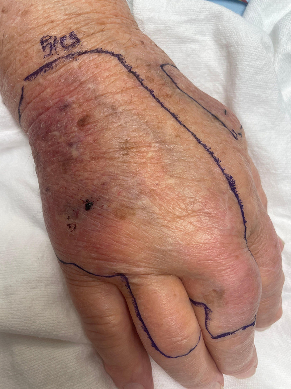
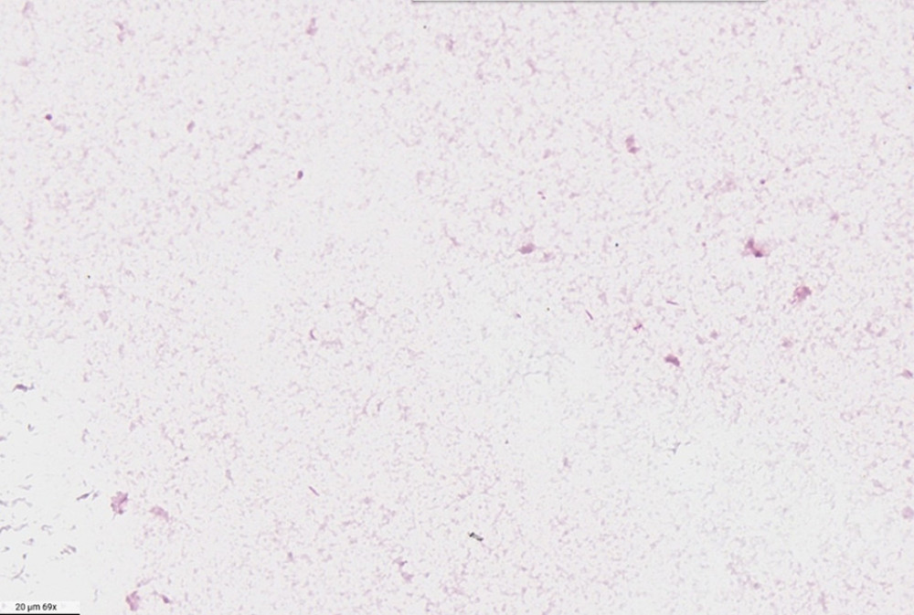
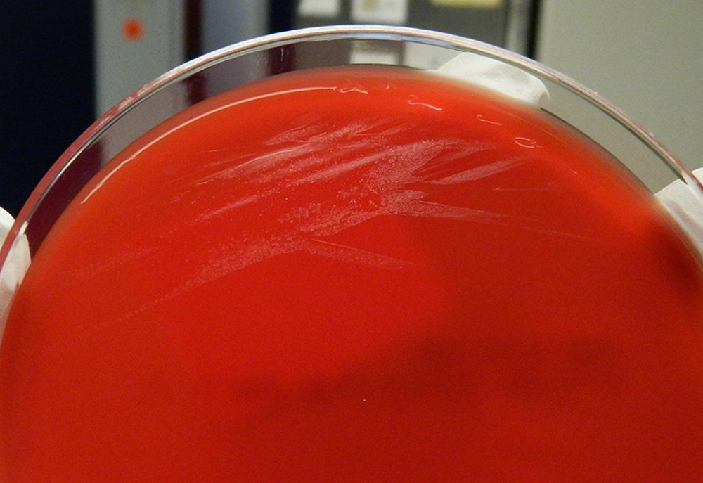
The patient began amoxicillin-clavulanate 875/125 mg twice daily in the emergency department for presumed cellulitis. His hand was placed in a Richard sling as recommended by the orthopedic consultation service. On the medical floor, he remained afebrile but reported generalized weakness and fatigue. The day after admission, his right hand became more erythematous and edematous (Figure 1). During this time, his leukocyte count increased to 21.0 × 10⁹/L and his CRP level increased to 173.3 mg/L (reference range, < 5.0 mg/L); however, concern for a necrotizing soft tissue infection remained low. Given the worsening hand swelling, erythema, and leukocytosis, antibiotic therapy was transitioned to intravenous ceftriaxone 2 g daily. At 30 hours, blood cultures showed positive results for gram-negative rods in 2 of 2 anaerobic bottles and 3 of 3 aerobic bottles. Gram staining (Figure 2) demonstrated gram-negative bacilli, and blood agar culture revealed nonhemolytic yellowish-beige colonies (Figure 3). While awaiting species identification, antibiotic therapy was changed to intravenous ampicillin-sulbactam, 3 g twice daily, as recommended by the infectious disease service.

The organism was subsequently identified as C. canimorsus, and ampicillin-sulbactam was continued while beta-lactamase testing was pending. Over the following 2 days, the patient’s leukocyte count normalized and his CRP level decreased to 97.1 mg/L. His hand erythema and edema improved; he was discharged with instructions to complete a 14-day course of oral amoxicillin-clavulanate 875/125 mg twice daily. After discharge, beta-lactamase test results were reportedly negative; however, his antibiotic regimen was not modified in the outpatient setting.

Discussion

Capnocytophaga species can be transmitted either through the human oral cavity or from zoonotic sources (eg, dogs and cats). A recent multicenter study of Capnocytophaga infections indicated that bacteremia was more commonly associated with human oral species, whereas bacteremia caused by zoonotic Capnocytophaga species was less frequent [5]. Human oral-associated Capnocytophaga bacteremia appears to occur more often in immunocompromised hosts; zoonotic infections are more commonly reported in patients without classic immunosuppressive risk factors. In addition to bacteremia, Capnocytophaga species have been implicated in respiratory infections, pleural empyema, septic arthritis, brain abscess, meningitis, and other invasive syndromes [5].

C. canimorsus is the zoonotic Capnocytophaga species most often associated with severe human disease. Although infection is uncommon, more than 70% of reported C. canimorsus infections present with bacteremia, and sepsis in high-risk hosts has been associated with mortality rates approaching 30% [6,7]. Severe disease is classically associated with asplenia, cirrhosis, alcohol use disorder, and other forms of immune compromise [5–7]. These associations likely reflect the organism’s ability to cause invasive infection when early innate immune control is impaired. Reported mechanisms of pathogenicity include evasion of innate immune recognition, resistance to complement-mediated killing, and impairment of phagocytic clearance, which may permit rapid progression from localized inoculation to bacteremia and sepsis in vulnerable hosts.

The present case is notable because the patient lacked the classic risk factors most often associated with severe C. canimorsus infection. In a retrospective review of C. canimorsus infections, most affected patients were either immunocompromised or had heavy alcohol use [8]. Intriguingly, approximately one-third reported a dog bite, although all had dog exposure, emphasizing that non-bite exposures can be clinically important [8]. Similar severe cases have been described in older adults without overt immunosuppression, including an 80-year-old woman who developed fatal septic shock after a dog bite and a 63-year-old man who developed septic shock after exposure to canine saliva [9,10]. The present case parallels these reports: all involved canine exposure, the absence of a clearly defined classic immunosuppressive condition, and age greater than 60 years. These observations suggest that advanced age represents an underrecognized contributor to host susceptibility.

Although age alone generally is not considered a major risk factor for C. canimorsus infection, immunosenescence provides a plausible biological explanation for severe disease in older adults. Aging is associated with declines in innate and adaptive immune function, including impaired neutrophil and macrophage activity, altered cytokine responses, and a reduced ability to contain invasive bacterial infections [11]. In this context, our patient’s advanced age may have lowered the threshold for progression from a localized wound infection to bacteremia. The lymphopenia observed on admission was likely a consequence of acute severe infection rather than a preexisting risk factor, but it further supports the presence of acute immune dysregulation during the illness.

In patients with recent canine or feline exposure through a bite, scratch, or lick, growth of small gram-negative bacilli in both aerobic and anaerobic culture bottles should raise clinical suspicion for Capnocytophaga infection [5]. Standard antimicrobial susceptibility guidance for Capnocytophaga species is limited, and beta-lactamase testing may require several days to become available [5]. Beta-lactamase production is more common among human oral Capnocytophaga species; therefore, empiric treatment with a beta-lactam/beta-lactamase inhibitor is appropriate in hospitalized patients while awaiting organism identification and susceptibility results [5]. Although beta-lactamase production appears less common in C. canimorsus, penicillin should be reserved for isolates with confirmed susceptibility. In the present case, clinical progression despite initial oral amoxicillin-clavulanate likely reflected the severity and natural evolution of the infection before effective intravenous therapy rather than true antimicrobial failure. Because beta-lactamase results were not available until well after the initial culture results and there was concern for polymicrobial soft tissue infection, the patient was treated with ampicillin-sulbactam and subsequently transitioned to amoxicillin-clavulanate for outpatient therapy.

Conclusions

C. canimorsus is a rare cause of serious human infection. Growth of gram-negative bacilli in aerobic and anaerobic cultures in the context of zoonotic exposure should raise clinical suspicion. Among hospitalized patients with suspected cases, including those who are immunocompetent or have few recognized risk factors, empiric treatment with ampicillin-sulbactam should be initiated while awaiting beta-lactamase testing and susceptibility results.

References

1. Suzuki M, Kimura M, Imaoka K, Yamada A: Vet Microbiol, 2010; 144(1–2); 172-76

2. Idate U, Bhat K, Kotrashetti V: J Oral Maxillofac Pathol, 2020; 24(2); 397

3. Gaastra W, Lipman LJ, Capnocytophaga canimorsus: Vet Microbiol, 2010; 140; 339-46

4. Van Dam AP, Jansz A: Clin Microbiol Infect, 2011; 17(2); 312-15

5. Chesdachai S, Tai DBG, Yetmar ZA: Open Forum Infect Dis, 2021; 8(7); ofab175

6. Butler T: Eur J Clin Microbiol Infect Dis, 2015; 34; 1271-80

7. Lion C, Escande F, Burdin JC: Eur J Epidemiol, 1996; 12; 521-33

8. Pfortmueller C, Efeoglou A, Furrer H, Dog bite injuries: Primary and secondary emergency department presentations—A retrospective cohort study: ScientificWorldJournal, 2013; 2013; 393176

9. Stephens MA, Silin N, Dakkak T: Am J Case Rep, 2025; 26; e946691

10. Mantovani E, Busani S, Biagioni E: Case Rep Crit Care, 2018; 2018; 7090268

11. Goyani P, Christodoulou R, Vassiliou E, Immunosenescence: Aging and immune system decline: Vaccines (Basel), 2024; 12(12); 1314

In Press

Case report  Canada

Radiation Associated Undifferentiated Pleomorphic Sarcoma of the Neck 2 Decades After External Beam Radiati...

Am J Case Rep In Press; DOI: 10.12659/AJCR.953580  

Case report  Colombia

Septic Spontaneous Abortion Associated With Intrauterine Fetal Death at 8 Weeks of Gestation Complicated by...

Am J Case Rep In Press; DOI: 10.12659/AJCR.953673  

Case report  Japan

Anterior Cutaneous Nerve Entrapment Syndrome in a 51-Year-Old Woman With Advanced Colon Cancer, Successfull...

Am J Case Rep In Press; DOI: 10.12659/AJCR.953662  

Case report  Japan

Combination of Multiple-Dose Activated Charcoal and Infusion Therapy That Led to Favorable Outcomes in Colc...

Am J Case Rep In Press; DOI: 10.12659/AJCR.953541  

Most Viewed Current Articles

07 Dec 2021 : Case report  USA 22,769,643

Edwardsiella tarda: A Classic Presentation of a Rare Fatal Infection, with Possible New Background Risk Fac...

DOI :10.12659/AJCR.934347

Am J Case Rep 2021; 22:e934347

06 Dec 2021 : Case report  Brazil 178,207

Lipedema Can Be Treated Non-Surgically: A Report of 5 Cases

DOI :10.12659/AJCR.934406

Am J Case Rep 2021; 22:e934406

21 Jun 2024 : Case report  China (mainland) 121,831

Intracranial Parasitic Fetus in a Living Infant: A Case Study with Surgical Intervention and Prognosis Anal...

DOI :10.12659/AJCR.944371

Am J Case Rep 2024; 25:e944371

0:00

07 Mar 2024 : Case report  USA 66,870

Neurocysticercosis Presenting as Migraine in the United States

DOI :10.12659/AJCR.943133

Am J Case Rep 2024; 25:e943133

0:00

Your Privacy

We use cookies to ensure the functionality of our website, to personalize content and advertising, to provide social media features, and to analyze our traffic. If you allow us to do so, we also inform our social media, advertising and analysis partners about your use of our website, You can decise for yourself which categories you you want to deny or allow. Please note that based on your settings not all functionalities of the site are available. View our privacy policy.

American Journal of Case Reports eISSN: 1941-5923