


26 August 2026: Articles 
Atypical Presentation of Hereditary Hemorrhagic Telangiectasia Without Recurrent Epistaxis Leading to Delayed Diagnosis
Unusual clinical course, Challenging differential diagnosis, Rare disease, Clinical situation which can not be reproduced for ethical reasons
Daniyaal Syed ABEF 1*, Farah Khan AEFG 2, Numair Khan AEF 3
DOI: 10.12659/AJCR.953158
Am J Case Rep 2026; 27:e953158


Abstract
BACKGROUND: Hereditary hemorrhagic telangiectasia (HHT) is a rare vascular disorder characterized by multisystem arteriovenous malformations (AVMs). The earliest symptoms often appear in childhood and typically encompass recurrent epistaxis. Severe outcomes, including cerebral hemorrhage and thrombotic complications, can increase morbidity and mortality. HHT is estimated to have near-complete penetrance, such that 97% of patients exhibit symptoms by age 60.
CASE REPORT: A 71-year-old man presented with a 1-month history of progressive shortness of breath, fatigue, dizziness, and lower-extremity edema. Further evaluation revealed severe iron-deficiency anemia (hemoglobin 6.4 g/dL, serum iron 21 µg/dL, total iron-binding capacity 462 µg/dL, transferrin saturation ~5%, and ferritin 13 ng/mL). He received 4 units of packed red blood cells, resulting in symptomatic improvement. Imaging did not identify any additional visceral malformations; follow-up esophagogastroduodenoscopy and colonoscopy findings were normal. Despite negative endoscopic findings, intermittent occult gastrointestinal blood loss remained the leading consideration given his prior history of bleeding gastrointestinal AVMs and laboratory findings consistent with iron-deficiency anemia. Capsule endoscopy—recommended to screen for small-bowel telangiectasias—was deferred. His medical history was notable for a delayed diagnosis of HHT. He remained clinically asymptomatic until age 67, when he developed spontaneous bilateral subdural hematomas and gastrointestinal bleeding.
CONCLUSIONS: This case highlights delayed recognition of HHT in the absence of recurrent epistaxis, followed by serious intracranial and gastrointestinal complications. Overreliance on classic mucocutaneous features may contribute to diagnostic delay. Clinicians should consider HHT in older adults with otherwise unexplained AVM-related hemorrhage or anemia to facilitate timely screening and management.
Keywords: Case Reports, Epistaxis, Hematology, Late-Onset Disorders, Phenotype
Introduction
Hereditary hemorrhagic telangiectasia (HHT), formerly known as Osler-Weber-Rendu syndrome, is a rare autosomal dominant vascular disorder characterized by blood vessel malformations throughout the body. These malformations typically present as mucocutaneous telangiectasias and visceral arteriovenous malformations (AVMs). HHT is estimated to affect 1 in 5000 to 10 000 individuals, with no increased incidence according to ethnicity or sex [1]. However, these estimates likely underestimate the true prevalence, given that diagnosis of an affected individual often leads to the identification of multiple relatives with manifestations of HHT who have not been formally diagnosed [2,3].
The pathophysiology of HHT results from mutations in genes involved in the transforming growth factor beta/bone morphogenetic protein signaling pathway. Such genes include
The most common and earliest clinical manifestation of HHT is spontaneous, recurrent epistaxis secondary to telangiectasias of the nasal mucosa, which occurs in up to 95% of cases [6]. On average, these episodes begin at age 12 and occur with a frequency of ~ 18 bleeding events per month [7]. Importantly, the presence and frequency of epistaxis are not predictive of disease severity—they serve as an early clinical marker that can aid diagnosis in suspected cases. Consistent with this early presentation, HHT demonstrates near-complete age-related penetrance (~ 97%) by age 60 [2]. In contrast to the often-early onset of epistaxis, cutaneous and oral telangiectasias typically become apparent later, frequently during the third decade of life. Commonly affected sites include the skin, particularly the distal digits and nose, as well as the lips, oral cavity, hard palate, and tongue. These vascular malformations may later involve the gastrointestinal tract, resulting in recurrent upper and/or lower gastrointestinal bleeding in 15% to 20% of individuals with HHT [3]. Given the substantial variability in clinical expression and absence of classic early mucocutaneous symptoms in some patients, recognition of HHT and appropriate screening for visceral AVMs are often delayed. High-risk individuals include first-degree relatives of patients with HHT and those with otherwise unexplained visceral AVMs. Genetic testing can confirm suspected HHT when clinical findings are incomplete and may facilitate cascade testing of relatives, particularly for pathogenic variants in
Overall, the spectrum of complications associated with HHT is broad, ranging from mild iron-deficiency anemia and recurrent epistaxis to ruptured AVMs that cause massive internal hemorrhage or seizures. The present case highlights a rare presentation of HHT in which the disease was not formally confirmed until age 68 in the absence of recurrent epistaxis. Through this report, we aim to demonstrate how reliance on classic mucocutaneous symptoms can delay the diagnosis of HHT and subsequent screening for associated complications.
Case Report
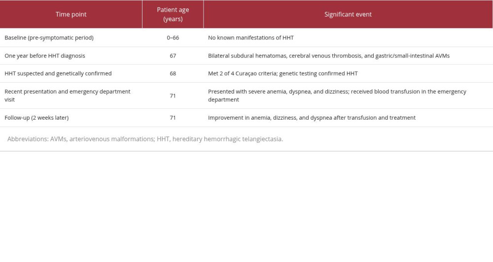
A 71-year-old White man with HHT and a medical history of iron-deficiency anemia, prostate cancer, bilateral subdural hematomas, femoral artery occlusion, and cerebral venous thrombosis reported experiencing 1 month of progressive shortness of breath, fatigue, dizziness, and bilateral lower-extremity edema (Table 1). His medications included clopidogrel bisulfate 75 mg once daily and calcium 500 mg twice daily. Initial laboratory evaluation was notable for severe microcytic, hypochromic anemia (hemoglobin 6.4 g/dL, hematocrit 26.2%, mean corpuscular volume 64.2 fL, mean corpuscular hemoglobin 15.7 pg, mean corpuscular hemoglobin concentration 24.4 g/dL, and red blood cell distribution width 21.0%). Iron studies were consistent with iron deficiency (serum iron 21 μg/dL, total iron-binding capacity 462 μg/dL, transferrin saturation ~ 5%, and ferritin 13 ng/mL). Peripheral blood smear demonstrated microcytosis and hypochromia without schistocytes. He denied signs of overt bleeding, including hematochezia, melena, coffee-ground emesis, or recurrent epistaxis. An electrocardiogram performed in the clinic showed unremarkable findings. Given the severity of anemia, the patient was advised to proceed to the emergency department for blood transfusion, where he received 4 units of packed red blood cells. A non-contrast computed tomography (CT) scan of the head and contrast-enhanced CT scans of the chest, abdomen, and pelvis were obtained to screen for intracranial pathology and potential visceral vascular malformations. Head CT showed no acute intracranial hemorrhage; no mesenteric or other vascular malformations were identified on cross-sectional imaging. Follow-up esophagogastroduodenoscopy (EGD) and colonoscopy findings were normal.
During the 2-week follow-up period, the patient reported substantial improvement in shortness of breath, with complete resolution of dizziness and fatigue. Repeat laboratory testing demonstrated pronounced improvement (hemoglobin 11.2 g/dL, hematocrit 40.8%, mean corpuscular volume 81.6 fL, mean corpuscular hemoglobin 22.4 pg, mean corpuscular hemoglobin concentration 31.2 g/dL, and red blood cell distribution width 18.4%). At that time, the patient began oral ferrous sulfate 325 mg once daily. Follow-up iron studies have not yet been performed. Although EGD findings were normal, intermittent bleeding from HHT-related gastrointestinal telangiectasias remained a leading consideration given the patient’s history. To localize the source of bleeding, video capsule endoscopy was recommended but has not yet been performed due to scheduling difficulties.
Notably, HHT was evaluated in this patient after family screening identified multiple affected relatives. At age 68, the patient met 2 of the 4 Curaçao criteria, suggesting possible HHT: (1) visceral lesions (gastrointestinal AVMs) and (2) a first-degree relative with definite HHT. These findings prompted genetic testing, which confirmed the diagnosis.
One year before his HHT diagnosis, the patient presented with spontaneous bilateral subdural hematomas and acute cerebral venous thrombosis. CT angiography identified a vascular malformation described as an AVM in the left parietal convexity, which was considered the likely source of hemorrhage. He underwent burr-hole evacuation of the subdural hematomas and received low-molecular-weight heparin for cerebral venous thrombosis. Later that year, further evaluation with EGD and colonoscopy identified AVMs in the stomach and small intestine requiring treatment. Before this series of events, the patient denied any history of anemia, recurrent epistaxis, visible telangiectasias, rectal bleeding, or other bleeding-related symptoms.
Discussion
HHT typically becomes clinically apparent earlier in life, most commonly due to recurrent epistaxis. However, the present case demonstrates that the absence of classic mucocutaneous symptoms can delay clinical suspicion and diagnosis, even in the presence of serious complications. This course is best interpreted as delayed clinical recognition and delayed clinical expression rather than true late onset, given that HHT-related vascular lesions may have been present for years before becoming symptomatic or detectable.
The diagnosis of HHT is based on the Curaçao criteria, which include: (1) spontaneous and recurrent epistaxis, (2) a first-degree relative with definite HHT, (3) mucocutaneous telangiectasias, and (4) visceral lesions (AVMs or telangiectasias). A definite diagnosis is established when 3 of the 4 criteria are met; the presence of 2 criteria suggests possible HHT and warrants further evaluation [8]. In the present case, the patient met 2 of the 4 criteria and was referred for genetic testing, which confirmed the diagnosis. Overall, the Curaçao criteria have demonstrated a sensitivity of 68% (95% confidence interval: 60%–76%) and specificity of 98% (95% confidence interval: 91%–100%) [9,10]. Genetic testing for HHT can be performed on either a confirmatory or preemptive basis, including in individuals who meet only 1 or 2 Curaçao criteria and in children of affected individuals who have not yet developed clinical manifestations. The sequence of events in our case highlights the practical limitations of solely relying on classic mucocutaneous symptoms when evaluating patients with visceral AVMs and iron-deficiency anemia.
Screening is recommended even in minimally symptomatic individuals due to the possibility of undetected AVMs. Screening for pulmonary AVMs typically begins with transthoracic contrast echocardiography (bubble echocardiography), followed by thoracic CT for lesion localization and consideration of embolization when indicated [11]. Screening for cerebral AVMs varies among guidelines and should be individualized. However, neurologic symptoms suggestive of a cerebral vascular lesion warrant evaluation with appropriate neuroimaging and specialist consultation [8].
Routine gastrointestinal endoscopy is not typically performed in asymptomatic patients. However, when HHT-related gastrointestinal bleeding is suspected, EGD is the first-line diagnostic test; colonoscopy is performed when otherwise indicated. If the initial EGD does not reveal clinically significant lesions and gastrointestinal bleeding remains suspected, further screening for small-bowel involvement with capsule endoscopy may be appropriate. During a cohort study of patients with HHT, video capsule endoscopy identified gastrointestinal involvement in 18.5% of patients with a normal EGD, emphasizing the potential for lesions to be missed on standard endoscopic evaluation [12]. In our patient, severe anemia developed in the absence of overt gastrointestinal bleeding; repeat EGD and colonoscopy findings were normal. However, his laboratory profile strongly supported iron-deficiency anemia. Given his history of gastric and small-intestinal AVMs, intermittent occult gastrointestinal bleeding remained a leading consideration. Definitive localization of the bleeding source was not possible because capsule endoscopy was deferred. Finally, because iron deficiency and anemia are common in HHT, all adults with the condition should be screened for iron deficiency and anemia, typically with a complete blood count and ferritin measurement. Children should be screened if they have recurrent bleeding and/or symptoms suggestive of anemia [13].
Conclusions
HHT is a rare and underdiagnosed condition that requires improved recognition to reduce diagnostic delays. Although recurrent childhood epistaxis and early clinical presentation are strongly associated with HHT, the present case demonstrates that the disease can remain clinically unrecognized in the absence of frequent nosebleeds. The absence of recurrent epistaxis or visible telangiectasias should not exclude HHT, particularly in patients with otherwise unexplained visceral AVMs, iron-deficiency anemia, or a suggestive family history. The unusual clinical course observed in our patient underscores the importance of maintaining suspicion for HHT throughout life and supports cascade evaluation of at-risk relatives.
References
1. Locke T, Gollamudi J, Chen P, Hereditary hemorrhagic telangiectasia (HHT): StatPearls [Internet], 2026, Treasure Island (FL), StatPearls Publishing Updated 2022 Dec 12. Available from:https://www.ncbi.nlm.nih.gov/books/NBK578186/
2. Guttmacher AE, Marchuck D, Trerotola SO, Pyeritz RE, Hereditary hemorrhagic telangiectasia: Emery and Rimoin’s Principles and Practice of Medical Genetics, 2013; Chap 49, Academic Press
3. Govani FS, Shovlin CL, Hereditary haemorrhagic telangiectasia: A clinical and scientific review: Eur J Hum Genet, 2009; 17(7); 860-71
4. Fernández-L A, Sanz-Rodriguez F, Blanco FJ, Hereditary hemorrhagic telangiectasia, a vascular dysplasia affecting the TGF-beta signaling pathway: Clin Med Res, 2006; 4(1); 66-78
5. Donaldson JW, McKeever TM, Hall IP, Complications and mortality in hereditary hemorrhagic telangiectasia: A population-based study: Neurology, 2015; 84(18); 1886-93
6. McDonald J, Gossage JR, Stevenson DA, Hereditary hemorrhagic telangiectasia: GeneReviews [Internet] Jun 26, 2000, Seattle (WA), University of Washington, Seattle [updated 2026 Feb 19]
7. Assar OS, Friedman CM, White RI, The natural history of epistaxis in hereditary hemorrhagic telangiectasia: Laryngoscope, 1991; 101(9); 977-80
8. Garg N, Khunger M, Gupta A, Kumar N, Optimal management of hereditary hemorrhagic telangiectasia: J Blood Med, 2014; 5; 191-206
9. Shovlin CL, Guttmacher AE, Buscarini E, Diagnostic criteria for hereditary hemorrhagic telangiectasia (Rendu-Osler-Weber syndrome): Am J Med Genet, 2000; 91(1); 66-67
10. Pahl KS, Choudhury A, Wusik K, Applicability of the Curaçao criteria for the diagnosis of hereditary hemorrhagic telangiectasia in the pediatric population: J Pediatr, 2018; 197; 207-13
11. Faughnan ME, Mager JJ, Hetts SW, Second international guidelines for the diagnosis and management of hereditary hemorrhagic telangiectasia: Ann Intern Med, 2020; 173(12); 989-1001
12. Mora-Luján JM, Iriarte A, Alba E, Gastrointestinal bleeding in patients with hereditary hemorrhagic telangiectasia: risk factors and endoscopic findings: J Clin Med, 2020; 9(182)
13. Grigg C, Anderson D, Earnshaw J, Diagnosis and treatment of hereditary hemorrhagic telangiectasia: Ochsner J, 2017; 17(2); 157-61
In Press
Case report 
Am J Case Rep In Press; DOI: 10.12659/AJCR.954074
Case report
Am J Case Rep In Press; DOI: 10.12659/AJCR.953592
Case report 
Am J Case Rep In Press; DOI: 10.12659/AJCR.954225
Case report 
Am J Case Rep In Press; DOI: 10.12659/AJCR.952870
Most Viewed Current Articles
07 Dec 2021 : Case report
28,712,481
DOI :10.12659/AJCR.934347
Am J Case Rep 2021; 22:e934347
06 Dec 2021 : Case report  180,580
DOI :10.12659/AJCR.934406
Am J Case Rep 2021; 22:e934406
21 Jun 2024 : Case report
124,319
DOI :10.12659/AJCR.944371
Am J Case Rep 2024; 25:e944371
08 May 2026 : Case report
95,142
DOI :10.12659/AJCR.952158
Am J Case Rep 2026; 27:e952158