


28 August 2026: Articles 
Fusarium solani Skin and Soft Tissue Infection in a Patient With Chronic Sclerosing Skin Graft-vs-Host Disease: A Case Report
Unusual or unexpected effect of treatment, Rare disease
Nathan J. Parker BDEF 1, Rod Quilitz DE 2, Hanaa I. Hussain EF 3, Jamie Caracciolo BDE 4, Olga Klinkova ADE 2*
DOI: 10.12659/AJCR.953522
Am J Case Rep 2026; 27:e953522


Abstract
BACKGROUND: Fusariosis is an exceedingly rare infection most often attributable to the fungus Fusarium solani. It is a rare but known cause of invasive mold infections in immunocompromised patients post-hematopoietic cell transplant (post-HCT). Fusariosis can be extremely difficult to treat due to its tendency to disseminate in the context of underlying immunosuppression, and its high in vitro resistance to mainline antifungal agents.
CASE REPORT: Here we present a post-HCT patient undergoing immunosuppressive therapy for diagnosed chronic graft-versus-host disease (GVHD) of the skin who presented with subacute skin and soft tissue infection of the right hand and arm with multi-drug-resistant F. solani. She initially presented with complaints of erythema and worsening wound exudation at her GVHD site, which had worsened over the course of days. The diagnosis was delayed due to chronic and diffuse scleroderma-like changes due to the underlying GVHD. Following fungal cultures demonstrating multi-drug resistance, the patient was successfully treated with an investigational antifungal drug, fosmanogepix.
CONCLUSIONS: This case demonstrates the importance of including fusariosis as a differential diagnosis in patients post-HCT, even long into the post-transplant period. Contrary to disseminated fusariosis, which arises most commonly in neutropenic patients, isolated skin and soft tissue fusariosis can occur in non-neutropenic post-transplant patients with chronic GVHD receiving immunosuppression. In addition, this case outlines the high complexity of fusariosis treatment due to the capacity of this organism to develop multi-drug-resistance to mainline antifungals. This report illustrates clinical and radiographic improvement following implementation of fosmanogepix therapy in a severely immunocompromised patient with GVHD and complex multi-drug-resistant Fusarium infection.
Keywords: Fusarium, multi-drug resistance, Antifungal Agents, Hematopoietic Stem Cell Transplantation
Introduction
Skin and soft tissue infections are common in transplant patients receiving chronic immunosuppression, and this population demonstrates a significantly higher risk of deep-space soft tissue infections and osteomyelitis [1,2]. In patients undergoing immunosuppression for the treatment of hematologic malignancies or after hematopoietic cell transplantation (HCT),
Case Report
A 64-year-old woman with myelodysplastic syndrome underwent matched unrelated donor allogeneic HCT prior to presentation (over 4.5 years earlier) and remained in remission (Figure 1). Her post-transplant course was complicated by chronic GVHD of multiple sites with ongoing ocular and sclerosing skin involvement. She therefore remained on immunosuppression with ruxolitinib (10 mg daily, oral) belumosudil (200 mg daily, oral), prednisone (5 mg daily, oral), and extracorporeal photopheresis (2 treatments every other week). In terms of chronic sclerosing skin GVHD, the patient experienced tightness of both hands, forearms, and posterior upper arms as well as scleroderma-like changes to her mid-torso area. Her hand mobility on both sides was limited due to skin tightness.
Several months prior to the current presentation, she developed swelling and erythema of the dorsum of her right hand and wrist and was diagnosed with cellulitis. Over the course of the next 3–4 months, she was diagnosed with multiple recurrences of cellulitis, which were treated with several courses of intravenous and oral antibiotics. She reported formation of blisters that would collapse and open up during several cellulitis episodes.
The patient presented to the extracorporeal photopheresis center at our institution for a scheduled treatment and was noted to have erythema and a draining wound on her right forearm. She was subsequently admitted to the hospital. She denied recent trauma, soil or plant exposure, exposure to fresh or salt water, or animal bites. The patient had previously worked in meat processing but had not worked for an extended period. From an infectious disease perspective, the patient was taking acyclovir (800 mg twice a day, oral), isavuconazole (372 mg daily, oral), trimethoprim-sulfamethoxazole (80 mg – 400 mg daily, oral), and letermovir (480 mg daily, oral) for prophylaxis.
On initial evaluation, the patient was afebrile and non-toxic appearing. She was noted to have a draining wound on the right forearm, with associated erythema, tenderness, and decreased range of motion over the volar aspect of the right wrist (Figure 2). The rest of the physical examination was unremarkable. Laboratory evaluation on admission revealed a white blood cell count of 13.51 k/μl (normal range 4.00–10.90) and a neutrophil count of 10.07 kμul (normal range 1.80–7.80). X-ray of the hand and arm demonstrated evidence of soft tissue edema over the volar aspect of the distal forearm, wrist, and hand, with no evidence of soft tissue mineralization. Blood cultures and superficial wound cultures were obtained on admission. Magnetic resonance imaging (MRI) demonstrated evidence of cellulitis with multifocal abscesses surrounding the extensor tendons and within the deep flexor compartment of the forearm, wrist, and hand (Figure 3A). Empiric treatment for cellulitis was initiated with intravenous vancomycin (1250 mg daily) and ceftriaxone (2 g daily). All antimicrobial prophylaxis medications listed above were continued on admission.
The superficial wound culture was positive for gram-positive skin flora and
On identification of
Fungal susceptibilities were completed (ARUP Laboratories, USA) and demonstrated high minimal inhibitory concentration (MIC) to voriconazole (MIC ≥ 8 μg/ml), isavuconazole (MIC ≥ 8 μg/ml), itraconazole (MIC ≥ 8 μg/ml), posaconazole (MIC ≥ 8 μg/ml), and terbinafine (MIC ≥ 2 μg/ml). Furthermore, the MIC for amphotericin B was 1 μg/ml. Based on the known high incidence of multidrug resistant (MDR)
Once fosmanogepix treatment (800 mg/day in a single oral dose) was initiated, voriconazole was discontinued. She reported nausea and retching associated with fosmanogepix therapy. To alleviate this, she was transitioned from a single oral 800-mg daily dose to 400 mg twice a day, and was prescribed ondansetron (4–8 mg every 8 hours as needed, oral), which she used intermittently for breakthrough symptoms. At 6-week follow-up after fosmanogepix initiation, the patient reported significant improvement in swelling and erythema of the right forearm. Repeat MRI of the right upper extremity at that point demonstrated near-resolution of the previously seen fluid collections (Figure 3B). The patient received a 3-month course of fosmanogepix. About 2 months after discontinuation, the patient experienced worsening of erythema and edema of the affected right upper extremity and underwent repeat evaluation and MRI. Repeat imaging revealed evidence of olecranon bursitis, superficial subcutaneous edema, and enhancement along the dorsal aspect of the distal forearm, wrist, and hand. Even though fungal work-up was negative, the patient was restarted on fosmanogepix (oral, 800 mg/day) as recurrent fusariosis could not be ruled out and secondary prophylaxis was recommended while she remains on immunosuppression for chronic GVHD. The patient’s symptoms improved, with resolution of the bursitis, and she was able to return back to work 4 months later.
Discussion
This patient demonstrated proven invasive fungal disease, with the growth of mold in cultures of samples obtained by sterile interventional radiology aspiration from deep soft tissue, which is usually a sterile site. This demonstrated abnormality was consistent with an infectious process [9].
In terms of treatment, until now, voriconazole has demonstrated the best effectiveness, with posaconazole and liposomal amphotericin B as potential second line therapy strategies in the case of refractory disease [10]. There is also no well-defined timeframe for the length of antifungal therapy against invasive disease in immunocompromised patients. Therefore, the decision for discontinuation is generally guided by many individual considerations, such as immune reconstitution, ongoing immunosuppression, and source control [10,11]. Even with extended duration, there is significant risk of recurrence if the patient remains on immunosuppression [11]. In addition to antifungal therapy, surgical debridement of necrotic wounds is also often necessary [7,8].
The emergence of MDR
Gastrointestinal irritation in the form of nausea, vomiting, and diarrhea have been reported to be one of the more common adverse effects of fosmanogepix therapy, with the transition of once daily dosing to twice daily dosing being the current recommended strategy for alleviation of gastrointestinal symptoms [17]. In this case, the implementation of fosmanogepix as therapy for
Conclusions
This case demonstrates the importance of considering
Figures
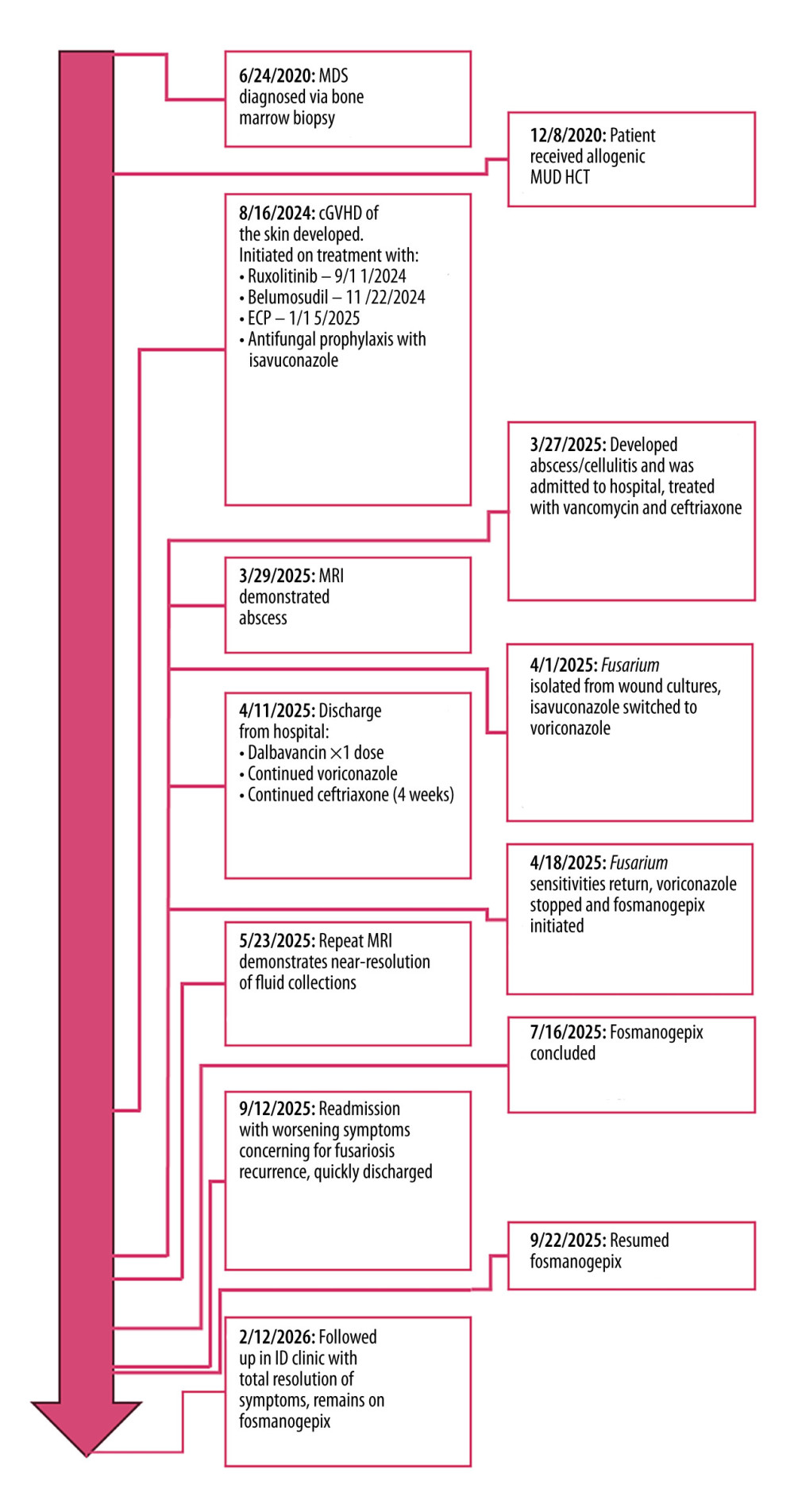 Figure 1. Timeline demonstrating the patient’s clinical course from original MDS diagnosis to final follow-up at an outpatient infectious disease clinic while receiving fosmanogepix therapy. MDS, myelodysplastic syndrome; MUD HCT, matched unrelated donor hematopoietic cell transplant; cGVHD, chronic graft-vs-host disease; ECP, extracorporeal photopheresis; MRI, magnetic resonance imaging.
 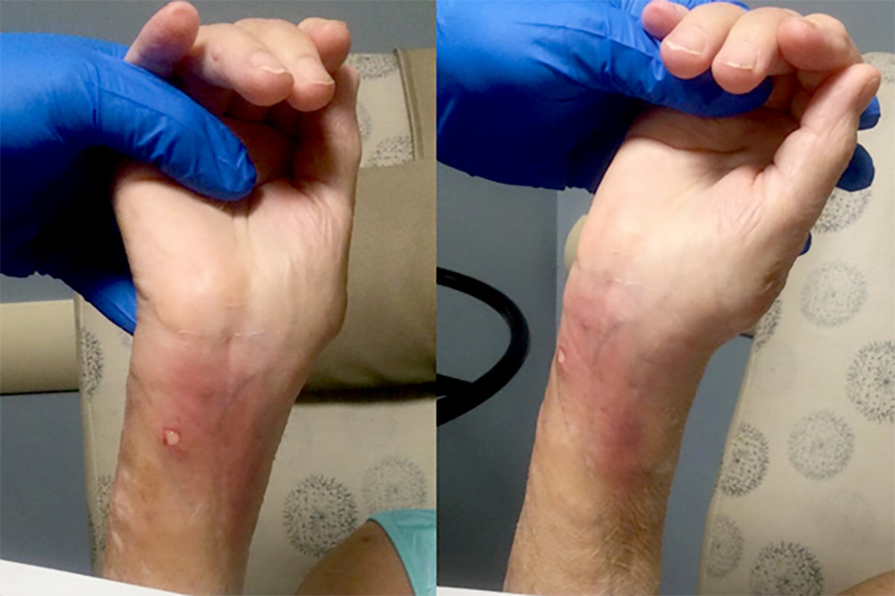 Figure 2. External appearance of affected forearm and wrist. Notable are the open draining wound site on the volar forearm and associated erythema radiating out into the rest of the forearm proximally and the wrist distally.
 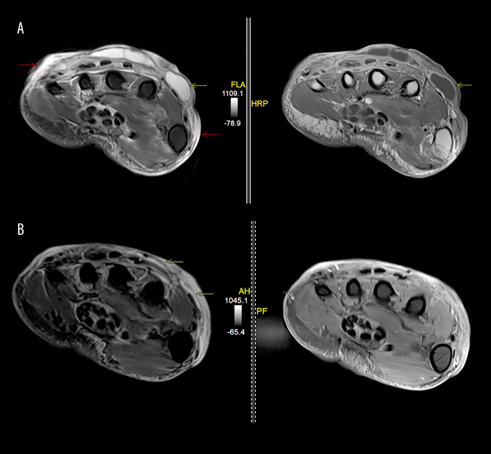 Figure 3. (A) Axial fat-suppressed T2-w and axial T1-w contrast-enhanced magnetic resonance images of the right hand at the level of the proximal metacarpals demonstrate extensive ill-defined subcutaneous edema (red arrows) and several rim-enhancing fluid collections (yellow arrows) involving the dorsal hand, suggesting cellulitis with abscesses. (B) Axial fat-suppressed T2-w and axial T1-w contrast-enhanced magnetic resonance images at the same level as in (A), which were obtained 8 weeks later, demonstrate near complete resolution of fluid collections (yellow arrows) with persistent but decreased subcutaneous edema of the dorsal hand.
 References
1. Gundlach BK, Sasor SE, Chung KC, Hand infections: Epidemiology and public health burden: Hand Clin, 2020; 36(3); 275-83
2. Klein MB, Chang J, Management of hand and upper-extremity infections in heart transplant recipients: Plast Reconstr Surg, 2000; 106(3); 598-601
3. Ungaro R, Mikulska M, The skin and soft tissue infections in hematological patients: Curr Opin Infect Dis, 2020; 33(2); 101-9
4. Burke VE, Lopez FA, Approach to skin and soft tissue infections in non-HIV immunocompromised hosts: Curr Opin Infect Dis, 2017; 30(4); 354-63
5. Nucci M, Anaissie E, Invasive fusariosis: Clin Microbiol Rev, 2023; 36(4); e0015922
6. Nucci M, Anaissie E: Clin Infect Dis, 2002; 35(8); 909-20
7. Gupta AK, Baran R, Summerbell RC: Curr Opin Infect Dis, 2000; 13(2); 121-28
8. Roberds A, Bobrov AG, Rautemaa-Richardson R, Walsh TJ, Invasive fungal diseases of combat wounds: Burden, epidemiology, and mycology: Mycopathologia, 2024; 189(6); 102
9. Donnelly JP, Chen SC, Kauffman CA, Revision and update of the consensus definitions of invasive fungal disease from the European Organization for Research and Treatment of Cancer and the Mycoses Study Group Education and Research Consortium: Clin Infect Dis, 2020; 71(6); 1367-76
10. Al-Hatmi AMS, Bonifaz A, Ranque S, Current antifungal treatment of fusariosis: Int J Antimicrob Agents, 2018; 51(3); 326-32
11. Fernández-Cruz A, Lewis RE, Kontoyiannis DP, How long do we need to treat an invasive mold disease in hematology patients? Factors influencing duration of therapy and future questions: Clin Infect Dis, 2020; 71(3); 685-92
12. Taj-Aldeen SJ: J Fungi (Basel), 2017; 3(2); 18
13. Kim JY, Kang CI, Lee JH, Clinical features and outcomes of invasive fusariosis: A case series in a single center with literature review: Infect Chemother, 2023; 55(2); 290-94
14. Winston DJ, Young PA, Schlamm HT, Schiller GJ: Clin Infect Dis, 2023; 77(6); 848-50
15. Wu G, Ganesh S, Forrester K, Khan S, Multi-drug resistant fusariosis treated with fosmanogepix in a lung transplant patient: J Heart Lung Transplant, 2024; 43(4); S463
16. Shaw KJ, Ibrahim AS, Fosmanogepix: A review of the first-in-class broad spectrum agent for the treatment of invasive fungal infections: J Fungi (Basel), 2020; 6(4); 239
17. Hodges MR, Tawadrous M, Cornely OA: Clin Infect Dis, 2025; 81(5); e302-e9
Figures
Figure 1. Timeline demonstrating the patient’s clinical course from original MDS diagnosis to final follow-up at an outpatient infectious disease clinic while receiving fosmanogepix therapy. MDS, myelodysplastic syndrome; MUD HCT, matched unrelated donor hematopoietic cell transplant; cGVHD, chronic graft-vs-host disease; ECP, extracorporeal photopheresis; MRI, magnetic resonance imaging.Figure 2. External appearance of affected forearm and wrist. Notable are the open draining wound site on the volar forearm and associated erythema radiating out into the rest of the forearm proximally and the wrist distally.Figure 3. (A) Axial fat-suppressed T2-w and axial T1-w contrast-enhanced magnetic resonance images of the right hand at the level of the proximal metacarpals demonstrate extensive ill-defined subcutaneous edema (red arrows) and several rim-enhancing fluid collections (yellow arrows) involving the dorsal hand, suggesting cellulitis with abscesses. (B) Axial fat-suppressed T2-w and axial T1-w contrast-enhanced magnetic resonance images at the same level as in (A), which were obtained 8 weeks later, demonstrate near complete resolution of fluid collections (yellow arrows) with persistent but decreased subcutaneous edema of the dorsal hand. In Press
Case report 
Am J Case Rep In Press; DOI: 10.12659/AJCR.953317
Case report 
Am J Case Rep In Press; DOI: 10.12659/AJCR.954643
Case report 
Am J Case Rep In Press; DOI: 10.12659/AJCR.954074
Case report
Am J Case Rep In Press; DOI: 10.12659/AJCR.953592
Most Viewed Current Articles
07 Dec 2021 : Case report
28,712,429
DOI :10.12659/AJCR.934347
Am J Case Rep 2021; 22:e934347
06 Dec 2021 : Case report  180,530
DOI :10.12659/AJCR.934406
Am J Case Rep 2021; 22:e934406
21 Jun 2024 : Case report
124,276
DOI :10.12659/AJCR.944371
Am J Case Rep 2024; 25:e944371
08 May 2026 : Case report
95,045
DOI :10.12659/AJCR.952158
Am J Case Rep 2026; 27:e952158