


16 September 2026: Articles 
Electrocardiographic and Arrhythmic Manifestations of End-Stage Atrial Myopathy in Patients With Longstanding Persistent or Permanent Atrial Fibrillation: A 3-Patient Case Series
Unusual clinical course
Frans Serpa ABDEF 1*, Nimesh Patel DE 1, Jose A. Joglar ADEG 1
DOI: 10.12659/AJCR.953661
Am J Case Rep 2026; 27:e953661


Abstract
BACKGROUND: Advanced atrial remodeling, often associated with atrial fibrosis, may be accompanied by severe atrial electrical dysfunction and a range of electrocardiographic and arrhythmic manifestations. Data on the clinical features of this process are limited.
CASE REPORT: We report 3 patients with end-stage atrial myopathy and longstanding persistent or permanent atrial fibrillation (AF). One patient with extensive biatrial scar developed recurrent low-amplitude atrial tachycardias after AF ablation. Two patients demonstrated progression from AF to atrial standstill with junctional rhythms. In both patients, serial ECGs showed loss of fibrillatory activity, and device interrogation demonstrated absent atrial sensing and capture. One patient underwent pacemaker implantation without atrial lead placement, due to the absence of atrial sensing or capture on electrophysiology mapping. Important coexisting substrates for atrial remodeling included hypertrophic cardiomyopathy, mitral valve disease, and prior cardiac surgeries.
CONCLUSIONS: Patients with longstanding persistent or permanent AF may demonstrate distinct findings suggestive of advanced atrial myopathy, including loss of fibrillatory waves on ECG, low-amplitude atrial tachycardias, atrial standstill, junctional rhythms, absent atrial sensing or capture on device interrogation, and extensive atrial scarring on electrophysiology mapping. Recognition of these clinical manifestations may provide insight into advanced atrial remodeling and potentially inform AF ablation and atrial pacing strategies. Further studies are needed to better define the mechanisms, prevalence, and clinical significance of these findings.
Keywords: Atrial Fibrillation, Atrial Remodeling, Myopathy, Central Core
Introduction
Atrial myopathy is defined as a complex of structural, architectural, contractile, or electrophysiological changes affecting the atria that can produce clinically relevant manifestations [1,2]. Atrial myopathy predisposes to atrial fibrillation (AF), while AF may further accelerate the development and progression of atrial myopathy [3].
Multiple electrocardiographic findings, together with imaging, histological, and biological markers, have been incorporated into proposed classification systems for atrial myopathy [4]. Some of the electrocardiographic features incorporated into these frameworks include prolonged P wave duration, low P wave voltage, abnormal P wave axis, increased P wave terminal force in V1, and low-amplitude fibrillatory waves [4]. Although these electrocardiogram (ECG) findings aid in staging this condition, the electrocardiographic and clinical manifestations of end-stage atrial myopathy in patients with AF remain poorly characterized and may be underrecognized in routine clinical practice.
In this 3-patient case series (Table 1), we use the term end-stage atrial myopathy to describe atrial electrical and structural remodeling characterized by absent or markedly diminished atrial electrical activity on ECG, absent atrial sensing or capture on device interrogation, and an extensive low-voltage atrial substrate on electroanatomic mapping.
Case Reports
PATIENT 1:
A 45-year-old man with longstanding persistent AF and prior embolic strokes was referred to our electrophysiology (EP) clinic for further evaluation. He had been treated with rate control, anticoagulation, and multiple prior electrical cardioversions at outside institutions. Baseline ECG showed AF with fibrillary waves (Figure 1).
At presentation, the patient reported palpitations and exertional dyspnea. Given persistent symptoms despite medical therapy, AF ablation was pursued after shared decision-making.
During the procedure, electroanatomic mapping demonstrated AF with intermittently organized atrial activity and extensive scar involving both atria. All 4 pulmonary veins showed electrical conduction and were successfully isolated with cryothermal ablation. Despite durable pulmonary vein isolation, the patient remained in AF. Subsequent mapping identified focal and micro-reentrant atrial tachycardias (ATs) involving the mitral isthmus and anterior septum, which were targeted with radiofrequency ablation. Sinus rhythm was restored after cardioversion and amiodarone administration.
After the procedure, the patient developed recurrent ATs. Subsequently, sinus arrest and clinically significant bradyarrhythmia were observed, prompting permanent pacemaker implantation (PPM). However, the contribution of amiodarone to the bradyarrhythmia cannot be excluded. Device interrogation confirmed the presence of ATs, and follow-up ECGs continue to show low-amplitude ATs (Figure 2).
This case illustrates advanced atrial remodeling manifesting as recurrent low-amplitude ATs after ablation in the setting of extensive atrial scar.
PATIENT 2:
A 42-year-old man with hypertrophic cardiomyopathy (HCM), mitral valve disease status after mechanical valve replacement and MAZE procedure, sick sinus syndrome with a dual-chamber PPM, and longstanding persistent AF established care in our EP clinic. Baseline ECG showed AF with fibrillary waves (Figure 3).
Approximately 1 year after initial assessment, he reported progressive dyspnea on exertion and chest pain. Transthoracic echocardiography demonstrated a decline in left ventricular ejection fraction (LVEF) from 51% to 30%–35%.
Device interrogation revealed a high ventricular pacing burden. To minimize the potential contribution of right ventricular pacing to left ventricular dysfunction, the device was reprogrammed to VVI 50. Subsequent follow-up showed symptom improvement, a reduced ventricular pacing burden, and partial recovery of LVEF. Several years later, the patient’s LVEF declined again to 35% or lower; therefore, an upgrade to a cardiac resynchronization therapy defibrillator was pursued. Follow-up interrogation showed 99% biventricular pacing and improved activity tolerance.
Serial ECGs demonstrated progression from longstanding AF to atrial standstill (Figure 4), likely multifactorial, in the setting of longstanding AF, other comorbidities, and cardiac history. The ECG showed periods of atrial pacing with apparent non-capture, and device interrogation revealed poor atrial sensing consistent with atrial electrical failure. AF ablation options were discussed, but the patient elected to continue medical management.
This case illustrates progression from longstanding AF to atrial electrical failure in a patient with multiple structural substrates for severe atrial remodeling.
PATIENT 3:
A 69-year-old woman with apical HCM and longstanding persistent AF established care at our EP clinic. She had been treated with rate control and anticoagulation.
She presented with symptomatic bradycardia, reporting heart rates in the 40s beats per minute. ECG showed a junctional rhythm without apparent atrial activity (Figure 5). An event monitor demonstrated baseline AF with 7 episodes of sustained premature ventricular contractions and a peak heart rate of 110 beats per minute, without critical events. She continued to report symptomatic bradycardia at follow-up visits.
Given persistent symptoms, PPM implantation was pursued. During the procedure, no atrial sensing or capture was detected despite assessment of multiple atrial sites, consistent with atrial standstill. The absence of atrial electrical activity was further supported by the pre-procedural ECG, which demonstrated junctional rhythm without discernible atrial activity. Because no atrial sensing or capture was identified, an atrial lead was not implanted. The patient subsequently underwent PPM implantation, with a right ventricular lead positioned in the left bundle branch area and programmed to VVIR mode.
This case illustrates atrial standstill recognized during PPM implantation, with absent atrial sensing and capture precluding atrial lead placement.
Discussion
In this 3-patient case series, we describe electrocardiographic and arrhythmic manifestations of advanced atrial remodeling in patients with longstanding persistent or permanent AF. In these patients, end-stage atrial myopathy was characterized by atrial electrical dysfunction manifested by absent or markedly diminished atrial electrical activity on ECG, absent atrial sensing or capture on device interrogation, and extensive atrial scar on electroanatomic mapping. These patients developed distinct arrhythmic manifestations, including recurrent ATs, atrial standstill, and junctional rhythms (Figure 6). Recognition of these presentations may help identify patients with severe atrial remodeling in routine clinical practice.
Fibrosis represents one of the central pathophysiological substrates of atrial myopathy [5]. Prior studies have demonstrated associations between atrial fibrosis and AF burden, as well as progressive changes in atrial structure and function across the spectrum of AF [5,6]. At the same time, atrial remodeling may result from multiple processes, including cardiomyopathies, valvular heart disease, prior cardiac surgery, and other structural abnormalities. In our series, advanced atrial remodeling likely arose from several factors, including AF and other coexisting substrates. Nonetheless, our goal was to highlight possible signs of end-stage atrial myopathy in patients with AF to help identify them during standard clinical evaluations.
Advanced atrial remodeling can be identified using imaging, device interrogation, and electroanatomic mapping. In our series, objective evidence of severe atrial disease included absent or markedly diminished atrial electrical activity on ECG, absent atrial sensing or capture on device interrogation, atrial standstill, and extensive atrial scar on electroanatomic mapping. These findings may represent distinct manifestations of advanced atrial electrical dysfunction, potentially aiding physicians in early detection. However, the clinical implications of these observations warrant further investigation.
Atrial myopathy has been increasingly recognized as an important risk factor for stroke [7], whereas atrial fibrosis has been associated with poor outcomes and recurrence following AF ablation [8]. Recognition of the arrhythmic manifestations presented here may provide additional insight into the severity of atrial disease and could inform management. In patient 1, extensive atrial scar was associated with recurrent low-amplitude ATs despite ablation. In patients 2 and 3, atrial standstill and atrial electrical failure were associated with impaired atrial sensing and capture, findings that influenced device management. Recognition of these features may help identify patients with advanced atrial disease in whom extensive atrial scar or absent atrial electrical activity could affect procedural considerations, including AF ablation and atrial lead implantation, respectively. Moreover, identification of atrial standstill has important implications for pacing strategy, as implantation of an atrial lead is often unsuccessful due to the absence of atrial electrical activity [9]. Given the small sample size and heterogeneous nature of this series, the prognostic and therapeutic implications of these associations remain uncertain and require validation in larger studies.
Collectively, this case series illustrates the clinical manifestations associated with advanced atrial remodeling. These observations suggest that loss of fibrillatory activity on ECG, atrial standstill, and progression from AF to low-amplitude ATs and junctional rhythms may serve as markers of atrial remodeling and severe electrical dysfunction in selected patients. Further studies are needed to better define the mechanisms, prevalence, and clinical significance of these findings.
This case series is limited by its small sample size, heterogeneous patient population, and lack of comprehensive atrial substrate characterization across all patients. Accordingly, these findings should be considered hypothesis-generating, educational, and descriptive and should be interpreted in the context of the clinical observations presented.
Conclusions
In this case series, patients with longstanding persistent or permanent AF demonstrated findings suggestive of advanced atrial myopathy, including loss of fibrillatory activity on ECG, low-amplitude ATs, atrial standstill, and junctional rhythms detected on device interrogation and EP mapping. These arrhythmic manifestations may reflect underlying advanced atrial remodeling; however, further studies are needed to validate these observations.
Figures
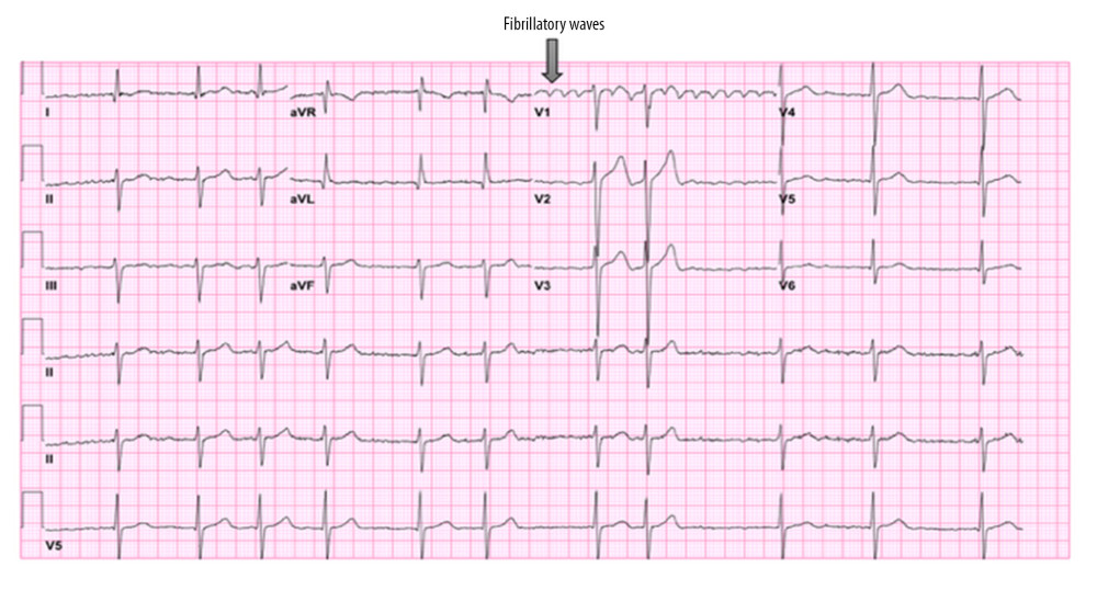 Figure 1. Baseline ECG from patient 1 demonstrating atrial fibrillation with fibrillatory waves in lead V1. The arrow indicates fibrillatory waves.
 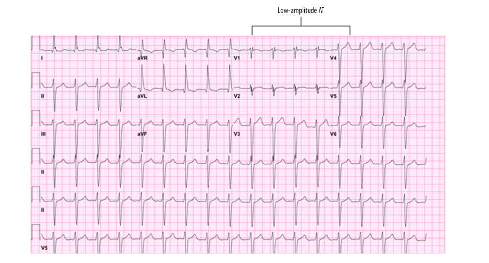 Figure 2. Follow-up ECG from patient 1 demonstrating atrial tachycardia (AT) following prior atrial fibrillation. The arrow indicates low-amplitude AT.
 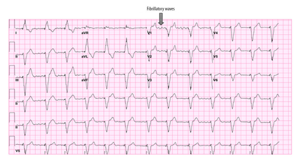 Figure 3. Baseline ECG from patient 2 demonstrating atrial fibrillation with fibrillatory waves in lead V1. The arrow indicates fibrillatory waves.
 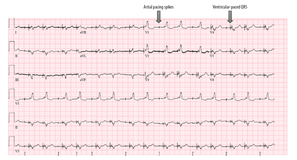 Figure 4. Follow-up ECG from patient 2 with ventricular-paced rhythm and absence of visible atrial activity. Atrial pacing spikes are present without clear evidence of atrial capture. Device interrogation demonstrated absent atrial sensing or capture.
 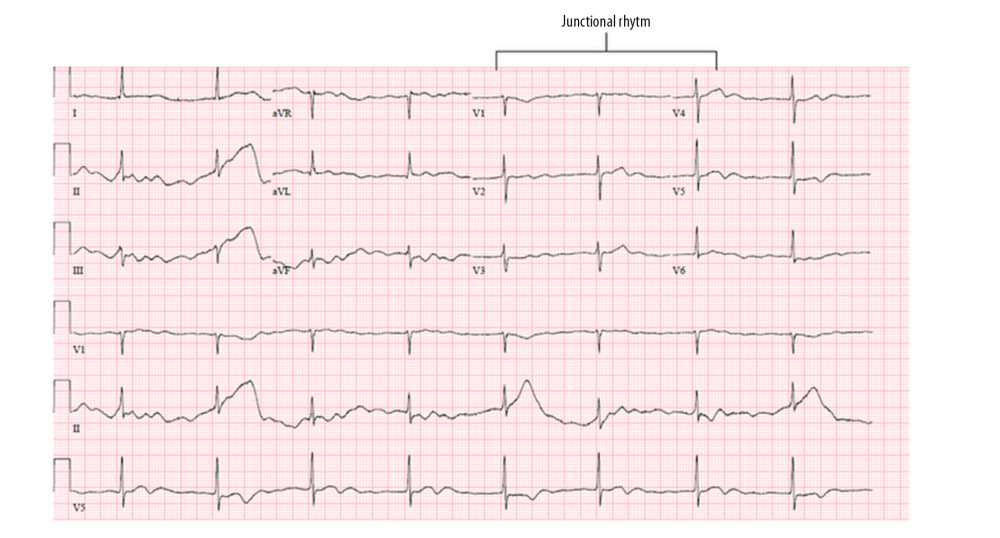 Figure 5. ECG from patient 3 demonstrating junctional rhythm without discernible atrial activity. At the time of permanent pacemaker implantation, no atrial sensing or capture could be demonstrated despite assessment of multiple atrial sites.
 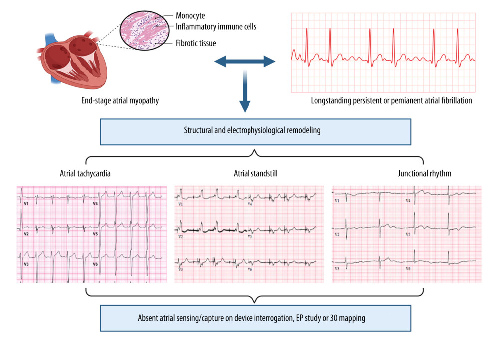 Figure 6. Spectrum of electrocardiographic and arrhythmic manifestations of end-stage atrial myopathy in patients with longstanding or permanent atrial fibrillation.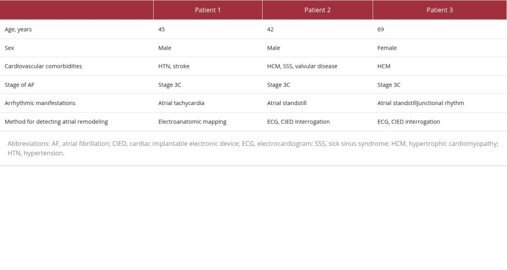
References
1. Goette A, Kalman JM, Aguinaga L, EHRA/HRS/APHRS/SOLAECE expert consensus on atrial cardiomyopathies: Definition, characterization, and clinical implication: EP Europace, 2016; 18(10); 1455-90
2. Shen MJ, Arora R, Jalife J, Atrial myopathy: JACC: Basic to Translational Science, 2019; 4(5); 640-54
3. Goldberger JJ, Arora R, Green D, Evaluating the atrial myopathy underlying atrial fibrillation: Circulation, 2015; 132(4); 278-91
4. Goette A, Kalman JM, Aguinaga L, EHRA/HRS/APHRS/SOLAECE expert consensus on atrial cardiomyopathies: Definition, characterization, and clinical implication: EP Europace, 2016; 18(10); 1455-90
5. Burstein B, Nattel S, Atrial fibrosis: Mechanisms and clinical relevance in atrial fibrillation: J Am Coll Cardiol, 2008; 51(8); 802-9
6. Hirsh BJ, Copeland-Halperin RS, Halperin JL, Fibrotic atrial cardiomyopathy, atrial fibrillation, and thromboembolism: Mechanistic links and clinical inferences: J Am Coll Cardiol, 2015; 65(20); 2239-51
7. Pastore MC, Campora A, Cameli M, Atrial fibrillation and myopathy predisposing to stroke and dementia: JACC: Advances, 2023; 2(5); 100427
8. Marrouche NF, Wilber D, Hindricks G, Association of atrial tissue fibrosis identified by delayed enhancement MRI and atrial fibrillation catheter ablation: the DECAAF study: JAMA, 2014; 311(5); 498-506
9. Bellmann B, Roser M, Muntean B, Atrial standstill in sinus node disease due to extensive atrial fibrosis: impact on dual chamber pacemaker implantation: Europace, 2016; 18(2); 238-45
Figures
Figure 1. Baseline ECG from patient 1 demonstrating atrial fibrillation with fibrillatory waves in lead V1. The arrow indicates fibrillatory waves.Figure 2. Follow-up ECG from patient 1 demonstrating atrial tachycardia (AT) following prior atrial fibrillation. The arrow indicates low-amplitude AT.Figure 3. Baseline ECG from patient 2 demonstrating atrial fibrillation with fibrillatory waves in lead V1. The arrow indicates fibrillatory waves.Figure 4. Follow-up ECG from patient 2 with ventricular-paced rhythm and absence of visible atrial activity. Atrial pacing spikes are present without clear evidence of atrial capture. Device interrogation demonstrated absent atrial sensing or capture.Figure 5. ECG from patient 3 demonstrating junctional rhythm without discernible atrial activity. At the time of permanent pacemaker implantation, no atrial sensing or capture could be demonstrated despite assessment of multiple atrial sites.Figure 6. Spectrum of electrocardiographic and arrhythmic manifestations of end-stage atrial myopathy in patients with longstanding or permanent atrial fibrillation. In Press
Case report 
Am J Case Rep In Press; DOI: 10.12659/AJCR.953317
Case report 
Am J Case Rep In Press; DOI: 10.12659/AJCR.954643
Case report 
Am J Case Rep In Press; DOI: 10.12659/AJCR.954074
Case report
Am J Case Rep In Press; DOI: 10.12659/AJCR.953592
Most Viewed Current Articles
07 Dec 2021 : Case report
28,712,429
DOI :10.12659/AJCR.934347
Am J Case Rep 2021; 22:e934347
06 Dec 2021 : Case report  180,530
DOI :10.12659/AJCR.934406
Am J Case Rep 2021; 22:e934406
21 Jun 2024 : Case report
124,276
DOI :10.12659/AJCR.944371
Am J Case Rep 2024; 25:e944371
08 May 2026 : Case report
95,045
DOI :10.12659/AJCR.952158
Am J Case Rep 2026; 27:e952158