


31 August 2026: Articles 
Septic Spontaneous Abortion Associated With Intrauterine Fetal Death at 8 Weeks of Gestation Complicated by Myometritis and Bilateral Psoas Muscle Abscess: A Case Report
Unknown etiology, Unusual clinical course, Challenging differential diagnosis, Management of emergency care
Jose L. Rojas-Oviedo ADE 1*, Nancy P. Ortega López BEF 1, Dauris Lineth Mejía Pérez AEF 1,2, Melissa Buitrago-Gomez BD 1, Juan C. Vallejo-Soto BE 3, Leidy D. Rojas Hernández BE 4
DOI: 10.12659/AJCR.953673
Am J Case Rep 2026; 27:e953673


Abstract
BACKGROUND: Septic abortion is a serious, life-threatening uterine infection that can occur before, during, or after a miscarriage or termination, and when it is associated with an intrauterine fetal death (IUFD), the retained fetal tissue can be a focus for bacterial infection. This report describes the case of a 34-year-old woman with septic abortion associated with IUFD at 8 weeks of gestation, complicated by myometritis and bilateral psoas muscle abscess.
CASE REPORT: A 34-year-old female patient presented with severe lower back pain radiating to the right lower extremity. Diagnostic workup initially ruled out neurological lesions but identified an impending septic spontaneous abortion due to IUFD at 8.7 weeks. Following cervical ripening with misoprostol and subsequent uterine curettage, she developed persistent fever and methicillin-resistant Staphylococcus aureus bacteremia. Abdominal and pelvic computed tomography (CT) documented fluid collections involving both iliopsoas muscles bilaterally at their distal insertion, alongside smaller collections in the right external obturator and thigh musculature. No surgical drainage was required due to the limited size of the abscesses. Targeted treatment consisting of a 28-day course of intravenous vancomycin resulted in full clinical and radiographic resolution.
CONCLUSIONS: Iliopsoas abscess is a complex clinical entity whose diagnosis can be challenging, even more in a pregnancy context. Very few cases of psoas abscess during pregnancy have been reported in the literature, and even fewer have been bilateral. Timely diagnosis and broad-spectrum antibiotic coverage are the cornerstones of treatment.
Keywords: Abortion, Septic, Psoas Abscess, Bacteremia, Pregnancy Complications
Introduction
Iliopsoas abscess is a very rare clinical condition with a reported incidence of approximately 0.4 in 100 000 [1]. It can be classified as primary, which is due to hematogenous or lymphatic spread, or secondary, which arises from intraabdominal inflammatory processes, particularly gastrointestinal, and is the most common cause [1]. Fewer than a dozen cases of psoas abscesses have been described during pregnancy, with the main causes being spinal tuberculosis, drug abuse, or pyelonephritis [2]. As a rare clinical entity, they should therefore be considered when a pregnant patient reports having persistent back or abdominal pain, weakness in the lower extremities, or fever of unknown origin [2].
Septic abortion is a severe, life-threatening variant of upper genital tract infection—including endometritis, myometritis, and parametritis—that occurs in the setting of a spontaneous miscarriage or a procedural pregnancy termination [3]. According to recent epidemiological reviews, septic abortion results primarily from the ascending proliferation of polymicrobial endogenous vaginal flora, including
The occurrence of an iliopsoas abscess during pregnancy is extremely rare, with fewer than a dozen cases documented in the medical literature [4]. Within this small pool of published data, the etiology is predominantly primary hematogenous spread or secondary extension from spinal tuberculosis or severe pyelonephritis [1]. Bilateral involvement of the iliopsoas muscles is an even greater clinical novelty, accounting for less than 3% of all documented secondary iliopsoas abscess cases [1]. Prior cases of bilateral iliopsoas abscess in obstetric patients have been almost exclusively tied to musculoskeletal or renal tuberculosis [2]. Because physiological lower back pain, pelvic girdle pain, and transient lower limb weakness are highly prevalent in normal pregnancies due to ligamentous changes, early identification of an underlying deep tissue abscess is highly challenging, frequently leading to delayed diagnoses and progression to life-threatening septic shock [2].
While previous literature has touched upon non-obstetric origins of psoas infections during pregnancy, a direct secondary dissemination pathway stemming from a primary uterine myometritis following a spontaneous miscarriage has not been thoroughly delineated [3].
This report describes the case of a 34-year-old woman with septic abortion associated with IUFD at 8 weeks of gestation, complicated by myometritis and bilateral psoas muscle abscess.
Case Report
A 34-year-old female patient, gravida 4, para 2, abortus 1, living 2 (G4P2C0A1V2), with an 8.7-week gestation documented through vaginal ultrasound, was admitted to the emergency department of our hospital with a 2-day history of lower back pain radiating to the lower right extremity, which was exacerbated by movement and limited mobility and gait. The first pregnancy, which was 17 years ago, was carried to term at 40 weeks, and there were no complications. However, 12 years ago, the patient had a spontaneous abortion with no apparent cause discovered. The third pregnancy, 9 years ago, was also carried to term without any complications. Initially, the patient did not consent to a lumbosacral X-ray because of radiation risk; therefore, analgesic management with non-steroidal anti-inflammatory drugs (NSAIDs) and opioids was initiated, but without improvement. The admission laboratory test results showed a complete blood count without leukocytosis or neutrophilia, a urinalysis within normal limits, and a urine Gram stain with no organisms.
She was initially evaluated by neurologists, who on physical examination found 4/5 strength in the lower right extremity, with positive Lasègue and Bragard’s signs (possibly due to irritation of the lumbar nerve roots since no neurological lesion was documented), and therefore requested lumbar spine magnetic resonance imaging (MRI), which showed only enlarged lumbar transverse processes in L5 without any other abnormal findings. Considering these findings, she was evaluated by our neurosurgery department. Since there were no signs of radiculopathy, there was no indication for prompt surgical management, and it was decided that any surgical procedure could be performed after hospitalization. Pain control continued to be a difficult issue; therefore, neuromodulator and topic analgesic management was initiated with pregabalin and diclofenac. An Doppler ultrasound of the lower right extremity showed no signs of deep vein thrombosis.
During hospitalization, the patient experienced a sudden episode of acute-onset dyspnea and desaturation; therefore, a chest X-ray was performed, showing blunting of the costophrenic angles and bilateral perihilar infiltrates. Given the suspicion of a pulmonary embolism, a D-dimer test was performed, yielding a positive result of 2.29 μg/mL, and full anticoagulation with low-molecular-weight heparin was initiated. Additionally, a transthoracic echocardiogram demonstrated a preserved left ventricular ejection fraction of 65%, and no segmentary alterations of contractility. Fetal wellness was confirmed with a transvaginal ultrasound, showing normal amniotic fluid.
Subsequently, the patient continued to experience a sluggish clinical course, with persistent lower back pain, 2 fever spikes, and leukocytosis reaching 26 330 cells/μL, with a differential count of 75% neutrophils, 14% lymphocytes, 5% monocytes, 4% eosinophils, and 2% basophils. Three days after the episode of dyspnea, the patient presented with rupture of membranes manifested as abundant amniorrhea; therefore, a cervicometry and obstetric ultrasound were performed, showing almost completely absent amniotic fluid (oligohydramnios), and embryo tachycardia, with a cervical length of 33 mm and an open internal cervical orifice (Figure 1).
Correlating the severe oligohydramnios, fetal distress, and systemic inflammatory response syndrome in the absence of an alternative infectious focus, an impending septic spontaneous abortion due to IUFD was diagnosed. The patient was immediately transferred to the labor and delivery unit. Empiric broad-spectrum antibiotic coverage with IV piperacillin/tazobactam (4.5 g every 6 hours) was initiated, and cervical ripening was induced with vaginal misoprostol to facilitate regular uterine contractions
Approximately 7 hours later, the patient expelled the fetus and placenta. She was then taken for obstetric curettage, during which fetid abundant ovular remnants were found in the cervical canal. The patient was transferred to the intensive care unit for continued monitoring. Over the next 48 hours, the patient continued to experience pain in her right lower extremity as well as asymmetry on physical examination, with grade II edema and a persistent systemic inflammatory response despite antibiotic management. An abdominal ultrasound was performed, revealing inflammatory changes in the myometrial wall, and a new venous Doppler ultrasound of the right lower extremity revealed an organized collection at the root of the right thigh measuring 25 × 16 × 23 mm, for a volume of 5 cm3.
Given the persistent fever and leukocytosis in 18 470 cells/μL, with a differential count of 81% neutrophils, 12% lymphocytes, 4% monocytes, 2% eosinophils, and 1% basophils, blood cultures were drawn and yielded gram-positive cocci at 20 hours, with identification of methicillin-resistant
With these imaging findings concomitant with bacteremia, a consultation was requested by the infectious disease service, who considered discontinuing clindamycin and initiated vancomycin at a dose of 1 g every 12 hours, with a full treatment duration of a minimum of 4 weeks. The patient was also evaluated by general surgery, who determined that there was no indication for surgical or percutaneous drainage of the iliopsoas abscess, given the size of the collections. A follow-up soft tissue ultrasound was requested, which revealed a predominantly hypoechoic collection in the upper third of the anterolateral thigh, with a volume of 5 cm3. A contrast-enhanced CT scan of the abdomen and pelvis showed the collections in the root of the right thigh and in the bilateral iliopsoas muscles previously documented, with a decrease in size compared with prior studies (Figure 3).
Follow-up clearance blood cultures were drawn at 96 hours, with persistent bacteremia; therefore, antibiotic therapy was continued, and new follow-up blood cultures were drawn 7 days later, which came back negative after 72 hours of incubation. Because of the presence of bacteremia, a transesophageal echocardiogram was performed to rule out endocarditis. It was negative for thrombi, vegetations, or shunts. Further studies were conducted to rule out a gastrointestinal or genitourinary origin of the psoas abscesses, and it was determined that the most plausible etiology was the myometritis secondary to the septic abortion. The patient completed a total of 28 days of vancomycin therapy, which was effective. Given the favorable clinical course, the patient was discharged without complications.
Discussion
The primary clinical lesson to be drawn from this case is that an obstetric pelvic infection, such as post-miscarriage myometritis, can serve as a direct contiguous source for the development of secondary bilateral iliopsoas and lower limb muscle abscesses. This condition is rare but can be potentially life-threatening due to complications associated with the infection [4]. As mentioned before, the etiology of this abscess can be classified as either primary or secondary, depending on its way of spread [1]. Primary iliopsoas abscess arises from hematogenous or lymphatic spread of infection, without an identifiable adjacent source, often occurring in immunocompromised individuals or those with chronic illnesses [4]. Secondary iliopsoas abscess results from direct extension of infection from adjacent structures, such as the gastrointestinal or genitourinary tract, or following trauma or surgical procedures in the retroperitoneal space [4]. The most common causative organisms that have been described are
Risk factors for developing iliopsoas abscess include diabetes, immunosuppression, IV drug use, and underlying intraabdominal or spinal pathology [6]. Clinical presentation is often nonspecific, with symptoms such as fever, back or flank pain, abdominal pain, and sometimes pain radiating to the hip or thigh [5]. The classic triad of fever, flank pain, and limitation of hip movement is present in only a minority of cases, making diagnosis challenging and often delayed, which can lead to septic shock [6]. The diagnosis relies on a high index of clinical suspicion and is confirmed with cross-sectional imaging, typically CT or MRI, which can delineate the extent of the abscess and guide management [6]. Management generally involves prompt initiation of broad-spectrum IV antibiotics, with percutaneous or surgical drainage indicated for larger size or refractory abscesses, or in cases complicated by sepsis or failure of medical therapy [6].
On the other hand, septic abortion is defined as any abortion (spontaneous or induced) complicated by upper genital tract infection, including endometritis or myometritis, and remains a significant cause of maternal morbidity and mortality globally [7]. Regarding etiology, septic abortion results from bacterial infection of retained products of conception within the uterus, with the most common being pathogens from endogenous vaginal flora, including aerobic and anaerobic bacteria such as
In our review of the sparse medical literature concerning iliopsoas abscess during pregnancy, we found almost all documented cases report a primary hematogenous origin or a secondary extension from severe pyelonephritis or spinal/renal tuberculosis [2]. Furthermore, bilateral psoas involvement is an extraordinary anomaly, representing less than 3% of all secondary psoas infections [2]. In the few prior obstetric cases in which bilateral iliopsoas abscesses were documented, such as those described by Veerappan et al and Nigam et al, the underlying etiologies were explicitly linked to acute destructive pyelonephritis or spinal tuberculosis [9,10]. Our patient’s case stands out significantly from these historical cohorts, as an exhaustive diagnostic and imaging workup successfully ruled out any spinal, renal, or gastrointestinal pathology, confirming a unique obstetric source—MRSA myometritis secondary to a spontaneous septic abortion.
Obstetric complications are less well documented, but the medical literature indicates that, with appropriate management, there is no increased risk of adverse fetal outcomes or higher cesarean delivery rates directly attributable to iliopsoas abscess [2,11]. However, untreated or severe infection may theoretically increase the risk of preterm labor or fetal compromise due to maternal systemic illness [2].
In the context of our case, the obstetric source appears to be the most plausible explanation of the etiology of the bilateral abscess, considering the timing of the symptoms, image findings, bacteremia, and exclusion of other causes with a thorough workup. However, there is no explicit way of acknowledging the dissemination pathway. This possible etiologic obstetric association observed in this case is very rare, and further studies need to be made to establish a clear relationship between these clinical entities. Clinicians must be aware that, in a similar clinical context, obstetric etiology must be considered.
Conclusions
As this case highlights, diagnosing and treating psoas abscesses during pregnancy can be challenging. Clinical suspicion should be heightened in cases of pregnant patients with nonspecific symptoms such as fever, back or flank pain, abdominal pain, and, sometimes, pain radiating to the hip or thigh, in which other causes have been ruled out. Iliopsoas abscess should emerge as a diagnostic possibility in patients that persist with clinical symptoms despite initial adequate management. Management requires interdisciplinary collaboration between prenatal physicians, intensive care units, and radiologists, since cross-sectional imaging is fundamental for definitive diagnosis.
Although some published articles have described the occurrence of iliopsoas abscess during pregnancy, evidence is very limited. In particular, the possible association between an obstetric infection and these abscesses has not been documented, representing a challenge for both the diagnosis and management of this pathology due to its low clinical suspicion.
Figures
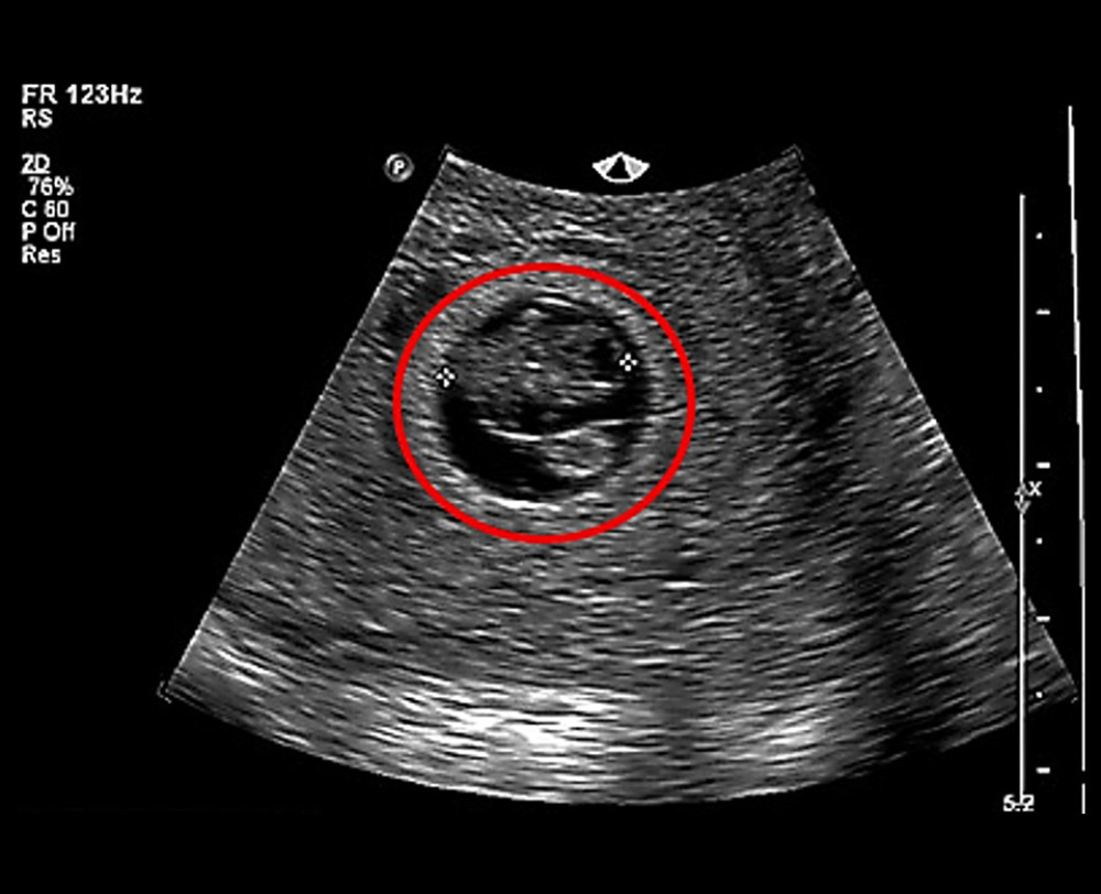 Figure 1. First-trimester diagnostic transvaginal ultrasound confirming structural parameters. The ultrasound demonstrates a restricted gestational sac accompanied by a severe presentation of oligohydramnios (highly deficient amniotic fluid volume).
 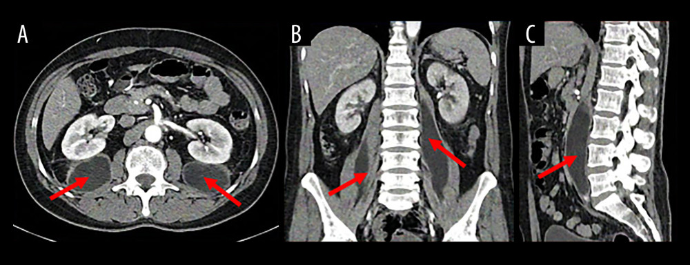 Figure 2. Baseline contrast-enhanced computed tomography angiogram of the abdomen and pelvis. Structural views displayed in the (A) axial, (B) coronal, and (C) sagittal imaging planes reveal well-defined hypodense fluid collections tracking within both the right and left iliopsoas muscles near their distal insertions (indicated by the solid red arrows). Concomitant inflammatory collections are visible within the right external obturator and surrounding regional thigh musculature.
 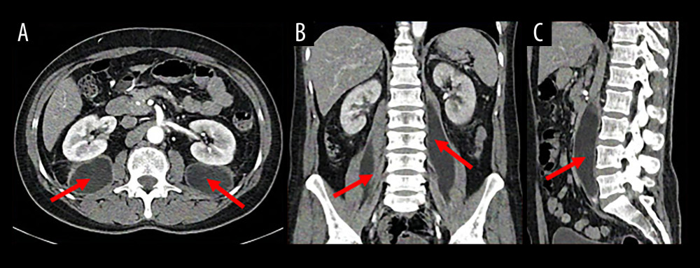 Figure 3. Follow-up contrast-enhanced computed tomography scan of the abdomen and pelvis. Follow-up imaging displayed in the (A) axial, (B) coronal, and (C) sagittal reconstruction planes demonstrate a significant reduction in the overall size, margins, and surrounding inflammatory stranding of the bilateral iliopsoas fluid collections (indicated within the solid red circles) following targeted intravenous antibiotic management.
 References
1. Shields D, Robinson P, Crowley TP, Iliopsoas abscess – A review and update on the literature: Int J Surg, 2012; 10(9); 466-69
2. Schröder C, Calite E, Böckenhoff P, Psoas abscess in pregnancy: A review of the literature and suggestion of minimally invasive treatment options: Arch Gynecol Obstet, 2024; 309(3); 987-92
3. Wall LL, Yemane A, Infectious complications of abortion: Open Forum Infect Dis, 2022; 9(11); ofac553
4. He W, Yuan Y, Huang J, A case report of iliopsoas abscess and literature review: Medicine (Baltimore), 2024; 103(33); e39356
5. Kumar B, Agarwal D, Meena DS: Am J Trop Med Hyg, 2024; 111(2); 297-99
6. Charalampopoulos A, Macheras A, Charalabopoulos A, Iliopsoas abscesses: Diagnostic, aetiologic and therapeutic approach in five patients with a literature review: Scand J Gastroenterol, 2009; 44(5); 594-99
7. Eschenbach DA, Treating spontaneous and induced septic abortions: Obstet Gynecol, 2015; 125(5); 1042-48
8. Rahangdale L, Infectious complications of pregnancy termination: Clin Obstet Gynecol, 2009; 52(2); 198-204
9. Veerappan I, Shanmugam A, Kumar S, Velayutham P, Bilateral psoas and bilateral perinephric abscesses complicating acute pyelonephritis in pregnancy: Indian J Nephrol, 2013; 23(1); 59-62
10. Nigam A, Prakash A, Pathak P, Abbey P, Bilateral psoas abscess during pregnancy presenting as an acute abdomen: Atypical presentation: BMJ Case Rep, 2013; 2013; bcr2013200860
11. Gezer A, Erkan S, Erzik C, Primary psoas muscle abscess diagnosed and treated during pregnancy: Case report and literature review: Infect Dis Obstet Gynecol, 2004; 12(3–4); 125-28
Figures
Figure 1. First-trimester diagnostic transvaginal ultrasound confirming structural parameters. The ultrasound demonstrates a restricted gestational sac accompanied by a severe presentation of oligohydramnios (highly deficient amniotic fluid volume).Figure 2. Baseline contrast-enhanced computed tomography angiogram of the abdomen and pelvis. Structural views displayed in the (A) axial, (B) coronal, and (C) sagittal imaging planes reveal well-defined hypodense fluid collections tracking within both the right and left iliopsoas muscles near their distal insertions (indicated by the solid red arrows). Concomitant inflammatory collections are visible within the right external obturator and surrounding regional thigh musculature.Figure 3. Follow-up contrast-enhanced computed tomography scan of the abdomen and pelvis. Follow-up imaging displayed in the (A) axial, (B) coronal, and (C) sagittal reconstruction planes demonstrate a significant reduction in the overall size, margins, and surrounding inflammatory stranding of the bilateral iliopsoas fluid collections (indicated within the solid red circles) following targeted intravenous antibiotic management. In Press
Case report 
Am J Case Rep In Press; DOI: 10.12659/AJCR.953543
Case report 
Am J Case Rep In Press; DOI: 10.12659/AJCR.954216
Case report 
Am J Case Rep In Press; DOI: 10.12659/AJCR.954932
Case report 
Am J Case Rep In Press; DOI: 10.12659/AJCR.954146
Most Viewed Current Articles
07 Dec 2021 : Case report
28,712,695
DOI :10.12659/AJCR.934347
Am J Case Rep 2021; 22:e934347
06 Dec 2021 : Case report  180,721
DOI :10.12659/AJCR.934406
Am J Case Rep 2021; 22:e934406
21 Jun 2024 : Case report
124,419
DOI :10.12659/AJCR.944371
Am J Case Rep 2024; 25:e944371
08 May 2026 : Case report
95,462
DOI :10.12659/AJCR.952158
Am J Case Rep 2026; 27:e952158