07 June 2021: Articles 
The Multifactorial Pathogenesis of Calciphylaxis: A Case Report
Challenging differential diagnosis, Rare disease
Marta Turek1ABCDEFG*, Joanna Stępniewska1ADE, Jacek Różański1DDOI: 10.12659/AJCR.930026
Am J Case Rep 2021; 22:e930026


Abstract
BACKGROUND: Calciphylaxis is a rare and lifethreatening syndrome characterized by small vascular calcifications, which lead to the occlusion of blood vessels and painful skin lesions with tissue necrosis. Although the disease can develop in a population without kidney failure, it is typically detected in patients receiving dialysis, with an increasing frequency ranging from 1% to 4%. Therefore, the disease is also known as calcific uremic arteriolopathy. The prognosis in patients with coexisting chronic kidney disease is very poor, with a 1-year mortality rate of up to 80%. Numerous risk factors for calciphylaxis have been described, such as obesity, diabetes mellitus, female sex, White race, overuse of calcium and vitamin D supplements, and vitamin K deficiency. The disease is often accompanied by disorders such as hyperphosphatemia, elevated parathyroid hormone level, and a deficiency of natural calcification inhibitors, such as fetuin-A and matrix Gla protein. However, not all patients with calciphylaxis have the abnormalities described above, suggesting that the pathogenesis of calciphylaxis is multifactorial and unfortunately still uncertain.
CASE REPORT: We report a case of calciphylaxis in a 52-year-old White woman with multiple comorbidities and on chronic hemodialysis treatment, who presented with severe subcutaneous painful nodules and necrotic ulcers on both legs.
CONCLUSIONS: Although the prognosis of this rare and underrecognized disease is poor, an early diagnosis and interdisciplinary treatment including pain relief, wound care, appropriate nutritional support, correction of mineral parameters, administration of sodium thiosulphate, and adequate hemodialysis therapy can improve patient quality of life.
Keywords: calciphylaxis, Renal Dialysis, renal insufficiency, Kidney Failure, Chronic, Quality of Life, vascular calcification
Background
Calciphylaxis is a rare and life-threatening syndrome characterized by small vascular calcifications, which lead to blood vessel occlusion and tissue necrosis [1]. The dominant clinical manifestation of this disorder involves painful skin lesions such as induration, nodules, purpura, and ulcers, but it can also involve other organs including the muscles, eye, or digestive tract and lead to dysfunction. The multiple bilateral nonhealing wounds appear on the extremities and are often secondary infections [2]. The wounds can cause a systemic inflammatory response, which can lead to sepsis and death in many patients [3]. In most cases, calciphylaxis affects patients who have advanced kidney failure and are being treated with renal replacement therapy, including hemodialysis and peritoneal dialysis, or have had kidney transplantation. However, the disease also occurs in individuals with adequate kidney function [4,5]. The other most common causes of calciphylaxis are cancer, chemotherapy, alcoholic liver disease, diabetes, POEMS syndrome, primary hyperparathyroidism, and connective tissue diseases [6]. The prognosis in patients with coexisting chronic kidney disease is very poor with a 1-year mortality rate of up to 80%, compared with that of patients without chronic kidney disease, which is up to 45%. Perhaps due to the complex and largely unknown pathogenesis of calciphylaxis, there have been no randomized controlled trials that have led to an approved therapy [7]. Therefore, an interdisciplinary approach is essential for the effective treatment of calciphylaxis [2].
Case Report
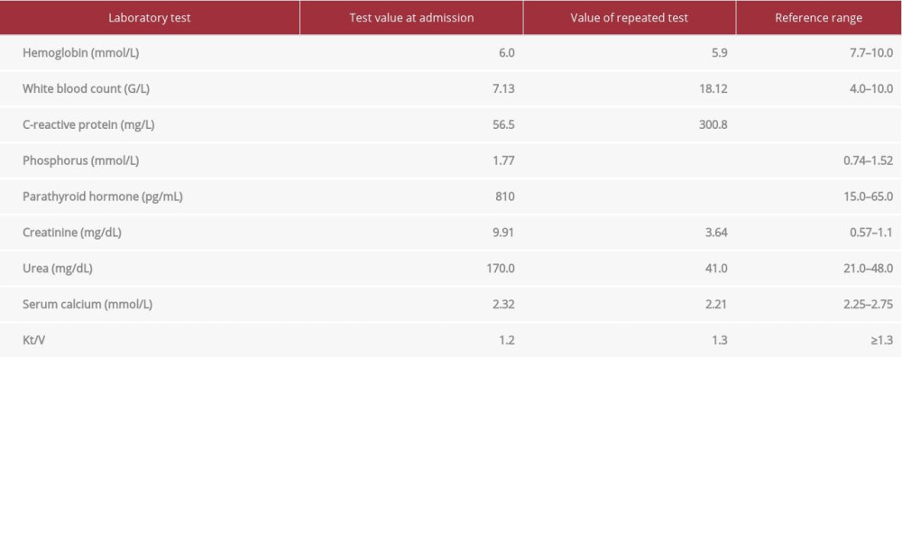
A 52-year-old White woman was admitted to the hospital for a calcium-phosphate imbalance and modification of therapy. The patient had a history of chronic kidney disease, for which she had been receiving hemodialysis treatment for 6 years, diabetes mellitus type 2, chronic atrial fibrillation treated with warfarin therapy, systemic lupus erythematosus, myasthenia gravis, obesity (body mass index, 32.5 kg/m2), and left knee arthroplasty. The patient reported having pain of the left lower extremity for 2 weeks prior to presentation. During the physical examination, palpable subcutaneous painful nodules were found in both legs (Figure 1), and the skin of the left leg had local signs of inflammation (redness, increased warmness). The left extremity was also pale and swollen. A peripheral pulse in this leg was not present, with a pulse palpable only on the femoral artery. The laboratory tests revealed normocytic, normochromic anemia, elevated C-reactive protein levels, parathormone levels, and plasma phosphorus concentration (Table 1). The Doppler ultrasound examination of the left leg showed numerous mid-tissue calcifications, features of superficial venous thrombosis, and critical stenosis of the left superficial femoral and popliteal artery due to atherosclerosis, which was confirmed by computer tomography angiography (Figure 2). The patient had a consultation with the vascular surgeon and was qualified for percutaneous transluminal angioplasty of the left popliteal artery. The day before the surgery, she presented with coffee-ground vomitus with deterioration of red cell parameters in the blood morphology. A gastroscopy was performed, but the localization of a bleeding source was not found. After the episode of gastrointestinal bleeding, the worsening of pain and gradual deterioration of the local left leg skin lesions were observed. A confluent blister and extensive ulceration covered with necrotic black eschar formed (Figure 3). The repeated laboratory tests revealed a significant increase in inflammatory parameters (Table 1). Owing to the overall clinical observation of the patient, we decided to perform a biopsy of a skin nodule, which showed extravascular soft tissue calcification and calcification of small vessels without angiitis. The patient was diagnosed with calciphylaxis. The initial treatment included administration of targeted antibiotics (piperacillin/tazobactam), cinacalcet, opioids, low-molecular-weight heparin (enoxaparin sodium), restoration of proper protein balance, discontinuation of calcium supplementation, intensification of hemodialysis treatment, and surgical debridement of the ulcers. Percutaneous transluminal angioplasty of the left popliteal artery was performed after the patient’s condition stabilized. During the intervention, 2 stents were implanted, and the blood flow was restored. As a result of the treatment, the pain, swelling, redness, and ulceration of the left extremity gradually decreased.
Discussion
The term “calciphylaxis” was first used in 1961 by Hans Selye to describe soft tissue calcification in rats caused experimentally by the administration of vitamin D, parathormone, and irritant factors such as calcium chloride [8]. The first case of calciphylaxis in a patient with end-stage chronic kidney disease was published by Bryant and White in 1898 [9]. Calciphylaxis is characterized by progressive necrotic skin lesions that lead to poorly healing ulcers. Initially, the skin lesions may resemble livedo reticularis or erythematous nodules, plaques, or blisters [10]. The involvement of the skin and subcutaneous tissue varies in intensity, and therefore the macroscopic picture includes a wide range of symptoms. As the lesion grows larger, it causes necrosis in the middle part of the lesion and ulceration [11]. In the present case, we observed such a gradual progression in skin and subcutaneous tissue changes. Surgical debridement was an important part of the management of the disease in our patient. The total duration of the illness was approximately 1 year and appeared after 6 years of hemodialysis treatment. The origin of chronic kidney disease in the present case was multifactorial. Most of our patient’s other conditions also posed a risk of calciphylaxis, such as lupus erythematosus, which had been treated with immunosuppressive drugs and corticosteroids in the past, diabetes mellitus, and obesity. No obvious trigger factor for calciphylaxis was found, but based on the animal model, the phosphorus-calcium imbalance with a high parathormone level and excessive doses of vitamin D might have provoked the manifestation of the disease. Therefore, the first-line treatment included the discontinuation of calcium and vitamin D supplements and intensification of dialysis therapy using a dialysate with a low calcium concentration. Parathyroidectomy or cinacalcet treatment are also established procedures for calciphylaxis. In our patient, the direct cause of the deterioration of skin lesions was an episode of gastrointestinal bleeding, most likely due to ischemia. The patient’s warfarin treatment also seemed to play an important role as a cause of calciphylaxis. The data in the literature have indicated that 50% of patients with chronic kidney disease and calciphylaxis were treated with warfarin, which increased mortality in this group. Early diagnosis is extremely important, but differential diagnosis is difficult because the list of other potential diagnoses is long and includes primary skin diseases as well as internal diseases with secondary skin manifestations [12]. Our patient was diagnosed with atherosclerosis of the lower extremities. The skin changes due to calciphylaxis needed to be differentiated from those due to chronic limb ischemia. The performed angioplasty with stent implantation resulted in the clinical improvement of pain and wound healing. The routine skin biopsy is not recommended because it often causes secondary deterioration of the local condition and can also contribute to infection [13], even though some authors consider it to be the criterion standard [14]. In calciphylaxis, histopathological examination reveals calcifications in the middle layer of the small and medium arteries, intimal fibrosis, and intravascular thrombus [15]. In the present patient, the biopsy of skin lesions enabled us to establish the final diagnosis. However, a simple X-ray of the affected area demonstrated the presence of calcifications of the medium and small vessels. Currently, there have been no randomized controlled trials that have led to an approved therapy for calciphylaxis [7]. The management is symptomatic and requires a multidisciplinary approach and comprehensive cooperation between an internist, nephrolo-gist, and surgeon. Management of calciphylaxis includes pain relief, wound care, appropriate nutritional support, antibiotic therapy in the case of infected wounds, correction of mineral parameters, hyperbaric oxygen therapy, local and systemic administration of sodium thiosulphate, and adequate hemodialysis therapy. It is also important to avoid and modify the risk factors [5].
Conclusions
Calciphylaxis is a rare disease, and making a diagnosis, especially in the early stages, is difficult. Initially, diagnosis should be guided by the clinical image. The purpose of this case report is to emphasize the need for considering calciphylaxis in the differential diagnosis of skin lesions such as induration, nodules, purpura, and ulcers, especially in patients with chronic kidney disease with additional risk factors. The predisposing conditions in our patient were female sex, White race, obesity, diabetes mellitus, warfarin therapy, hyperphosphatemia, hyperparathyroidism, and autoimmune disorders. Although the prognosis of this rare and underrecognized disease is poor, an early diagnosis and interdisciplinary treatment can improve a patient’s quality of life.
References:
1.. Salmhofer H, Franzen M, Hitzl W, Multi-modal treatment of calciphylaxis with sodium-thiosulfate, cinacalcet and sevelamer including long-term data: Kidney Blood Press Res, 2013; 37; 346-59
2.. Nigwekar SU, Thadhani R, Brandenburg VM, Calciphylaxis: N Engl J Med, 2018; 378; 1704-14
3.. Bhambri S, Bhambri A, Del Rosso JQ, Calciphylaxis: A review: J Clin Aesthet Dermatol, 2008; 1; 38-41
4.. Weenig RH, Sewell LD, Davis MDP, Calciphylaxis: Natural history, risk factor analysis, and outcome: J Am Acad Dermatol, 2007; 56(4); 569-79
5.. Nigwekar SU, Kroshinsky D, Nazarian RM, Calciphylaxis: Risk factors, diagnosis, and treatment: Am J Kidney Dis, 2015; 6; 133146
6.. Goel SK, Bellovich K, McCullough PA, Treatment of severe metastatic calcification and calciphylaxis in dialysis patients: Int J Nephrol, 2011; 2011; 701603
7.. Bryant JH, White WH, A case of calcification of the arteries and obliterative endarteritis, associated with hydronephrosis, in a child aged six months: Guys Hosp Rep, 1898; 55; 17-20
8.. Selye H, Gentile G, Jean P, An experimental model of “dermatomyositis” induced by calciphylaxis: Can Med Assoc J, 1961; 85(14); 770-76
9.. Nigwekar SU, Wolf M, Sterns RH, Hix JK, Calciphylaxis from nonuremic causes: A systematic review: Clin J Am Soc Nephrol, 2008; 3(4); 1139-43
10.. Khanna U, Dominguez A, Keller J, Update on calciphylaxis etiopatho-genesis, diagnosis, and management: Cutis, 2018; 102(6); 395-400
11.. Cohen GF, Vyas NS, Sodium thiosulfate in the treatment of calciphylaxis: J Clin Aesthet Dermatol, 2013; 6; 41-44
12.. Brandenburg VM, Cozzolino M, Ketteler M, Calciphylaxis: A still unmet challenge: J Nephrol, 2011; 24(2); 142-48
13.. Kumar VA, Calcific uremic arteriolopathy: An underrecognized entity: Perm J, 2011; 15; 85-87
14.. Tindni A, Gaurav K, Panda M, Non-healing painful ulcers in a patient with chronic kidney disease and role of sodium thiosulfate: Case report: Cases J, 2008; 1; 178
15.. Sowers KM, Hayden MR, Calcific uremic arteriolopathy: Pathophysiology, reactive oxygen species and therapeutic approaches: Oxid Med Cell Longev, 2010; 3(2); 109-21
Figures
In Press
Case report 
Am J Case Rep In Press; DOI: 10.12659/AJCR.952926
Case report 
Am J Case Rep In Press; DOI: 10.12659/AJCR.949566
Case report 
Am J Case Rep In Press; DOI: 10.12659/AJCR.952005
Case report 
Am J Case Rep In Press; DOI: 10.12659/AJCR.952443
Most Viewed Current Articles
07 Dec 2021 : Case report
22,246,998
DOI :10.12659/AJCR.934347
Am J Case Rep 2021; 22:e934347
06 Dec 2021 : Case report  174,005
DOI :10.12659/AJCR.934406
Am J Case Rep 2021; 22:e934406
21 Jun 2024 : Case report
119,629
DOI :10.12659/AJCR.944371
Am J Case Rep 2024; 25:e944371
07 Mar 2024 : Case report
64,542
DOI :10.12659/AJCR.943133
Am J Case Rep 2024; 25:e943133