09 December 2022: Articles 
Pediatric Patient with Rhabdomyosarcoma Involving Temporal Bone: Case Report and Overview of Recent Cases
Challenging differential diagnosis, Management of emergency care, Patient complains / malpractice, Rare disease, Educational Purpose (only if useful for a systematic review or synthesis)
Khalid Suwayyid AlomarDOI: 10.12659/AJCR.937307
Am J Case Rep 2022; 23:e937307
Abstract
BACKGROUND: In the pediatric age group, middle ear tumors are rare. Rhabdomyosarcoma is considered the most common soft-tissue sarcoma in children. It comprises 5% of all pediatric malignant tumors. It is hypothesized to originate from embryonic mesenchymal cells of striated skeletal muscles. These malignant lesions display an aggressive behavior with local and distant metastasis and can be staged as per the Intergroup Rhabdomyosarcoma Study Group, depending on the organs involved, such as the orbit, head, neck, or genitourinary tract.
CASE REPORT: In this study, a 2-year-old boy with no medical ailments was presented with a history of ear pain and on/off bleeding from the right ear for 1 year. The patient’s case was initially managed medically, for the clinical picture of an ear infection. However, clinical improvement was not seen. Therefore, radiological imaging was done. After further investigations, the diagnosis was confirmed, and a rare case of embryonal rhabdomyosarcoma of the temporal bone was reported.
CONCLUSIONS: Rhabdomyosarcoma is an uncommon tumor in which delayed diagnosis could cause a significant fatality rate in children. Physicians need a strong index of suspicion to make an early diagnosis. The presented case is of a 2-year-old boy with a clinical picture of a complicated ear infection who was found to have rhabdomyosarcoma of the temporal bone. Early detection and multimodal treatment are critical for a positive outcome.
Keywords: Cholesteatoma, Middle Ear, Rhabdomyosarcoma, Temporal Bone, Child, Humans, Child, Preschool, Face, Otitis
Background
Middle ear tumors in the pediatric age group are rare [1–3]. Rhabdomyosarcoma is considered the most prevalent soft-tissue sarcoma in children and comprises 5% of all pediatric malignant tumors [2]. It is hypothesized to originate from embryonic mesenchymal cells of striated skeletal muscles. These malignant lesions usually display an aggressive behavior, with local and distant metastasis, and can be staged as per the Intergroup Rhabdomyosarcoma Study Group, depending on the organs involved, such as the orbit, head, neck, or genitourinary tract [4,5].
The clinical presentation of rhabdomyosarcoma includes ear discharge with granulation tissue, aural polyps, facial nerve palsy, abducens nerve palsy, hearing impairment, headache, and bleeding [6]. However, other disease entities might present with similar symptoms that could be attributed to benign conditions, such as chronic suppurative otitis media and cholesteatoma [7]. Therefore, any presumed otologic infection not responsive to medical therapy for an average of 2 weeks, especially when facial nerve palsy and auricular polyps are present, should raise suspicion of a middle ear tumor. In such cases, auricular polyps warrant further investigation [8]. In this report, the clinical, radiographic, and histological findings of a patient with rhabdomyosarcoma arising from the mastoid have been reported. Additionally, a literature review on similar cases was conducted. To the best of our knowledge, similar cases have not been reported in Saudi Arabia.
Case Report
A 2-year-old boy, not known to have any chronic disease, presented to the Emergency Department with a 1-month history of right ear on/off bleeding. His parents noticed a right external auditory canal mass, which, in that time, had progressive in size and was associated with a bloody discharge, foul smell, and mild otalgia. The patient received a 1-week course of oral antibiotics and topical antibiotic drops, with no improvement. A physical examination revealed that the patient’s vital signs were stable and he was afebrile. The right ear had a 3×3 cm external auditory canal mass, which was fragile and actively bleeding. Neck examination showed a right level 2 neck mass, which was hard and not tender. The patient was started on intravenous antibiotics of ceftriaxone and vancomycin.
A computed tomography (CT) scan of the temporal bone and neck with contrast was performed and showed features of right otomastoiditis, with bony destruction in the right mastoid air cells and right petrous apex. Additionally, a fluid collection was noted in the right parapharyngeal region, with a small intracranial component (Figure 1A). Magnetic resonance imaging of the brain and internal auditory canal was done, revealing an enhancing mass at the right mandibular angle with a small intracranial extension (Figure 1B, 1C). Dural venous sinus thrombosis was also excluded. The patient underwent a right ear mastoidectomy and external auditory canal mass biopsy 3 days after the initial presentation. The intraoperative findings were sclerotic mastoid with extensive granulation tissue, minimal discharge from mastoid, intact tegmen tympani and sigmoid, and intact and mobile ossicles. Furthermore, the facial nerve was preserved and stimulated through the procedure. The patient was noted to have a grade 2 right facial nerve palsy (House-Brackmann) on the second day after surgery. Macroscopically, the specimen consisted of a fragment of brown tan hemorrhagic tissue, measuring 1.0×0.5×0.5 cm.
Microscopic examination revealed stratified squamous epithelium with areas of surface ulceration in the underlying lamina propria (Figure 2A). A cellular population of small round blue cells within a myxoid stroma was seen within the lamina propria, with condensation under the surface epithelium “cambium layer” (Figure 2B). The cells had scant cytoplasm and atypical pleomorphic and hyperchromatic nuclei with coarse chromatin (Figure 2C). A panel of immunohistochemical staining was done that revealed the neoplastic cells were positive for desmin, CD10, and MyoD1 (Figure 2D), with patchy staining for myogenin and MYF-4. The histomorphological and immunohistochemical features were consistent with the diagnosis of rhabdomyosarcoma. The fluorescent in situ hybridization study was negative for PAX-FOXO1 fusion. This confirmed a botryoid variant of embryonal rhabdomyosarcoma. The tumor board advised starting with chemotherapy. CT scans of the chest, abdomen, and pelvis were performed for staging and revealed no evidence of metastases. The bone scan, bone marrow aspiration, and cerebral spinal fluid were negative. During the follow-up visits, the patient developed a hoarse voice with liquid aspiration 6 weeks after initiating chemotherapy.
Discussion
Historically, rhabdomyosarcoma was initially described by Weber in the mid-1800s [3]. Moreover, Soderberg described the first case of rhabdomyosarcoma of the middle ear and mastoid in the early 1900s [4]. Rhabdomyosarcoma is the most common soft-tissue tumor in the pediatric age group [5]. Most cases are diagnosed in patients under the age of 10 years [6]. Approximately 77% and 43.5% of cases of temporal bone rhabdomyosarcoma occur in patients younger than 12 and 5 years of age, respectively [7]. It is classified based on histology into embryonal, alveolar, pleomorphic, and mixed histologic sub-types [8]. The most common histologic variant is embryonal rhabdomyosarcoma [8]. Predominantly, rhabdomyosarcoma occurs in head and neck regions, orbits, skull base, nasal cavity, and nasopharynx [9]. About 30% to 40% of cases occur in the head and neck regions [9]. However, rhabdomyosarcoma rarely occurs in the ear and the temporal bone [10].
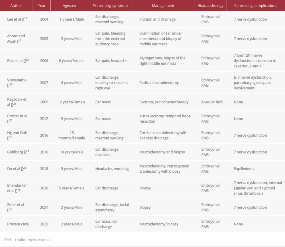
We searched for all the cases presented in the English-language literature between 2000 and 2022. The results are summarized in Table 1. Due to the evolution in imaging technology and oncological treatment, early identification and better management of these cases are seen today, in comparison to the circumstances that were seen a few decades ago, when rhabdomyosarcoma was always fatal. Most cases presented with coexisting complications, such as facial nerve dysfunction. In all cases except 1, the pathological diagnosis was consistent with embryonal rhabdomyosarcoma. The clinical presentation of these tumors originating from the ear might mimic otitis media with a polypoidal mass [11]. Additionally, the patient might present with palpable lymph nodes [12]. Patients could have facial nerve involvement due to the erosive nature of the disease [9]. Usually, at the time of diagnosis, the actual site of origin cannot be determined. Hence, in most cases, there is widespread local invasion throughout the petrous bone. Moreover, up to 67% of cases exhibit extensive bone erosion [13].
In 1 article, a child presented with acute onset of unilateral erythematous swelling over the mastoid after 10 days of a purulent right ear discharge. It was presumed to be acute mastoiditis, complicated by a subperiosteal abscess. Therefore, physicians attempted incision and drainage. The mass started to grow rapidly after 5 days, and the child developed facial palsy on the affected side [14]. Although uncommon, temporal bone rhabdomyosarcoma should be considered a differential diagnosis in patients failing to respond to multiple courses of antibiotics or in patients with multiple cranial nerve involvement.
In a case report published in 2006, the patient developed acute facial weakness, accompanied by a history of ear pain and headache for 5 days. After visiting the primary physician’s office, a diagnosis of Bell’s palsy was made and she was referred to the Emergency Department to withdraw blood for a Lyme titer. However, the receiving physician examined the cranial nerves, which revealed 7th and 12th cranial nerve palsy. This was considered alarming for a non-idiopathic cause of facial nerve paralysis; hence, imaging was done to rule out space-occupying lesions [15]. Pediatric rhabdomyosarcoma that arises in the temporal bone is generally considered to be an aggressive neoplasm due to the anatomical proximity of nerves and vessels. Moreover, the tumors located here are more likely to spread intracranially. Most reported cranial nerve palsies with temporal bone rhabdomyosarcoma are palsies of the 5th, 7th, 6th, 9th, 11th, and 12th cranial nerves, with the facial nerve being the earliest to be affected at presentation. In 12% to 50% of cases of head and neck rhabdomyosarcoma, radiological evidence of metastasis to the cervical lymph nodes is present [14,15]. In 1977, a study was conducted that revised the presenting symptoms of 50 patients with temporal bone rhabdomyosarcoma. The results showed a mass in the area of the ear was seen in 56% of patients, a polyp in the ear canal in 54%, aural discharge in 40%, aural bleeding in 30%, earache in 22%, deafness in 14%, and 12th cranial nerve palsy in 14% of patients [16].
Currently, the first line of treatment for rhabdomyosarcoma is chemotherapy. The standard regimens include vincristine, actinomycin D, and cyclophosphamide. The most recent agents include etoposide, ifosfamide, and melphalan [8]. Radiotherapy is limited to locoregional control. In most cases, surgical excision is not a good treatment option because of the involvement of vital structures and intracranial extension [6]. The overall 5-year survival for localized disease in patient’s aged 1 to 4 years with the embryonal histology type is up to 80% [12].
Conclusions
Overall, rhabdomyosarcoma is an uncommon tumor that is associated with a high fatality rate in children, when diagnosis is delayed. Hence, physicians should have a strong index of suspicion to make an early diagnosis. In this study, we presented the case of a young patient with a clinical picture of a complicated ear infection who was found to have rhabdomyosarcoma of the temporal bone. Early detection and multi-modal treatment are critical for ensuring a positive outcome in these patients.
Figures
References:
1.. Barnes P, Maxwell M, Embryonal rhabdo-myosarcoma of middle ear: J Laryngol Otol, 1972; 86(11); 1145-54
2.. Shirani S, Alizadeh F, Sharif S, Rhabdomyosarcoma of the middle and inner ear: Iran J Radiol, 2003; 1(3–4); 157-59
3.. Durve D, Kanegaonkar R, Albert D, Levitt G, Paediatric rhabdomyosarcoma of the ear and temporal bone: Clin Otolaryngol Allied Sci, 2004; 29(1); 32-37
4.. Söderberg P, Rhabdomyome épipharyngé ayant envahi l’oreille et les méninges: Acta Oto-Laryngologica, 1933; 18(4); 453-59
5.. Abbas A, Awan S, Rhabdomyosarcoma of the Middle Ear and Mastoid: A case report and review of the literature: Ear Nose Throat J, 2005; 84(12); 780-84
6.. Ulutin C, Bakkal H, Kuzhan O, A cohort study of adult rhabdomyosarcoma: A single institution experience: World J Med Sci, 2008; 3(2); 54-59
7.. Viswanatha B, Embryonal rhabdomyosarcoma of the temporal bone: Ear Nose Throat J, 2007; 86(4); 218-22
8.. Chao CK, Sheen TS, Shau WY, Treatment, outcomes, and prognostic factors of ear cancer: J Formos Med Assoc, 1999; 98(5); 314-18
9.. Potter GD, Embryonal rhabdomyosarcoma of the middle ear in children: Cancer, 1966; 19(2); 221-26
10.. Khatami F, Bazrafshan E, Monajemzadeh M, Seyed M, Congenital embryonal rhabdomyosarcoma with prenatal onset: Iran J Pediatr, 2008; 18(1); 62-66
11.. Malogolowkin MH, Ortega JA, Rhabdomyosarcoma of childhood: Pediatr Ann, 1988; 17(4); 251-68
12.. Lee C, Aga L, Yong Lie Q, Lin Z, Rhabdomyosarcoma presenting as an acute suppurative mastoiditis: Otolaryngol Head Neck Surg, 2004; 131(5); 791-92
13.. Reid S, Hetzel T, Losek J, Temporal bone rhabdomyosarcoma presenting as acute peripheral facial nerve paralysis: Pediatr Emerg Care, 2006; 22(10); 743-45
14.. Carter RL, Tumors of the head and neck: Clinical and pathological considerations: J R Soc Med, 1982; 75(1); 57
15.. Punyko J, Mertens A, Baker K, Long-term survival probabilities for childhood rhabdomyosarcoma: Cancer, 2005; 103(7); 1475-83
16.. Prat J, Gray GF, Massive neuraxial spread of aural rhabdomyosarcoma: Arch Otlaryngol, 1977; 103; 301-3
17.. Zubir FS, Saniasiaya J, Abdul Gani H, Aural polyp with facial asymmetry in an unfortunate infant: Malays Fam Physician, 2021; 16(1); 133-35
18.. Bhandarkar A, Menon A, Kudva R, Pujary K, Embryonal rhabdomyosarcoma – a mimicker of squamosal otitis media: Iran J Otorhinolaryngol, 2020; 32(108); 57-61
19.. Sri Ravali P, Jakkula A, Gogineni T, Damera S, Embryonal rhabdomyosarcoma of external ear – a rare case report: Ann. Maxillofac Surg, 2021; 11(2); 317
20.. Viswanatha B, Embryonal rhabdomyosarcoma of the temporal bone: Ear Nose Throat J, 2007; 86(4); 218-22
21.. Ragsdale B, Lee J, Mines J, Alveolar rhabdomyosarcoma on the external ear: A case report: J Cutan Pathol, 2009; 36(2); 267-69
22.. Crozier E, Rihani J, Koral K, Cope-Yokoyama S, Embryonal rhabdomyosarcoma of the auricle in a child: Pediatr Int, 2012; 54(6); 945-47
23.. Ng S, Goh B, A Toddler with rhabdomyosarcoma presenting as acute otitis media with mastoid abscess: Chin Med J, 2016; 129(10); 1249-50
24.. Goldberg M, Pediatric temporal bone rhabdomyosarcoma: JAAPA, 2016; 29(8); 1-3
25.. Do T, Linabery A, Patterson R, Tu A, Cranial rhabdomyosarcoma masquerading as infectious mastoiditis: Case report and literature review: Pediatr Neurosurg, 2018; 53(5); 317-21
Figures
In Press
06 Mar 2024 : Case report 
Am J Case Rep In Press; DOI: 10.12659/AJCR.942937
12 Mar 2024 : Case report 
Am J Case Rep In Press; DOI: 10.12659/AJCR.943244
13 Mar 2024 : Case report 
Am J Case Rep In Press; DOI: 10.12659/AJCR.943275
13 Mar 2024 : Case report 
Am J Case Rep In Press; DOI: 10.12659/AJCR.943411
Most Viewed Current Articles
07 Mar 2024 : Case report 
DOI :10.12659/AJCR.943133
Am J Case Rep 2024; 25:e943133
10 Jan 2022 : Case report 
DOI :10.12659/AJCR.935263
Am J Case Rep 2022; 23:e935263
19 Jul 2022 : Case report 
DOI :10.12659/AJCR.936128
Am J Case Rep 2022; 23:e936128
23 Feb 2022 : Case report 
DOI :10.12659/AJCR.935250
Am J Case Rep 2022; 23:e935250


