


18 August 2026: Articles 
Seizure-Induced Rhabdomyolysis Complicated by Acute Kidney Injury: A Case Report
Unusual or unexpected effect of treatment, Educational Purpose (only if useful for a systematic review or synthesis)
Harsimranjit Kaur ABCDEF 1*, Huma Hasnain Kennedy EF 1, Niloofar Nobakht ABCDEFG 1
DOI: 10.12659/AJCR.951097
Am J Case Rep 2026; 27:e951097


Abstract
BACKGROUND: Tonic-clonic seizures can precipitate severe rhabdomyolysis due to sustained muscle activity, leading to release of intracellular contents, including proteins, that can pose an elevated risk for acute kidney injury (AKI). Rhabdomyolysis management centers on aggressive intravenous (IV) fluid resuscitation while the risk of volume overload is vigilantly minimized. Nephrologists regularly assess the need for dialysis while addressing the nuances in each case. This case illustrates the clinical decision-making involved in managing extreme biochemical derangements in seizure-induced rhabdomyolysis with conservative medical management.
CASE REPORT: A 32-year-old man with a history of seizure disorder presented following a generalized tonic-clonic seizure. Laboratory studies reflected marked abnormalities, including a creatine kinase level of 83 261 u/L, peaking at 133 300 u/L shortly after admission, creatinine of 9.3 mg/dL from baseline of 1.0 mg/dL, aspartate aminotransferase of 819 u/L, and alanine aminotransferase of 192 u/L. Despite significantly elevated creatine kinase and creatinine levels, the patient remained hemodynamically stable, with preserved urine output and no life-threatening electrolyte or acid-base disturbances. He received aggressive IV fluids to enhance kidney perfusion and prevent further myoglobulin induced injury, with intermittent diuretics use to maintain euvolemia. Ultimately, kidney function and laboratory markers improved.
CONCLUSIONS: Rhabdomyolysis can cause AKI by various mechanisms. This report highlights the importance of timely and appropriate nephrology-guided management. Although aggressive hydration remains the standard of care, careful monitoring is required to prevent fluid overload. Close monitoring and conservative management alone may be sufficient in select patients without life-threatening biochemical derangements, thereby preventing the need for kidney replacement therapy.
Keywords: Acute Kidney Injury, Dialysis, rhabdomyolysis
Introduction
Rhabdomyolysis is a clinical condition that consists of the breakdown of skeletal muscle at the cellular level [1]. This results in release of intracellular contents, including proteins, that can pose an elevated risk for acute kidney injury (AKI) [2]. The etiologies of rhabdomyolysis include trauma, immobilization, high-intensity exercise, substance use, and, less commonly, seizures [1,2]. In seizure-induced rhabdomyolysis, prolonged tonic-clonic activity results in sustained muscle contraction and hypoxia, which precipitate muscle necrosis and myoglobin release. There are several challenges that nephrologists encounter when approaching severe cases of rhabdomyolysis. Management involves the careful balance of aggressive resuscitation efforts with intravenous (IV) fluids while avoiding iatrogenic fluid overload. Nephrologists also regularly assess the need for kidney replacement therapy while addressing the nuances in each case. This report aims to describe the clinical presentation, management and outcome of a patient with seizure-induced rhabdomyolysis complicated by AKI, highlighting the challenges of maintaining fluid balance and medication dose adjustment.
Case Report
A 32-year-old man with a 3-year history of seizure disorder presented after having a generalized tonic-clonic seizure. Prior to this, the patient was followed by neurology, with reported excellent compliance to seizure medications, including levetiracetam 1000 mg twice a day. He denied any precipitating significant event, illness, sleep deprivation, trauma, illicit substance use, or excessive physical exertion. Initial evaluation revealed no evidence of infection or other metabolic derangement to suggest alternative seizure. The patient initially presented to an outside hospital; however, he opted to leave against medical advice to arrive at our institution for further care. The seizure was estimated to have lasted over 5 minutes and required intubation for airway protection. He was extubated after less than 24 hours. Computed tomography of the head without contrast revealed no acute intracranial abnormalities.
Electroencephalography performed at the outside hospital was abnormal, indicating mild diffuse cerebral dysfunction. There was excessive beta activity; however, it was attributed to the benzodiazepines the patient had received upon arrival in the emergency department.
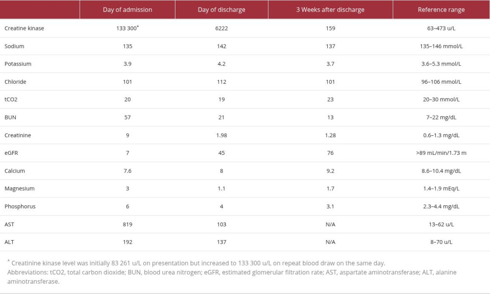
Upon presentation at our institution, the patient’s vital signs were stable. Physical examination revealed normal heart and lung sounds. Once extubated, the patient was alert and oriented without any apparent neurologic deficits or signs of peripheral edema. Initial laboratory studies reflected marked abnormalities, including creatine kinase (CK) level of 83 261 u/L which increased to 133 300 u/L shortly after admission, creatinine level of 9.3 mg/dL from baseline 1.0 per electronic medical record, aspartate aminotransferase level of 819 u/L, and alanine aminotransferase level of 192 u/L (Table 1). Urinalysis revealed colorless urine and was notable for 3+ blood, 1+ protein, and 1 red blood cell per high-power field. Upon presentation, the patient was started on aggressive IV fluids with normal saline at 200 mL/h to enhance kidney perfusion and prevent further myoglobulin induced injury. This rate was chosen to maintain adequate urine output while accounting for body composition lean muscle mass, preserved urine production, and absence of cardiopulmonary disease. Fluid balance was monitored by strict input and output records and daily weights, along with daily physical examinations. The patient required 2 mg of IV bumetanide twice during admission to maintain euvolemia, as he began developing lower extremity edema. He did not report shortness of breath and did not require supplemental oxygen; oxygen saturation remained above 98%, and lung examination was normal. Despite initial severe AKI, kidney replacement therapy was deferred due to the patient’s overall hemodynamic stability, continued and adequate urine production, lack of uremic symptoms, and lack of marked electrolyte and acid-base imbalance. No nephrotoxic agents, such as non-steroidal anti-inflammatory drugs and IV contrast, were administered. Urine alkalization was considered but not implemented given the observed improvement with only IV fluids. Over the course, our patient’s CK level improved to less than 10 000 u/L. Creatinine also improved to 1.98 mg/dL (Table 1). Our neurology colleagues were consulted and recommended levetiracetam at a renally adjusted dose. The dose was decreased from 1000 mg twice a day to 500 mg twice a day. The patient was discharged with instructions to maintain adequate oral hydration and close follow-up with his primary care provider and with outpatient monitoring of CK, creatinine, and liver function tests. About 3 weeks after discharge, repeat laboratory tests revealed a CK level of 159 u/L and creatinine level of 1.28 mg/dL (Table 1).
Discussion
Rhabdomyolysis can lead to AKI by several different pathophysiologic mechanisms. Myoglobin, released from cells during muscle breakdown, is nephrotoxic due to direct tubular toxicity, leading to vasoconstriction and renal ischemia [2]. Myoglobin and other precipitates, such as uric acid, can cause tubular obstruction and therefore direct tubular injury [2]. Furthermore, intravascular volume depletion, either from a dehydrated state or from third spacing with possible muscle edema, can further potentiate kidney ischemia [4,5]. Unliganded, free heme is toxic, and its lipophilic properties enable it to intercalate into plasma and cellular membranes, granting it access to various cellular compartments [3]. The effect is often amplified in acidic urine [2]. It should be noted that the decision to initiate kidney replacement therapy in clinical practice should not be based solely on serum myoglobin or CK concentrations [6].
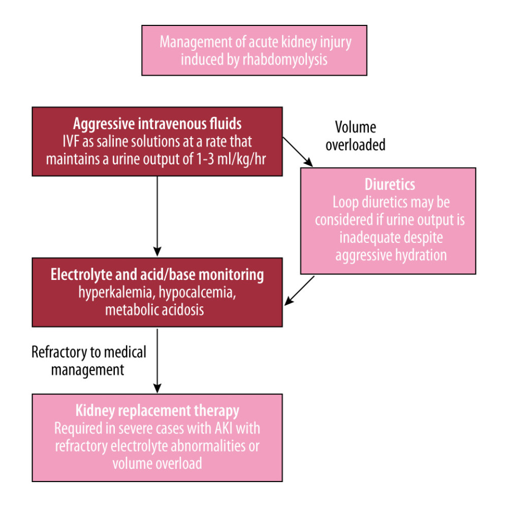
The current literature lacks high-quality randomized controlled trial for many therapeutic options; however, the standard of care entails prompt and aggressive hydration with IV fluids (Figure 1) [5]. In our case, the patient received normal saline at 200 mL/h. Due to the high rate of IV fluids, the patient began to develop peripheral edema. This uncovered one of the challenges often faced with treating rhabdomyolysis. Maintaining the balance of hydration without leading to significant peripheral edema and/or pulmonary edema requires cautious monitoring. As in this case, intermittent diuretic use to prevent fluid overload permits continuing hydration to prevent further kidney function compromise. These cases can become quite complex in older adult patients and those with heart failure with reduced ejection fraction.
Furthermore, the presence of kidney dysfunction should prompt review of current medications to consider dose adjustment. Levetiracetam is a renally excreted medication [7]. In our case, the dose was decreased to account for the decreased creatinine clearance. Initially, the patient was understandably hesitant to decrease an anti-epileptic dose in fear of risking another seizure. The patient later agreed after understanding that the concerted recommendations by nephrology and neurology to adjust the dose resulted in a similar effective dose due to the decreased clearance of the drug. Continuing at a higher than renally adjusted dose would potentially lead to toxicity of the drug and other undesirable consequences [7]. Timely resumption of full-dose medication when creatinine returns to normal is pivotal.
While our patient’s laboratory data revealed promising recovery of kidney function without the need of kidney replacement therapy, close monitoring for electrolyte, acid base, and creatinine in the weeks following discharge was essential. Severe kidney injury may result in chronic kidney disease due to ischemia and loss of glomerular filtration rate in a permanent manner a few weeks after an AKI [8]. Patients should be encouraged to maintain adequate hydration, compliance with anti-epileptics, and avoidance of rigorous exercise. Outpatient nephrology follow-up should be individually based and especially considered if kidney function has not recovered to normal or baseline levels.
Although this patient initially exhibited severe laboratory abnormalities, specifically elevated CK and creatinine levels, these values do not independently mandate dialysis. Dialysis initiation should be based on clinical parameters such as electrolyte stability, acid-base balance, and urine production. Therefore, conservative management may not be considered appropriate in patients with oliguria, hyperkalemia, and severe academia, or even in those with underlying cardiopulmonary disease, making aggressive IV hydration alone challenging.
Our case reinforces established rhabdomyolysis management principles while emphasizing nuances in severe presentations. Similar findings have been detailed in other rare causes of rhabdomyolysis-induced AKI. For example, Vural et al describe a case of hypothermia-associated rhabdomyolysis requiring hemodialysis, underlining the importance of early recognition and multidisciplinary intervention [9]. This report is limited in its single-patient design, absence of long-term follow-up, lack of kidney biopsy or myoglobin quantification, and the inability to identify predictive factors for renal recovery. As such, the causality cannot be established, and findings should not be generalized beyond carefully selected patients.
Conclusions
Seizure-induced rhabdomyolysis poses unique challenges in the management of AKI. The present case underscores the importance of a multidisciplinary approach with integral nephrology involvement, allowing for the early identification of candidates suitable for medical management despite alarming laboratory values. Early recognition to provide prompt medical management not only prevents or improves current kidney injury but also reduces the risk of chronic kidney disease. Optimization of patient risk factors is paramount to prevent recurrence of rhabdomyolysis. This case reinforces the importance of vigilant monitoring of volume status, electrolytes, and renal function to mitigate the risk of irreversible kidney injury. Key clinical lessons include early nephrology consultation, prompt but balanced fluid repletion to prevent fluid overload, and careful reassessment of renally cleared medications.
References
1. Huerta-Alardin AL, Varon J, Marik PE, Bench-to-bedside review: Rhabdomyolysis – An overview for clinicians: Crit Care, 2005; 9(2); 158-69
2. Bosch X, Poch E, Grau JM, Rhabdomyolysis and acute kidney injury: N Engl J Med, 2009; 361(1); 62-72
3. Nath KA, Singh RD, Croatt AJ, Adams CM, Heme proteins and kidney injury: Beyond rhabdomyolysis: Kidney360, 2022; 3(11); 1969-79
4. Malinoski DJ, Slater MS, Mullins RJ, Crush injury and rhabdomyolysis: Crit Care Clin, 2004; 20(1); 171-92
5. Rout P, Chippa V, Adigun R, Rhabdomyolysis: StatPearls [Internet], 2025, Treasure Island (FL), StatPearls Publishing Updated July 7, 2025. Available from: https://www.ncbi.nlm.nih.gov/books/NBK448168
6. Petejova N, Martinek A, Acute kidney injury due to rhabdomyolysis and renal replacement therapy: A critical review: Crit Care, 2014; 18(3); 224
7. Kumar A, Maini K, Kadian R, Levetiracetam: StatPearls [Internet], 2025, Treasure Island, FL, StatPearls Publishing Updated December 3, 2023. Available from: https://www.ncbi.nlm.nih.gov/books/NBK499890/
8. Zager RA, Progression from acute kidney injury to chronic kidney disease: Clinical and experimental insights and queries: Nephron Clin Pract, 2014; 127(1–4); 46-50
9. Vural A, Ekiz M, Günaydın M, A case of acute renal failure requiring emergency hemodialysis due to hypothermia-associated rhabdomyolysis: J Surg Med, 2020; 4(11); 1092-94
In Press
Case report 
Am J Case Rep In Press; DOI: 10.12659/AJCR.953543
Case report 
Am J Case Rep In Press; DOI: 10.12659/AJCR.954216
Case report 
Am J Case Rep In Press; DOI: 10.12659/AJCR.954932
Case report
Am J Case Rep In Press; DOI: 10.12659/AJCR.954146
Most Viewed Current Articles
07 Dec 2021 : Case report
28,712,695
DOI :10.12659/AJCR.934347
Am J Case Rep 2021; 22:e934347
06 Dec 2021 : Case report  180,721
DOI :10.12659/AJCR.934406
Am J Case Rep 2021; 22:e934406
21 Jun 2024 : Case report
124,419
DOI :10.12659/AJCR.944371
Am J Case Rep 2024; 25:e944371
08 May 2026 : Case report
95,462
DOI :10.12659/AJCR.952158
Am J Case Rep 2026; 27:e952158