


19 August 2026: Articles 
Incidental Discovery of a Fallopian Tube Mesenteric Borderline Serous Tumor During Cesarean Section: A Case Report and Literature Review
Rare disease
Yingmei Tuo ABCDEFG 1, Dongyan Chen BE 2, Ying Geng CDE 3, Lianming Liao ACDEF 4*DOI: 10.12659/AJCR.952204
Am J Case Rep 2026; 27:e952204


Abstract
BACKGROUND: Borderline serous tumors (BSTs) are epithelial neoplasms of low malignant potential, accounting for approximately 10% to 15% of all ovarian epithelial tumors and are generally associated with a favorable prognosis. Primary fallopian tube BSTs are uncommon and usually detected incidentally. Fallopian tube mesenteric BST arising during pregnancy is extremely rare, with limited literature available. This case highlights the diagnostic challenges of this rare disease and offers practical insights into perioperative management.
CASE REPORT: A 29-year-old woman (gravida 2, para 1) at 37 weeks of gestation with a twin pregnancy underwent elective cesarean delivery. Intraoperatively, a 4×3×3 cm cystic mass originating from the mesentery of the ampulla of the left fallopian tube was identified and excised. The lesion was first detected on ultrasonography 18 months before delivery, but serial imaging during the antepartum period failed to yield a definitive diagnosis. Postoperative histopathological assessment, including gross and microscopic examination, confirmed the diagnosis of BST. Follow-up at 6 and 9 months postoperatively showed no signs of recurrence, and serum CA-125 levels remained within the normal range.
CONCLUSIONS: Fallopian tube mesenteric BST in pregnancy presents substantial diagnostic challenges due to its rarity and non-specific imaging features. Fertility-sparing resection is safe and effective for preserving reproductive function. The complexity of twin gestation requires careful intraoperative decision-making and highlights the value of multidisciplinary teamwork. Thorough intraoperative evaluation and long-term postoperative surveillance are essential to improve long-term outcomes.
Keywords: Case Reports, Fallopian Tube Neoplasms, Cesarean Section, Pregnancy Complications, Neoplastic, CA-125 Antigen
Introduction
Borderline serous tumors (BSTs) are a unique category of epithelial neoplasms with low-grade malignant behavior, representing roughly 10% to 15% of all epithelial ovarian tumors. Primary fallopian tube BSTs are exceedingly uncommon, with only 12 cases [1] documented in the literature to date, and those originating from the ampullary mesentery of the fallopian tube near the fimbrial end are particularly rare. These tumors predominantly affect women of reproductive age (30–40 years), which is consistent with our patient’s age.
When occurring during pregnancy, preoperative diagnosis of these tumors becomes particularly challenging, as pregnancy-related hormonal changes and anatomical obscuration caused by uterine enlargement can mask their typical features [2]. Studies have demonstrated that the majority of pregnancy-associated adnexal tumors are not definitively diagnosed before surgery, with most identified incidentally during cesarean section or other gynecological procedures [3]. Fallopian tube BSTs in pregnancy are no exception; their rare location in the fallopian tube ampullary mesentery further complicates preoperative diagnosis, making them more prone to incidental discovery, as observed in our case.
Our case involves a 29-year-old G2P1 woman at 36+6 weeks of twin pregnancy, whose left adnexal lesion was first detected on pelvic ultrasound in June 2023 when she presented with 2 months of irregular menstruation. Despite over 18 months of serial imaging follow-up, a definitive preoperative diagnosis could not be established, primarily due to the lesion’s small size, fallopian tube ampullary mesenteric location, and non-specific imaging characteristics that overlapped with benign pregnancy-associated adnexal lesions (eg, corpus luteum cysts). The lesion was ultimately identified and completely excised during cesarean section on November 21, 2024, with postoperative pathology confirming fallopian tube mesenteric borderline serous tumor (BST) without stromal invasion.
By describing this case in detail and reviewing the relevant literature, we aim to raise clinical awareness of pregnancy-associated fallopian tube ampullary mesenteric BSTs and share our experience of managing this rare entity during pregnancy. Of note, the term ‘fallopian tube mesenteric BST’ is used in the literature to describe this entity; we use ‘borderline serous tumor (BST)’ throughout this report in accordance with the current WHO classification.
Case Report
PATIENT DEMOGRAPHICS AND CLINICAL HISTORY:
A 29-year-old woman (G2P1) was admitted to the Department of Obstetrics at our hospital on November 21, 2024, for delivery preparation at 36+6 weeks of twin pregnancy. There was no family history of gynecological malignancy.
PAST MEDICAL HISTORY:
Her obstetric history included a full-term vaginal delivery of a healthy male infant at our hospital on June 29, 2022. On June 12, 2023, she presented with 2 months of irregular menstruation. Pelvic ultrasound revealed a 19 × 14 mm left adnexal mixed-echo mass with a thin, well-defined wall and internal hypoechoic protrusions. Color Doppler flow imaging showed minimal blood flow signals in the peripheral and internal hypoechoic regions. A follow-up ultrasound on July 10, 2023, demonstrated slight enlargement of the lesion to 19 × 18 mm, with increased flow signals within the solid component. She was admitted for further evaluation on July 17, 2023. Serum CA-125 was 22.51 U/mL (reference range: 0–35 U/mL). Pelvic magnetic resonance imaging showed a 1.6 × 1.7 cm oval lesion with long T1 and long T2 signals adjacent to the left uterine border, featuring heterogeneous internal signals and small nodules; it was diagnosed as a cystic lesion with intracystic nodules, and close follow-up was recommended. However, follow-up imaging was not performed as the patient conceived shortly afterward.
Her last menstrual period was March 8, 2024. First-trimester ultrasound confirmed a twin pregnancy, and both fetuses were in cephalic presentation by the third trimester. An ultrasound on April 24, 2024, showed that the left adnexal anechoic mass had increased to 30 × 21 mm with poor internal sound transmission. Throughout the pregnancy, she remained asymptomatic, with no vaginal bleeding, abdominal pain, or pelvic discomfort, and routine prenatal ultrasound scans showed no other abnormal findings.
On admission, vital signs were stable (temperature 36.5°C, pulse 82 beats/min, blood pressure 125/80 mm Hg). Abdominal examination revealed a uterine height of 33 cm and an abdominal circumference of 94 cm. Fetal heart rate monitoring showed normal baseline rates (140–150 beats/min) for both twins. Gynecological examination was limited by uterine enlargement and failed to detect definite abnormalities. Preoperative laboratory test results—including complete blood count, coagulation profile, and liver and renal function—were within normal limits; CA-125 was not rechecked preoperatively. Despite over 18 months of serial imaging follow-up (from June 2023 to November 2024), a definitive preoperative diagnosis could not be established. This was primarily due to the lesion’s small size, fallopian tube mesenteric location [4], and non-specific imaging characteristics (mixed/anechoic cyst with a mild solid component), which overlapped considerably with benign pregnancy-associated adnexal lesions such as corpus luteum cysts or benign serous cysts.
SURGICAL PROCEDURE:
After comprehensive evaluation, the obstetric team performed a lower uterine segment cesarean section at 37 weeks of gestation. Intraoperatively, the uterus was consistent with 37 weeks of gestation. The left adnexal lesion, which had been monitored for over 18 months, was localized to the ampullary mesentery of the left fallopian tube near the fimbrial end; a 4 × 3 × 3 cm cyst was palpable, with a smooth surface, transparent wall, clear demarcation from the ovarian tissue, and no pelvic adhesions.
The patient and her family were immediately informed of the need for pathology confirmation and the potential risk of malignancy, and concurrent cystectomy was recommended. Following signed informed consent, the lesion was completely excised along the cyst capsule. The excised specimen was shown to the family before being sent for pathological examination.
Due to the weekend, frozen section pathology was unavailable. The patient and her family were informed intraoperatively that a second operation or adjuvant chemoradiotherapy might be required if postoperative pathology indicated malignancy or a borderline tumor, and they acknowledged this possibility.
The cesarean section then proceeded uneventfully, delivering 2 live female infants weighing 2680 g and 1850 g, with Apgar scores of 9 and 10 at 1 and 5 minutes, respectively. The placenta and fetal membranes were completely delivered, uterine contractions were adequate, and estimated intraoperative blood loss was approximately 500 mL.
PATHOLOGICAL AND IMMUNOHISTOCHEMICAL FINDINGS:
Histopathological examination of the resected specimen showed slender micropapillary structures lined by cuboidal to columnar epithelium, with moderate cellular atypia and no stromal invasion (Figure 1). Immunohistochemical staining revealed ER (diffuse +), PR (focal +), AE1/AE3 (diffuse +), WT-1 (nuclear +), p53 (+, 40% positive rate), p16 (patchy +), PAX-8 (nuclear +), and inhibin-α (weak +). The definitive diagnosis was fallopian tube mesenteric borderline serous tumor (BST) without stromal invasion. The micropapillary structures were considered reactive hyperplasia, with no definitive neoplastic micropapillae identified.
POSTOPERATIVE FOLLOW-UP:
In this case, transient postoperative elevation of CA-125 followed by normalization was attributed to surgical stress—a benign, self-limiting change with no clinical significance for tumor recurrence. 18F-FDG PET/CT at 81 days postoperatively showed no evidence of recurrence, and the patient remains in good clinical condition on follow-up.
At approximately 6 months postoperatively (May 9, 2025), B-ultrasound re-examination revealed no enlarged lymph nodes in the bilateral neck, supraclavicular fossa, or axilla, and no space-occupying lesions in the abdominal organs, uterus, or adnexa; CA-125 measured 14.60 U/mL. At approximately 9 months postoperatively (August 10, 2025), B-ultrasound showed no obvious abnormalities in the uterus or bilateral adnexa, with CA-125 stable at 15.05 U/mL.
Of note, the tumor was ER (diffuse +) and PR (focal +) positive in this case. The potential role of postoperative endocrine therapy and its implications for recurrence risk are discussed below.
Discussion
CASE CHARACTERISTICS:
We report a case of pregnancy-associated fallopian tube mesenteric borderline serous tumor, an exceedingly rare entity in a young woman of reproductive age. In the present case, a left adnexal cystic mass was initially noted during routine prenatal ultrasonography in June 2023 and followed serially; nevertheless, precise preoperative characterization proved elusive, with final identification occurring fortuitously at the time of cesarean delivery.
Several factors contributed to the diagnostic difficulty. First, the tumor was small (maximum preoperative diameter of 3 cm) and arose in an atypical location—the ampullary mesentery of the fallopian tube—which is not a site traditionally associated with BSTs. This anatomical rarity rendered the lesion prone to misdiagnosis as a physiological ovarian cyst (eg, corpus luteum cyst) or benign serous cyst, as imaging features were non-specific for borderline malignancy. Second, pregnancy-related uterine enlargement altered pelvic anatomy, obscuring the lesion on cross-sectional imaging and hindering sonographic characterization. Third, serum CA-125 levels remained persistently within the normal range throughout the preoperative monitoring period and stabilized postoperatively (with consistent normal values up to August 10, 2025), providing no auxiliary diagnostic clues preoperatively but confirming favorable short-term recovery. Fourth, the rarity of this anatomical site itself limited clinical suspicion preoperatively.
Consistent with the literature, the majority of pregnancy-associated adnexal tumors are not definitively diagnosed before surgery and are identified incidentally during cesarean section [5] or other obstetric procedures.
FALLOPIAN TUBE MESENTERIC BORDERLINE SEROUS TUMOR: SUMMARY OF PUBLISHED CASES:
To our knowledge, this case represents 1 of 12 primary fallopian tube BSTs reported worldwide. A review of 11 reported cases is available in the literature [6]. Patient age ranged from 3 to 43 years, and lesion size varied from 1.7 to 23 cm. Clinical presentations included abdominal or pelvic pain, abdominal fullness, or incidental detection. Surgical interventions encompassed partial salpingectomy, salpingo-oophorectomy, hysterectomy, and partial omentectomy, among others.
This case is distinguished from previously reported cases by 3 distinctive features: occurrence in a twin pregnancy, incidental discovery during cesarean section, and successful fertilitysparing cystectomy without radical surgery.
DIFFERENTIAL DIAGNOSIS:
Histopathological examination combined with immunohistochemistry remains the gold standard for diagnosing BSTs, including the micropapillary subtype. Core diagnostic criteria include tumor cells forming slender micropapillary structures lacking fibrovascular cores, exhibiting moderate-to-severe cellular atypia and mitotic figures, and demonstrating absence of stromal invasion (Figure 2) [7].
Immunohistochemistry plays a critical role in differential diagnosis. In this case, both WT-1 and PAX-8—specific markers for female reproductive tract epithelial tumors—were positive, supporting a Müllerian-derived origin. Positive ER/PR expression is consistent with hormonal regulation of the tumor, which aligns with our patient’s reproductive age and pregnancy status.
The pathological diagnosis in this case was not without contention, particularly regarding the nature of the micropapillary structures. After careful review, these were considered reactive hyperplasia secondary to pregnancy-related hormonal stimulation, rather than a neoplastic component. This highlights that pathological interpretation of borderline tumors, particularly during pregnancy, involves a degree of subjectivity, underscoring the importance of review by an experienced gynecological pathologist [8].
Yin et al [8] reported successful management of an adnexal mass during pregnancy using laparoendoscopic single-site surgery, demonstrating that minimally invasive approaches can be considered in selected cases when appropriate surgical expertise is available. This further supports the feasibility of conservative surgical management of adnexal lesions during pregnancy.
TREATMENT STRATEGY AND PREGNANCY OUTCOME:
Management of pregnancy-associated BSTs requires individualized planning based on tumor stage, pathological subtype, and the patient’s reproductive goals, with maternal and fetal safety as the overriding principle.
Current guidelines recommend conservative surgery (tumor resection or ipsilateral adnexectomy) without routine chemotherapy for young reproductive-aged patients with localized, non-metastatic BSTs [9]. In our case, intraoperative assessment confirmed a localized lesion unrelated to the ovary, and complete cystectomy was performed. This approach preserved ovarian function and fertility while avoiding overtreatment. Adequate intraoperative communication with the patient and family regarding the potential need for reoperation ensured flexibility in subsequent management.
From an obstetric perspective, the uneventful cesarean delivery of healthy twins, combined with the patient’s stable postoperative recovery and favorable follow-up outcomes (up to August 10, 2025), demonstrates that concurrent tumor resection during the second or third trimester is safe and feasible when surgical indications are carefully met. Conservative resection (cystectomy in this case) is the preferred approach for localized fallopian tube mesenteric borderline serous tumor (BST) in pregnancy, as it preserves fertility and ovarian function while avoiding overtreatment, consistent with our follow-up results showing no adverse effects on maternal recovery or long-term health. Published cases have also not reported adverse pregnancy outcomes with this strategy, further supporting its clinical applicability for similar rare cases.
POSTOPERATIVE FOLLOW-UP AND PROGNOSIS:
In this case, the postoperative course was favorable, with CA-125 levels normalizing after an initial transient elevation attributed to surgical stress. Serial imaging at 81 days, 6 months, and 9 months postoperatively confirmed no evidence of recurrence. These findings support the efficacy of conservative resection for pregnancy-associated fallopian tube mesenteric BST and underscore the importance of long-term surveillance, as recommended by current guidelines [9].
Of note, the tumor in this case was ER/PR positive, which implies potential hormonal dependence of the lesion, consistent with the patient’s reproductive age and pregnancy status, during which estrogen and progesterone levels are significantly elevated. Whether postoperative endocrine therapy can further reduce the recurrence risk of ER/PR-positive fallopian tube mesenteric borderline serous tumor (BST) remains uncertain, as current clinical guidelines lack specific recommendations for fallopian tube mesenteric BST, especially those occurring during pregnancy. This uncertainty underscores the need for longer-term follow-up (beyond the current 9 months) and more accumulated case data to clarify the value of adjuvant endocrine therapy. Given the low but existing long-term recurrence risk of BSTs, regular follow-up (as recommended, pelvic contrast-enhanced MRI and tumor marker rechecks every 3 months) remains essential for all patients with this rare entity, and our extended follow-up data (up to August 10, 2025) further support the necessity of continuous surveillance to monitor for potential late recurrence.
LIMITATIONS OF THE STUDY:
Several limitations of this study should be acknowledged, which also provide directions for future research.
First, this study is limited by its singlecenter, retrospective design and relatively small sample size. Although the follow-up period has been extended to approximately 9 months (up to August 10, 2025), it is still relatively short for a low-malignant potential tumor like BST. Borderline serous tumors may have a long-term recurrence risk, and short-to-medium-term follow-up cannot fully confirm long-term prognosis. Longer-term data (≥ 1 year, ideally 3–5 years) are needed to further verify the effectiveness of conservative cystectomy.
Second, the number of published cases of fallopian tube mesenteric borderline serous tumor (BST) is extremely limited [4] (~ 12 cases), and there is a lack of large-sample, prospective studies. This makes it difficult to establish consistent diagnostic criteria, treatment protocols, or prognostic factors. More case reports and multicenter studies are warranted.
Third, we did not conduct in-depth analysis of pathological characteristics, such as the detailed distribution of immunohistochemical markers, the degree of cellular atypia, or the relationship between reactive hyperplasia of micropapillary structures and pregnancy-related hormonal changes. Further pathological exploration could clarify the pathogenesis and improve diagnostic accuracy.
Fourth, we did not compare the diagnostic value of different imaging modalities (eg, pelvic ultrasound vs MRI) for this rare lesion during pregnancy. A detailed comparison of imaging findings with pathological results could provide more targeted guidance for preoperative evaluation of similar lesions.
Conclusions
Thorough intraoperative assessment of bilateral adnexal regions during cesarean delivery is imperative for establishing the pathological diagnosis of preoperatively detected but undiagnosed adnexal lesions in pregnancy. Fertility-sparing conservative resection is appropriate for young reproductive-aged patients with localized fallopian tube mesenteric BST, yielding favorable maternal–fetal outcomes. Definitive diagnosis requires postoperative pathological examination supplemented by immunohistochemical analysis, and sustained regular follow-up is essential for early detection of potential recurrence.
This case, together with the summarized published cases, provides a reference for the diagnosis and management of fallopian tube mesenteric BST in pregnancy. Multicenter studies with larger sample sizes and extended follow-up are warranted to establish evidence-based guidelines for this rare entity.
Figures
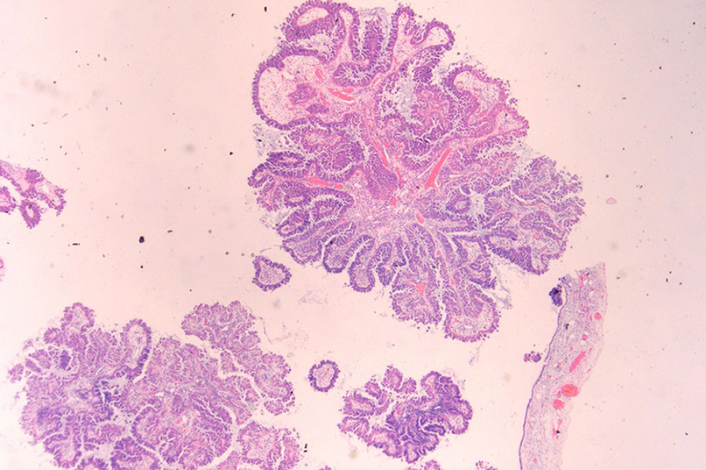 Figure 1. A complex branched papillary tumor is observed within the cyst. The papillary stroma contains fibrovascular cores, with no definite invasive foci identified. The epithelial cells lining the papillae show micropapillary changes in some areas. (HE staining, low magnification).
 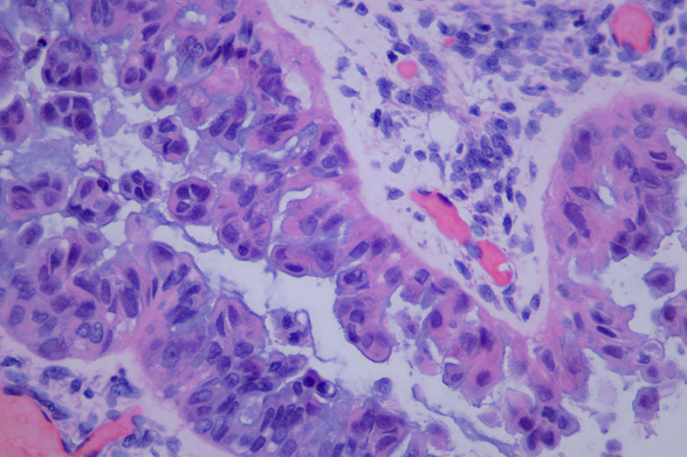 Figure 2. The tumor cells lining the papillae are arranged in multiple layers, with eosinophilic cytoplasm showing serous features, mild to moderate nuclear atypia, no definite nucleoli identified, and rare mitotic figures. (HE staining, high magnification).
 References
1. Matsumoto A, Ito T, Hamaguchi F, Primary and recurrent serous borderline tumors during pregnancy: A case report and literature review: Int Cancer Conf J, 2021; 10(3); 160-69
2. Gaughran J, Magee C, Mitchell S, Adnexal masses in pregnancy: A single-centre prospective observational cohort study: Diagnostics (Basel), 2024; 14(19); 2182
3. Wang M, Li Y, Xu T, Clinical analysis of 17 cases of borderline ovarian tumors during pregnancy: Front Oncol, 2022; 12; 934751
4. Limaiem F, Halouani A, An uncommon paratubal cyst: Serous borderline tumor: Clin Case Rep, 2023; 11(3); e7140
5. Yüksel S, Alan C, Retrospective evaluation of incidental adnexal masses encountered during cesarean section: Bağcılar Medical Bulletin, 2022; 7(1); 38-42
6. Choi SM, Choi MY, Kang WD, Serous borderline tumor of the fallopian tube: Obstet Gynecol Sci, 2014; 57(4); 334-37 [published erratum appears in Obstet Gynecol Sci. 2014;57:424]
7. Kurman RJ, Carcangiu ML, Herrington CS, Young RH: WHO classification of tumours of the female reproductive organs, 2014, Lyon, IARC
8. Yin M, Yang J, Zhou H, Zhang X, Laparoendoscopic single-site surgery for adnexal disease during pregnancy: A single-center preliminary experience. Front: Surg, 2022; 9; 994360
9. National Comprehensive Cancer Network: NCCN clinical practice guidelines in oncology: ovarian cancer. Version 1.2023, 2023, Plymouth Meeting (PA), National Comprehensive Cancer Network [cited 2026 May 18]. Available from: https://www.nccn.org/professionals/physician_gls/pdf/ovarian.pdf
Figures
Figure 1. A complex branched papillary tumor is observed within the cyst. The papillary stroma contains fibrovascular cores, with no definite invasive foci identified. The epithelial cells lining the papillae show micropapillary changes in some areas. (HE staining, low magnification).Figure 2. The tumor cells lining the papillae are arranged in multiple layers, with eosinophilic cytoplasm showing serous features, mild to moderate nuclear atypia, no definite nucleoli identified, and rare mitotic figures. (HE staining, high magnification). In Press
Case report 
Am J Case Rep In Press; DOI: 10.12659/AJCR.953543
Case report 
Am J Case Rep In Press; DOI: 10.12659/AJCR.954216
Case report
Am J Case Rep In Press; DOI: 10.12659/AJCR.954932
Case report 
Am J Case Rep In Press; DOI: 10.12659/AJCR.954146
Most Viewed Current Articles
07 Dec 2021 : Case report
28,712,695
DOI :10.12659/AJCR.934347
Am J Case Rep 2021; 22:e934347
06 Dec 2021 : Case report  180,721
DOI :10.12659/AJCR.934406
Am J Case Rep 2021; 22:e934406
21 Jun 2024 : Case report
124,419
DOI :10.12659/AJCR.944371
Am J Case Rep 2024; 25:e944371
08 May 2026 : Case report
95,462
DOI :10.12659/AJCR.952158
Am J Case Rep 2026; 27:e952158