


16 August 2026: Articles 
Severe Tricuspid Regurgitation: Traumatic Papillary Muscle Rupture Mimicking Infective Endocarditis—A Case Report
Challenging differential diagnosis, Management of emergency care, Rare disease
Soichiro Kobayashi E 1,2, Taku Omori E 1*, Daisuke Izumi E 2,3, Toshiya Tokui E 4, Atsunobu Kasai E 2, Kaoru Dohi E 1
DOI: 10.12659/AJCR.952267
Am J Case Rep 2026; 27:e952267


Abstract
BACKGROUND: Differentiating traumatic tricuspid regurgitation (TR) from infective endocarditis (IE) is clinically crucial in patients presenting with abnormal tricuspid valve findings after blunt chest trauma. Particularly, papillary muscle rupture caused by trauma can produce mobile valvular structures that closely resemble vegetations, making accurate diagnosis challenging in the acute setting.
CASE REPORT: A man in his 60s with pulmonary sarcoidosis was admitted after a 5-m fall, sustaining multiple fractures. Preoperative transthoracic echocardiography revealed a 7-mm mobile structure on the anterior tricuspid leaflet with flail motion and severe TR. Because of fever, IE was suspected; however, blood cultures and further investigations excluded IE. A diagnosis of traumatic TR due to papillary muscle rupture was made. Although the patient was hemodynamically stable under medical therapy, the heart team recommended surgery to prevent long-term heart failure. Four months after fracture repair, tricuspid valve repair with artificial chordae and annuloplasty was performed. The surgical findings were fully consistent with the preoperative diagnosis, and pathological examination of the ruptured papillary muscle demonstrated coagulative necrosis without significant inflammatory infiltration, supporting a traumatic etiology. At 4-year follow-up, he remains stable without signs of heart failure exacerbation.
CONCLUSIONS: Accurate differentiation between traumatic TR due to papillary muscle rupture and IE is essential, as misclassification may lead to unnecessary antimicrobial therapy or delayed surgical intervention. This case highlights how mobile traumatic lesions can closely mimic vegetations and underscores the importance of comprehensive evaluation to guide appropriate management and optimize outcomes.
Keywords: Cardiovascular Diseases, Case Reports, Heart Valve Diseases, Traumatology
Introduction
Traumatic tricuspid regurgitation (TR) is rare, and its acute-phase management, including surgical intervention, has not been standardized. Diagnosis can be challenging because clinical attention is often directed toward more obvious traumatic injuries, and acute TR is frequently overlooked due to the absence of signs or symptoms in the early phase [1–3]. In severe cases—particularly those involving papillary muscle rupture—abnormal valvular structures can mimic vegetations, necessitating careful differentiation from infective endocarditis (IE). Furthermore, traumatic TR can progress over time and lead to heart failure symptoms years after the initial injury [4]. We present a case of traumatic TR due to papillary muscle rupture, initially mimicking IE and successfully treated with early surgical repair. This case emphasizes the importance of the prompt recognition, accurate diagnosis, and timely surgical management of traumatic TR.
Case Report
A man in his 60s with pulmonary sarcoidosis, interstitial pneumonia, hyperuricemia, dyslipidemia, and benign prostatic hyperplasia was transported to the emergency department after falling 5 m from a ladder onto concrete, sustaining multiple traumatic injuries. He had fractures of the distal left radius, right calcaneus, and the seventh left rib (Figure 1A, 1B). On admission, he had fever (37.7 °C), oxygen saturation 94% on room air, blood pressure 104/70 mm Hg, and heart rate 98 beats per minute. Physical examination revealed a grade 1/6 systolic murmur, best heard at the fourth left sternal border. The abdomen was flat and soft, without rash or edema. Laboratory test results showed a white blood cell count of 7100/μL (reference range: 3300–8600/μL), hemoglobin level of 13.6 g/dL (13.7–16.8 g/dL), creatinine level of 1.23 mg/dL (0.65–1.07 mg/dL), C-reactive protein level of 0.29 mg/dL (< 0.14 mg/dL), and N-terminal pro-brain natriuretic peptide level of 1787 pg/mL (< 125 pg/mL). Chest radiography revealed cardiomegaly with a cardiothoracic ratio of 53%, without pulmonary edema or pleural effusion (Figure 1C). Electrocardiography showed sinus tachycardia without ST-T changes. Transthoracic echocardiography on day 2, prior to orthopedic surgery, revealed a 7-mm mobile mass-like structure on the anterior tricuspid leaflet with pronounced flail motion accompanied by a TR jet. The TR jet was defined as severe grade because of a vena contracta width of 8.3 mm, a color Doppler jet area of 14.7 cm2, and a ratio of color jet area to right atrial area of 68%. No pericardial effusion was observed (Figure 2, Videos 1, 2). The echocardiogram results are shown in Table 1.
Because of fever and the abnormal anterior tricuspid leaflet structure, IE was suspected. Blood cultures were obtained, and empiric ceftriaxone (2 g/day) was initiated. By day 6, cultures remained negative, and no Duke criteria were met aside from the structural anomaly. IE was therefore ruled out, and the flail leaflet was attributed to blunt chest trauma, with acute TR considered secondary to that injury.
The patient remained hemodynamically stable, and acute TR was managed with oral diuretics (azosemide 15 mg/day). Orthopedic surgeries were performed on days 8 and 10 without complications. Regarding the traumatic TR, the heart team recommended surgical intervention, considering long-term heart failure prevention.
Four months later, the patient underwent tricuspid valvuloplasty with artificial chordae reconstruction and ring annuloplasty. Intraoperative inspection revealed papillary muscle rupture with complete detachment of all chordae tendineae connected to the anterior tricuspid leaflet, resulting in flail motion (Figure 3). Pathological examination of the ruptured papillary muscle demonstrated loss of myocardial nuclei and increased cytoplasmic eosinophilia, with only scarce inflammatory cell infiltration, consistent with coagulative necrosis (Figure 4). Postoperative transthoracic echocardiography confirmed satisfactory valve function, with only mild residual TR. He was discharged on postoperative day 10.
At 4 years postoperatively, the patient remains stable without heart failure exacerbation.
Discussion
Traumatic TR is rare and often undiagnosed in the early stages, partly because of the discrepancy between regurgitant severity and murmur intensity, and because clinical attention is typically focused on more obvious injuries, such as fractures or hemorrhage [5]. Consequently, knowledge regarding the acute-phase management, surgical indications, and optimal timing of intervention for traumatic TR remains limited.
Fever is frequently observed during the acute phase of trauma due to elevations in cytokines, such as tumor necrosis factor-alpha [6]. In cases of traumatic TR due to papillary muscle rupture, the resulting abnormal structures can mimic vegetations. Careful differentiation from IE is therefore essential, as demonstrated in this case. A thorough evaluation, including blood cultures, is warranted.
The pathological findings suggest a direct traumatic avulsion of the papillary muscle caused by external mechanical force, rather than an inflammatory process, with resultant ischemic changes in the tissue. In blunt thoracic trauma, such external forces may also be transmitted to other cardiac structures. Thus, clinicians should be alert to acute complications that may accompany valvular injury, including myocardial contusion, cardiac tamponade from associated cardiac chamber injury, and lifethreatening arrhythmias. In this patient, intraoperative and histopathological findings argued against IE, supporting the diagnosis of traumatic papillary muscle rupture.
Recent reports indicate that the natural history of traumatic TR varies according to the extent and components of valvular and subvalvular injury. Limited injuries to the chordal and leaflet apparatus are associated with a more benign clinical course. In contrast, multimobility injuries, for example traumatic TR involving papillary muscle rupture, are associated with a higher risk of persistent severe TR, progressive right ventricle dilatation, and late heart failure [3,7]. Clinically relevant triggers for surgical consideration include flail leaflet or ruptured subvalvular apparatus, echocardiographic markers of severe TR, progressive right ventricle dilatation or dysfunction, and the development of symptoms or biomarkers of volume overload. In our patient, the TR met quantitative criteria for severe grade, and the anterior leaflet demonstrated large flail motion due to papillary muscle rupture. Although the patient was hemodynamically stable initially, these findings raised concern for future right ventricle remodeling and reduced likelihood of durable conservative management. Additionally, delayed surgical intervention can complicate repair, as papillary muscles, chordae, and affected leaflets often became atrophic or degenerative [8]. For these reasons, the heart team recommended surgical repair to maximize the chance of durable valve reconstruction and to prevent late right ventricular dysfunction.
Conclusions
This case highlights the clinical importance of early recognition and careful differentiation of traumatic severe TR from IE in patients presenting with abnormal tricuspid valve findings after blunt chest trauma. Accurate diagnosis based on the multidisciplinary approach that integrates clinical assessment, microbiological evaluation, and detailed echocardiographic examination is essential for appropriate management.
Figures
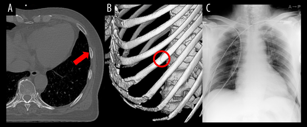 Figure 1. Chest X-ray and computed tomography findings in the emergency department.Computed tomography revealed the fracture of the left seventh rib (A, B), and chest X-ray revealed mild cardiomegaly without pleural effusion or pulmonary congestion (C). A red arrow and a circle indicate the fracture site.
 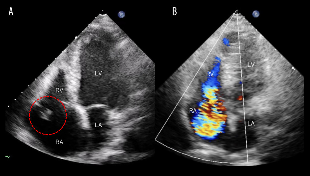 Figure 2. Transthoracic echocardiography findings on hospital day 2.Transthoracic echocardiography demonstrated a 7-mm mobile, mass-like structure attached to the flail anterior tricuspid leaflet (A, red dotted circle) with a severe eccentric regurgitant jet (B). Abbreviations: LA, left atrium; LV, left ventricle; RA, right atrium; RV, right ventricle.
 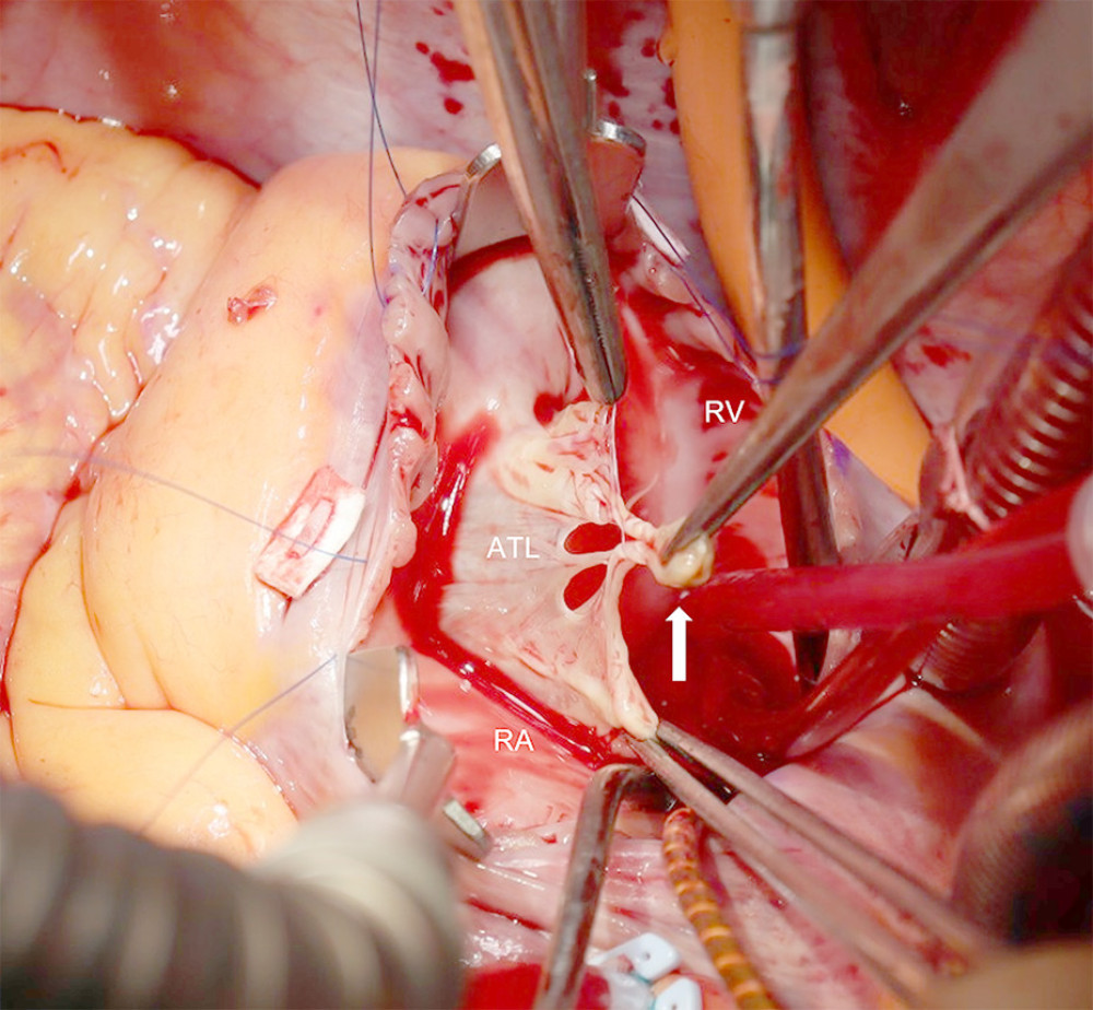 Figure 3. Intraoperative Image.The white arrow indicates the ruptured tip of the papillary muscle connected to the chordae tendineae of the anterior tricuspid leaflet, resulting in prolapse of the leaflet. Abbreviations: ATL, anterior tricuspid leaflet; RA, right atrium; RV, right ventricle.
 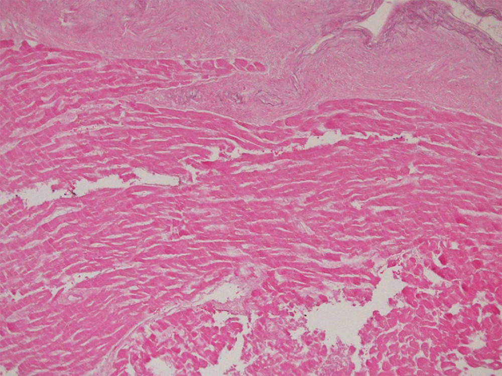 Figure 4. Microscopic image of the ruptures papillary muscle.Pathological examination showed coagulative necrosis.
 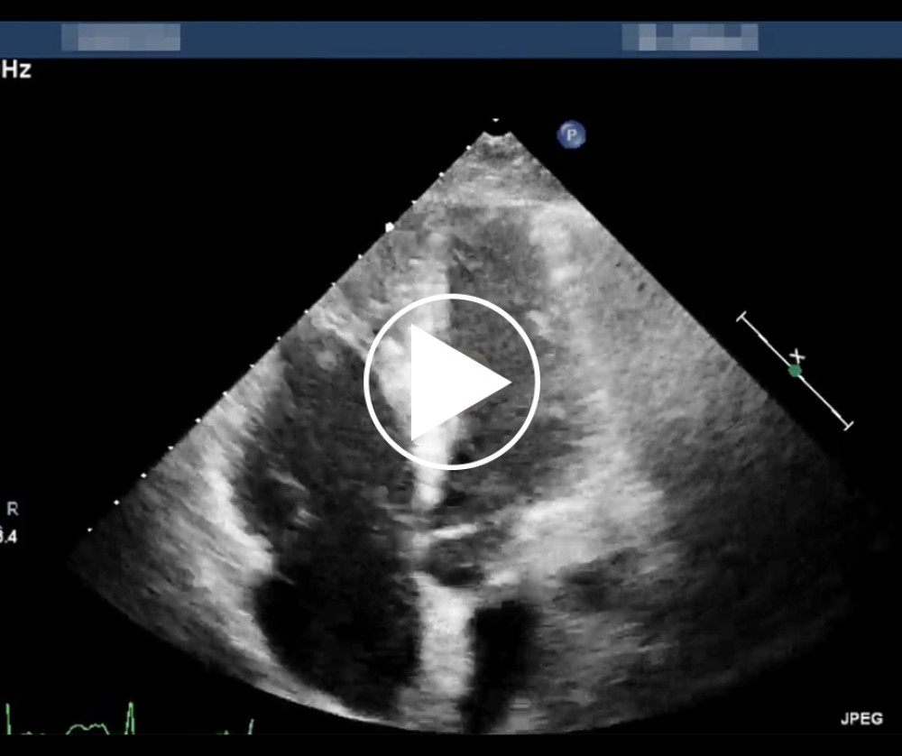 Video 1. The apical 4-chamber view on transthoracic echocardiography shows a 7-mm mobile mass-like structure attached to the anterior tricuspid leaflet, demonstrating pronounced flail motion.
 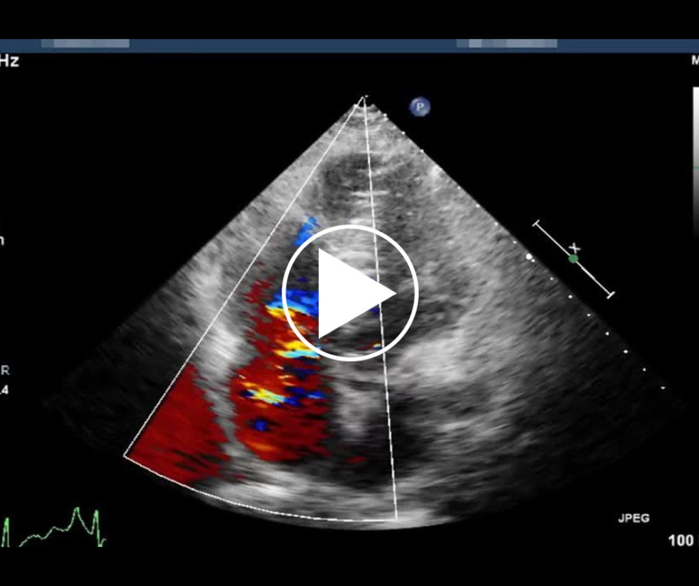 Video 2. A color Doppler image demonstrating pronounced tricuspid regurgitant.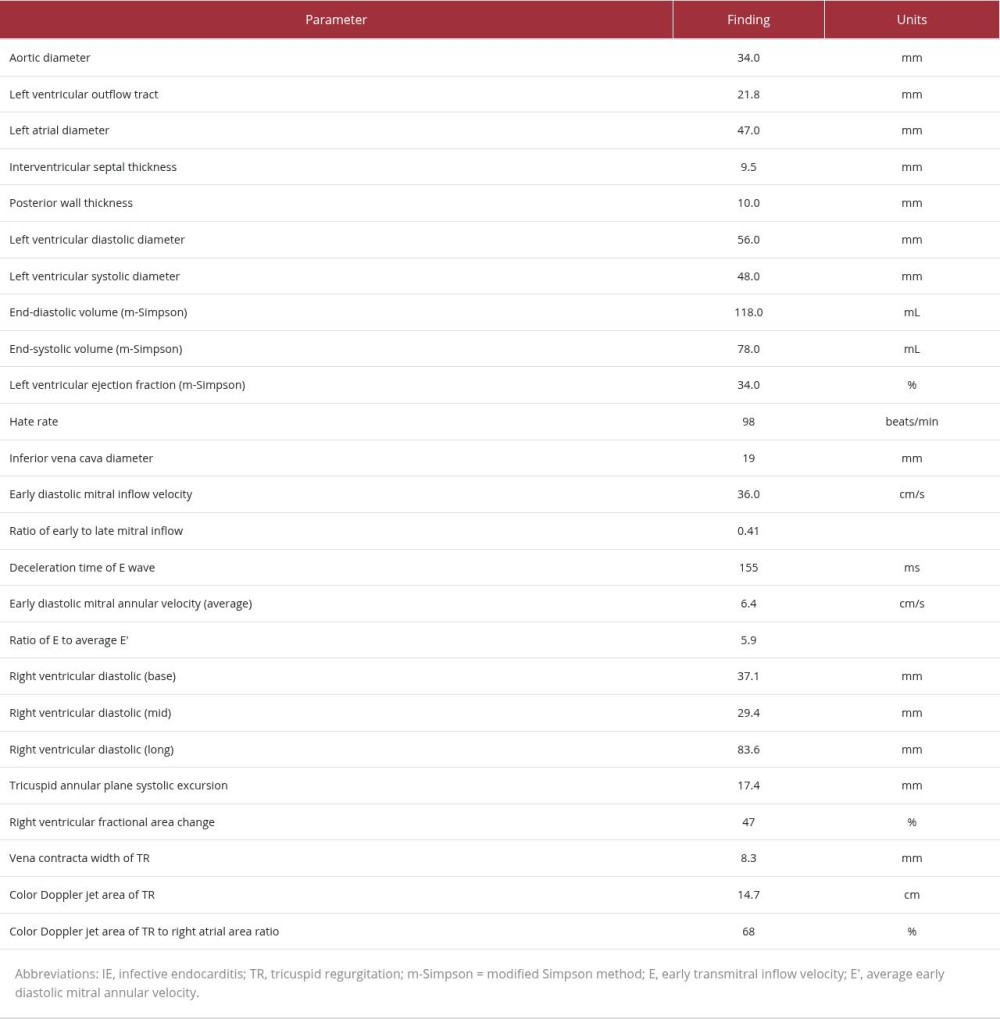
References
1. Ismailov RM, Ness RB, Lawrence BA, Miller TR, Blunt cardiac injury associated with cardiac valve insufficiency: Trauma links to chronic disease?: Injury, 2005; 36(9); 1022-28
2. Kakuda N, Nakayama A, Ishizuka M, Delayed diagnosis of severe traumatic tricuspid regurgitation after traffic injury: J Cardiol Cases, 2022; 25(4); 204-6
3. Buttiglione G, Hofer D, Hangler H, Bonaros N, Surgical management of traumatic tricuspid regurgitation: A case report: Eur Heart J Case Rep, 2025; 9(1); ytae676
4. Eranki A, Villanueva C, Wilson-Smith A, Seah P, Traumatic tricuspid valve regurgitation: A two case series: Trauma Case Rep, 2022; 37; 100593
5. Gayet C, Pierre B, Delahaye JP, Traumatic tricuspid insufficiency. An underdiagnosed disease: Chest, 1987; 92; 429-32
6. Kany S, Vollrath JT, Relja B, Cytokines in Inflammatory Disease: Int J Mol Sci, 2019; 20; 6008
7. Longfellow E, Aberle C, Lamelas J, Traumatic injury of the tricuspid valve-navigating the challenges in diagnosis and management: J Cardiothorac Vasc Anesth, 2022; 36; 906-14
8. Ma WG, Luo GH, Sun HS, Surgical treatment of traumatic tricuspid insufficiency: Experience in 13 cases: Ann Thorac Surg, 2010; 90; 1934-38
Figures
Figure 1. Chest X-ray and computed tomography findings in the emergency department.Computed tomography revealed the fracture of the left seventh rib (A, B), and chest X-ray revealed mild cardiomegaly without pleural effusion or pulmonary congestion (C). A red arrow and a circle indicate the fracture site.Figure 2. Transthoracic echocardiography findings on hospital day 2.Transthoracic echocardiography demonstrated a 7-mm mobile, mass-like structure attached to the flail anterior tricuspid leaflet (A, red dotted circle) with a severe eccentric regurgitant jet (B). Abbreviations: LA, left atrium; LV, left ventricle; RA, right atrium; RV, right ventricle.Figure 3. Intraoperative Image.The white arrow indicates the ruptured tip of the papillary muscle connected to the chordae tendineae of the anterior tricuspid leaflet, resulting in prolapse of the leaflet. Abbreviations: ATL, anterior tricuspid leaflet; RA, right atrium; RV, right ventricle.Figure 4. Microscopic image of the ruptures papillary muscle.Pathological examination showed coagulative necrosis.Video 1. The apical 4-chamber view on transthoracic echocardiography shows a 7-mm mobile mass-like structure attached to the anterior tricuspid leaflet, demonstrating pronounced flail motion.Video 2. A color Doppler image demonstrating pronounced tricuspid regurgitant. In Press
Case report 
Am J Case Rep In Press; DOI: 10.12659/AJCR.953543
Case report
Am J Case Rep In Press; DOI: 10.12659/AJCR.954216
Case report 
Am J Case Rep In Press; DOI: 10.12659/AJCR.954932
Case report 
Am J Case Rep In Press; DOI: 10.12659/AJCR.954146
Most Viewed Current Articles
07 Dec 2021 : Case report
28,712,695
DOI :10.12659/AJCR.934347
Am J Case Rep 2021; 22:e934347
06 Dec 2021 : Case report  180,721
DOI :10.12659/AJCR.934406
Am J Case Rep 2021; 22:e934406
21 Jun 2024 : Case report
124,419
DOI :10.12659/AJCR.944371
Am J Case Rep 2024; 25:e944371
08 May 2026 : Case report
95,462
DOI :10.12659/AJCR.952158
Am J Case Rep 2026; 27:e952158