


08 September 2026: Articles 
Vogt-Koyanagi-Harada Disease Presenting With Progressive Visual Loss and Multisystem Involvement: A Case Report
Challenging differential diagnosis, Rare disease, Educational Purpose (only if useful for a systematic review or synthesis)
Samah A. Elshweikh ABCDEFG 1,2, Atheer G. Almutairi ABCDEFG 3, Husna Irfan Thalib ABCDEFG 4*, Reema Waleed Alolayan ABCDEFG 3, Mona M. Aly ABCDEFG 5,6
DOI: 10.12659/AJCR.952534
Am J Case Rep 2026; 27:e952534


Abstract
BACKGROUND: Vogt-Koyanagi-Harada (VKH) disease is a rare autoimmune disorder targeting melanocyte-rich tissues that often presents with bilateral ocular inflammation and multisystem involvement. Early manifestations, such as headache and optic disc edema, can mimic other conditions, leading to diagnostic delays. Clinician awareness is essential, particularly in high-risk populations, to ensure timely intervention.
CASE REPORT: We report the case of a 55-year-old Indian man who presented with a 20-day history of progressive bilateral visual loss, headache, and hearing impairment. Ophthalmologic examination revealed hand motion vision in both eyes, anterior chamber inflammation, disc edema, and serous retinal detachments. Spectral-domain optical coherence tomography confirmed neurosensory detachment and disc edema, whereas B-scan ultrasonography demonstrated pronounced choroidal thickening. Brain and orbital magnetic resonance imaging revealed bilateral focal nodular choroidal lesions. Laboratory investigations excluded infectious, malignant, and systemic inflammatory etiologies. A diagnosis of VKH disease was established based on the clinical presentation, multimodal imaging findings, and revised diagnostic criteria. The patient was treated with high-dose intravenous methylprednisolone followed by a prolonged oral taper; azathioprine was initiated early as a steroid-sparing agent to prevent relapse.
CONCLUSIONS: This case underscores the importance of prompt ophthalmologic evaluation and multimodal imaging in the diagnosis of VKH disease. Early initiation of intensive immunosuppressive therapy may help limit inflammatory progression and reduce long-term complications.
Keywords: Autoimmune Diseases, Case Reports, Ophthalmology, Vision Disorders, Vogt-Koyanagi-Harada Disease
Introduction
Vogt-Koyanagi-Harada (VKH) disease is a rare multisystem autoimmune disorder that primarily affects melanocyte-rich tissues in the eyes, ears, and skin, often resulting in bilateral panuveitis, auditory involvement, and integumentary changes. It predominantly affects individuals of Asian, Middle Eastern, Hispanic, or Native American descent and has a recognized genetic predisposition linked to the HLA-DR4, HLA-DR53, and HLA-DQ4 alleles [1,2]. The disease typically progresses through 4 phases: a prodromal phase with flu-like or neurologic symptoms; an acute uveitic phase with bilateral panuveitis and serous retinal detachments; a convalescent phase with vitiligo, poliosis, and sunset glow fundus; and a chronic recurrent phase characterized by relapsing anterior uveitis [1,2].
VKH disease is responsible for a substantial proportion of uveitis cases worldwide [1,2]. Diagnosis is primarily clinical and based on revised international diagnostic criteria that incorporate ocular, neurologic, auditory, and integumentary findings. Early treatment involving high-dose systemic corticosteroids, often combined with immunomodulatory therapy, is the cornerstone of management to reduce chronic recurrence and prevent visual complications [3–5].
Despite its characteristic clinical course, early diagnosis can be challenging. Initial symptoms such as headache, neck stiffness, and optic disc edema often mimic other neurologic or ophthalmic conditions (eg, optic neuritis, idiopathic intracranial hypertension, and brain tumors), contributing to a reported misdiagnosis rate of approximately 9% [3,4]. Accurate and timely diagnosis relies on careful ophthalmologic evaluation, multimodal imaging (including optical coherence tomography and B-scan ultrasonography), and systemic assessment to exclude alternative causes. Early recognition and aggressive immunosuppressive therapy are essential to prevent disease progression [1,5,6].
In this report, we describe a 55-year-old Indian man who presented with rapidly progressive bilateral visual loss, headache, auditory symptoms, and ocular inflammation consistent with VKH disease. This case highlights the importance of prompt ophthalmologic evaluation and comprehensive systemic assessment in patients with suspected VKH disease.
Case Report
A 55-year-old Indian man with no significant medical history presented to the emergency department reporting 20 days of blurred vision in both eyes, accompanied by persistent headache, neck stiffness, and decreased hearing. The visual decline was gradual and progressive. The patient denied systemic symptoms such as fever, nausea, vomiting, or photophobia; there was no history of recent infection, ocular trauma, or initiation of new medications. He was admitted to the Internal Medicine Department for further investigation and management, including evaluation of possible intracranial hypertension or pseudotumor cerebri.
Upon initial presentation, ophthalmologic examination revealed hand motion visual acuity in both eyes and an intraocular pressure of 9 mm Hg bilaterally. Anterior segment evaluation demonstrated nonpigmented keratic precipitates, iris congestion with loss of the normal iris pattern, Koeppe nodules, sluggish pupillary reactions, and 2+ cells and flare in the anterior chamber of both eyes. Spectral-domain optical coherence tomography (Figure 1) showed multiple subretinal fluid compartments with fibrin-like septa and optic disc edema in both eyes. Fundus examination revealed 2+ vitreous haze, hyperemic optic disc edema, and serous retinal detachment in both eyes (Figure 2). B-scan ultrasonography demonstrated pronounced choroidal thickening, serous retinal detachment, and vitreous opacities. Indocyanine green angiography was not available at our facility.
On the day of presentation (hospital day 1), the patient underwent a comprehensive evaluation. Laboratory investigations ruled out infectious, malignant, and systemic inflammatory causes. Multidisciplinary consultations were obtained from the neurology, immunology, and otolaryngology services. HLA typing and genetic testing were not performed due to limited availability at our institution.
On hospital day 2, brain and orbital magnetic resonance imaging revealed bilateral focal nodular choroidal lesions, further supporting the diagnosis of VKH disease (Figure 3). The patient continued to experience persistent blurred vision and conjunctival hyperemia, prompting close monitoring.
Following the diagnosis, the patient received high-dose intravenous methylprednisolone (1 g/day for 3 days), followed by a slow oral steroid taper. Azathioprine was initiated early as a steroid-sparing immunomodulatory therapy. Topical 1% prednisolone acetate and 1% atropine sulfate eye drops were also administered.
After initiation of systemic corticosteroid therapy, ocular inflammation appeared to stabilize; however, visual recovery remained limited. Hand motion visual acuity was documented in both eyes at the last local follow-up visit. The patient subsequently requested a medical report to continue treatment in India, and no further follow-up data were available.
Discussion
This case highlights the diagnostic challenges of VKH disease when the initial presentation is dominated by neurologic symptoms (eg, headache, neck stiffness, and optic disc edema) before overt bilateral panuveitis becomes apparent. Early ophthalmologic evaluation and multimodal imaging were essential to establish the diagnosis and initiate prompt immunosuppressive therapy.
Ophthalmologic evaluation demonstrated classic findings of acute VKH disease, including bilateral granulomatous panuveitis, optic disc edema, vitreous haze, and serous retinal detachments. Spectral-domain optical coherence tomography confirmed multilobulated neurosensory retinal detachments, whereas B-scan ultrasonography demonstrated diffuse choroidal thickening. Although indocyanine green angiography was unavailable, the combination of multimodal imaging findings and the characteristic clinical presentation strongly supported the diagnosis of VKH disease.
This case illustrates the classic chronological progression of VKH disease. The initial prodromal phase was dominated by neurologic symptoms, followed by acute uveitis with serous retinal detachment and choroidal thickening [1,6]. Awareness of this temporal pattern, combined with careful ophthalmologic and systemic evaluation, was essential for an accurate diagnosis. VKH disease has been reported more frequently in Asian, Middle Eastern, Hispanic, and Native American populations, supporting the recognized role of genetic susceptibility [5,7,8].
Treatment was initiated with high-dose intravenous methylprednisolone (1 g/day for 3 days) to rapidly control inflammation, followed by a slow, prolonged oral taper to prevent relapse. Azathioprine was introduced early as a steroid-sparing immunomodulatory agent. Topical therapy with 1% prednisolone acetate and 1% atropine sulfate controlled anterior segment inflammation. This regimen is consistent with current recommendations to prevent chronic disease progression and irreversible visual loss [1,6,9–11]. Although long-term follow-up data for this patient were limited to the initial recovery period, the present case is consistent with previous reports indicating that early aggressive treatment is critical to prevent irreversible structural damage.
In our patient, the prodromal and acute uveitic phases appeared to substantially overlap. Although neurologic symptoms were the primary presenting complaint, the presence of bilateral hand motion vision upon admission suggested rapid transition to the acute uveitic phase.
The present case emphasizes that VKH disease should be considered in patients who present with bilateral ocular inflammation accompanied by neurologic or auditory symptoms [6,12,13]. Limitations include the unavailability of indocyanine green angiography, HLA typing, and cerebrospinal fluid analysis, which could have further clarified central nervous system involvement and genetic predisposition. Long-term follow-up data were not available, preventing evaluation of recurrence risk and sustained visual recovery.
Despite these limitations, our case demonstrates the educational and clinical value of early recognition of VKH disease, particularly in genetically predisposed populations; it underscores the importance of integrating detailed ophthalmologic examination, multimodal imaging, systemic evaluation, and multidisciplinary management.
Differential diagnoses included idiopathic intracranial hypertension, optic neuritis, sympathetic ophthalmia, sarcoidosis, and infectious uveitis. However, the presence of bilateral granulomatous panuveitis, serous retinal detachments, diffuse choroidal thickening, and associated neurologic and auditory manifestations supported a diagnosis of VKH disease based on the revised diagnostic criteria [14].
Conclusions
This case highlights the rapid progression of VKH disease from early ocular inflammation to multisystem involvement, including auditory and neurologic manifestations. The initial diagnostic ambiguity underscores the challenge of early detection when optic disc edema and headache precede overt panuveitis. Early recognition and prompt initiation of systemic immunosuppressive therapy remain essential to control inflammation and reduce the risk of irreversible ocular complications. We emphasize the need for rapid ophthalmologic assessment, multidisciplinary collaboration, and awareness of high-risk populations to improve clinical outcomes.
Figures
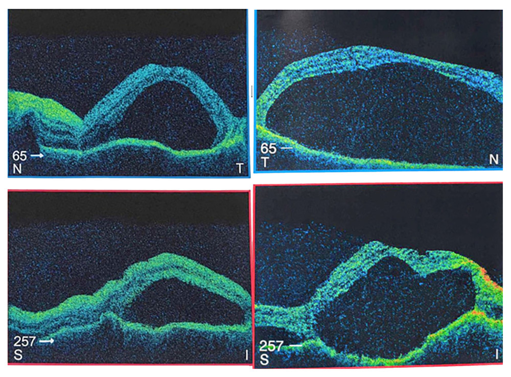 Figure 1. Spectral-domain optical coherence tomography of both eyes demonstrating multilobulated serous retinal detachments with subretinal fluid septations and optic disc edema, consistent with acute Vogt-Koyanagi-Harada disease.
 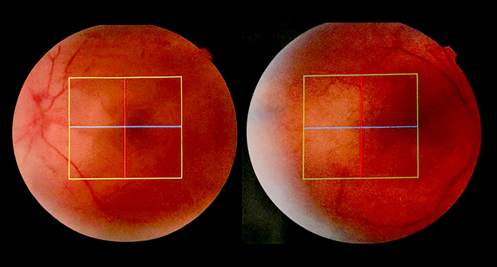 Figure 2. Color fundus photographs of both eyes demonstrating optic disc hyperemia, diffuse retinal elevation, and loss of the foveal contour associated with serous retinal detachment in acute Vogt-Koyanagi-Harada disease.
 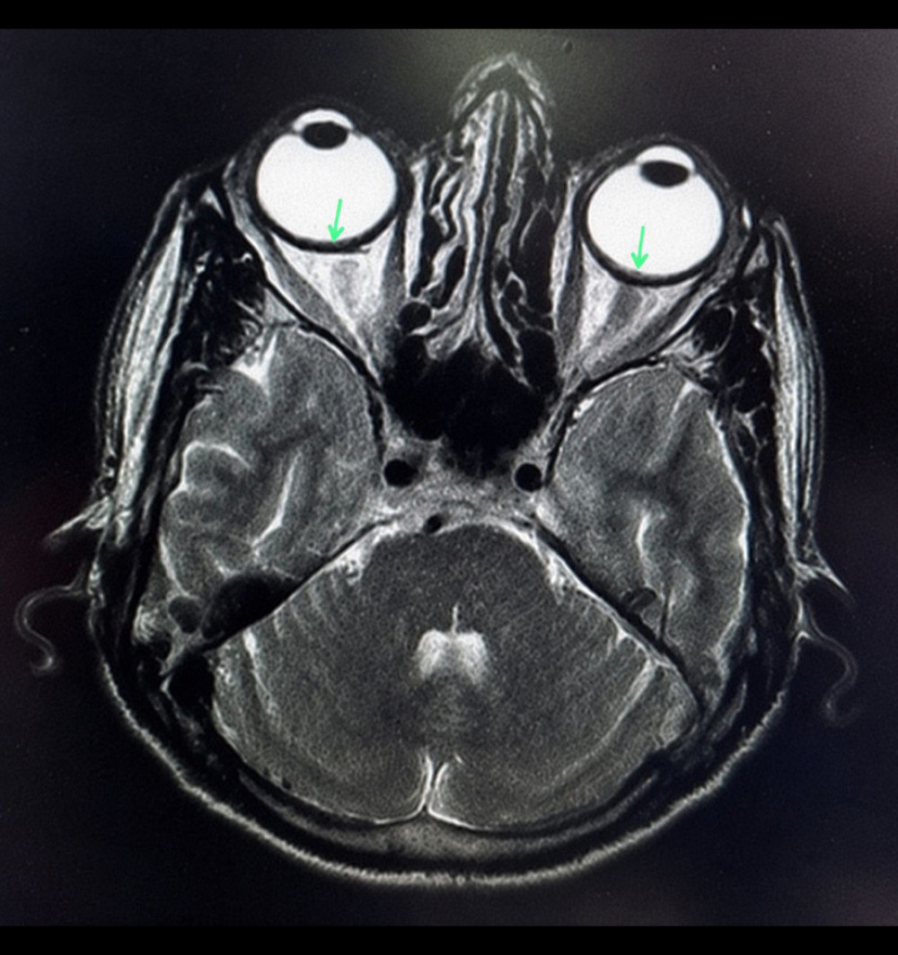 Figure 3. Magnetic resonance imaging of the brain and orbits demonstrating bilateral focal nodular choroidal lesions (arrows), supporting the presence of active choroidal inflammation in Vogt-Koyanagi-Harada disease.
 References
1. Stern EM, Tripathy K, Vogt-Koyanagi-Harada syndrome. [Updated 2026 Mar 1]: StatPearls [Internet], 2026, Treasure Island (FL), StatPearls Publishing
2. Moorthy RS, Inomata H, Rao NA, Vogt-Koyanagi-Harada syndrome: Surv Ophthalmol, 1995; 39(4); 265-92
3. Shoughy SS, Tabbara KF, Initial misdiagnosis of Vogt-Koyanagi-Harada disease: Saudi J Ophthalmol, 2019; 33(1); 52-55
4. Hussain A, Khurana R, Vogt-Koyanagi-Harada syndrome: A diagnostic conundrum: Cureus, 2021; 13(12); e20138
5. Prathyusha T, Asif M, Gadde ST, Vogt-Koyanagi-Harada syndrome: A case report: Cureus, 2024; 16(7); e64702
6. Bezci Aygun F, Akgoz Koyuncuoglu M, Kadayıfcılar S, Ozen S, Clinical characteristics and long-term outcomes of Vogt-Koyanagi-Harada disease in pediatric age group: BMC Ophthalmol, 2025; 25(1); 509
7. Zou H, Zhang K, Chen X, Sha S, Vogt-Koyanagi-Harada disease after SARS-CoV-2 infection: Case report and literature review: Immun Inflamm Dis, 2024; 12(4); e1250
8. Manni P, Saturno MC, Accorinti M, Vogt-Koyanagi-Harada disease and COVID: J Clin Med, 2023; 12(19); 6242
9. Hwang GE, Lee JW, Jeon S, Vogt-Koyanagi-Harada syndrome-like uveitis after nivolumab administration as a treatment for ovarian cancer: Doc Ophthalmol, 2022; 144(2); 153-62
10. Sood AB, O’Keefe G, Bui D, Jain N, Vogt-Koyanagi-Harada disease associated with hepatitis B vaccination: Ocul Immunol Inflamm, 2019; 27(4); 524-27
11. Kurono Y, Takeda T, Kunimatsu Y, Vogt-Koyanagi-Harada disease during chemoimmunotherapy for non-small cell lung cancer: Respirol Case Rep, 2020; 8(3); e00545
12. Al Hashmi S, Al Habsi N, Al Abrawi S, Vogt-Koyanagi-Harada syndrome (VKHS): First two cases reported in pediatric age group in Oman: Case Rep Pediatr, 2023; 2023; 1745603
13. Hernandez C, LePoole C, Tessler HH, Vogt-Koyanagi-Harada syndrome in a 6-year-old Hispanic boy: Pediatr Dermatol, 2012; 29(2); 191-94
14. Read RW, Holland GN, Rao NA, Revised diagnostic criteria for Vogt-Koyanagi-Harada disease: Report of an international committee on nomenclature: Am J Ophthalmol, 2001; 131(5); 647-52
Figures
Figure 1. Spectral-domain optical coherence tomography of both eyes demonstrating multilobulated serous retinal detachments with subretinal fluid septations and optic disc edema, consistent with acute Vogt-Koyanagi-Harada disease.Figure 2. Color fundus photographs of both eyes demonstrating optic disc hyperemia, diffuse retinal elevation, and loss of the foveal contour associated with serous retinal detachment in acute Vogt-Koyanagi-Harada disease.Figure 3. Magnetic resonance imaging of the brain and orbits demonstrating bilateral focal nodular choroidal lesions (arrows), supporting the presence of active choroidal inflammation in Vogt-Koyanagi-Harada disease. In Press
Case report 
Am J Case Rep In Press; DOI: 10.12659/AJCR.953555
Case report
Am J Case Rep In Press; DOI: 10.12659/AJCR.953169
Case report 
Am J Case Rep In Press; DOI: 10.12659/AJCR.954027
Case report
Am J Case Rep In Press; DOI: 10.12659/AJCR.954380
Most Viewed Current Articles
07 Dec 2021 : Case report  28,713,109
DOI :10.12659/AJCR.934347
Am J Case Rep 2021; 22:e934347
06 Dec 2021 : Case report  180,916
DOI :10.12659/AJCR.934406
Am J Case Rep 2021; 22:e934406
21 Jun 2024 : Case report
124,581
DOI :10.12659/AJCR.944371
Am J Case Rep 2024; 25:e944371
08 May 2026 : Case report
95,842
DOI :10.12659/AJCR.952158
Am J Case Rep 2026; 27:e952158