


13 August 2026: Articles 
Neonatal Endogenous Endophthalmitis Complicated by Spontaneous Globe Rupture: A Case Report
Unusual clinical course, Challenging differential diagnosis, Rare disease
Mary Robinson AE 1*, Apurwa Naik AE 2, Cigdem Ozturk AE 3, Kenneth Alexander A 4
DOI: 10.12659/AJCR.952748
Am J Case Rep 2026; 27:e952748


Abstract
BACKGROUND: This report presents a case of endophthalmitis complicated by spontaneous globe rupture to raise awareness concerning early signs of these extremely rare complications of sepsis in the neonatal population and to highlight key clinical management considerations, including prompt ophthalmologic evaluation, B-scan ultrasonography, intravitreal antibiotics, systemic antimicrobial therapy, and expedited planning for vitreoretinal and oculoplastic surgical interventions.
CASE REPORT: A neonate born at 22 weeks of gestation experienced clinically significant deterioration during week 7 of life due to sepsis and multisystem organ failure. During this period, drainage from the right eye was observed and initially suspected to represent conjunctivitis, prompting topical antimicrobial therapy. However, closer examination indicated that the extruded material likely represented the crystalline lens, vitreous humor, and choroidal tissue. B-scan ultrasonography demonstrated suspected bilateral intraocular infection with globe rupture of the right eye. Infectious studies revealed negative blood cultures, negative ocular specimen cultures, and positive cytomegalovirus testing. The infant ultimately succumbed to the systemic disease process before vitreoretinal surgical intervention could be performed.
CONCLUSIONS: Signs of endogenous endophthalmitis may be difficult to recognize in premature or low-birthweight infants; however, this diagnosis should be considered and prompt ophthalmologic evaluation pursued in the setting of systemic infection accompanied by new ocular manifestations. Comprehensive reviews of endogenous endophthalmitis cases with rare complications, including spontaneous globe rupture, are needed to promote earlier recognition and intervention in the neonatal population.
Keywords: Case Reports, Cytomegalovirus Infections, Endophthalmitis, Infant, Premature, Ophthalmology, sepsis
Introduction
Most pediatric open globe injuries occur after ocular trauma, and penetrating injury represents the most common etiology [1]. Trauma may be accidental, non-accidental, or iatrogenic in nature [2,3]. Additional etiologies of globe rupture in neonates include congenital anomalies and infections. One such anomaly, Peters anomaly, is characterized by anterior segment mesenchymal dysgenesis. Ocular findings associated with this genetic disorder include central corneal opacities, glaucoma, and serious defects of Descemet’s membrane and the corneal endothelium [4]. From an infectious disease perspective, corneal perforation is a documented but rare complication of endophthalmitis [5,6].
Endophthalmitis, an inflammatory process involving the intraocular vitreous cavity, may arise from exogenous or endogenous infections. Exogenous sources of infectious organisms include penetrating trauma, intraocular foreign bodies, ocular procedures, and infection of adjacent tissues [7]. In neonates, endogenous bacterial and fungal seeding of the eye can occur in the setting of systemic infection [5,6]. Systemic viral infections, including congenital and acquired cytomegalovirus (CMV) and dengue fever, have also been associated with ocular manifestations [8,9]. Congenital CMV acquired via transplacental transmission can result in chorioretinitis with associated scarring, optic nerve atrophy, and cortical visual impairment. CMV is a recognized cause of retinitis in immunocompromised hosts, as well as a cause of iridocyclitis and corneal endotheliitis in both immunocompromised and immunocompetent individuals [8]. A recent case report by Kaur et al described dengue hemorrhagic shock syndrome complicated by spontaneous globe rupture in a pediatric patient [9].
Neonates hospitalized in the neonatal intensive care unit (NICU) have numerous risk factors for sepsis and systemic infection, including prematurity, low birth weight, immature immune systems, use of indwelling and central catheters, endotracheal intubation, need for invasive procedures, exposure to multidrug-resistant nosocomial organisms, and prolonged hospitalization [10]. These perinatal infectious risk factors, combined with compromise of other organ systems, may increase the risk of endogenous endophthalmitis in the NICU population [5].
This case involving a preterm neonate illustrates the presentation and clinical evaluation of endogenous endophthalmitis complicated by spontaneous globe rupture in the setting of shock and CMV viremia.
Case Report
A female neonate born at 22 weeks of gestation was delivered to a 25-year-old mother after spontaneous preterm labor. The pregnancy was complicated by twin gestation and absence of prenatal care. This infant, Twin B, was delivered vaginally; positive-pressure ventilation and endotracheal intubation were required immediately after birth. She was transferred to the NICU due to prematurity, extremely low birth weight (490 g), and respiratory failure.
The infant’s perinatal course was complicated by multiple respiratory, cardiac, neurologic, hematologic, and infectious conditions. Respiratory status was severely compromised by hypoxic respiratory failure warranting surfactant administration, systemic corticosteroid therapy, and trials of multiple ventilatory support modalities. Cardiac findings included a secundum atrial septal defect and large patent ductus arteriosus. The patent ductus arteriosus was treated with acetaminophen, resulting in documented closure on subsequent echocardiograms. Neurologic complications included severe bilateral intraventricular hemorrhages. Anemia of prematurity and thrombocytopenia required multiple blood product transfusions throughout the NICU course. Notably, Twin A died during the first week of life.
From an infectious disease perspective, maternal laboratory studies obtained at delivery did not show hepatitis B, human immunodeficiency virus, or syphilis; the mother displayed rubella immunity. The infant received hepatitis B immunoglobulin and hepatitis B vaccination while awaiting maternal laboratory results, along with empiric intravenous (IV) ampicillin and gentamicin until blood cultures demonstrated no growth. CMV testing upon NICU admission showed negative results. On day 9 of life, a sepsis evaluation was initiated due to persistent hyperglycemia requiring insulin infusion. The evaluation was notable for
During week 7 of life, the infant developed bilateral ocular discharge initially suspected to represent conjunctivitis (Figure 1), prompting initiation of topical polymyxin therapy. While attempting to clean drainage from the right eyelid, a clear, round tissue-like structure was extruded from the eye (Figure 2A, 2B). This was followed by continued extrusion of a clear gel-like substance and dark, string-like material (Figure 2C). An ophthalmology consultation was obtained; based on evaluations of the patient and extruded material, there was strong suspicion that the crystalline lens had been expelled, followed by vitreous humor and choroidal tissue. Unfortunately, specimens were unable to be collected for pathological analysis.
Physical examination revealed enophthalmos of the right eye, which was softer on palpation relative to the left eye. Due to pronounced bilateral chemosis, anterior and posterior segment structures could not be visualized. A more detailed examination was deferred given the risk of iatrogenic injury. Bedside B-scan ultrasonography demonstrated bilateral intravitreal condensations and distortion of the right globe structure (Figure 3). Clinical and radiographic findings were consistent with right globe rupture and suspected bilateral intraocular infection.
Considering the urgency of the ophthalmologic findings, expedited transfer to a facility with vitreoretinal and oculoplastic surgical capabilities was arranged so that the infant could undergo evaluation for potential vitreoretinal procedures and/or evisceration. Recommended intravitreal antibiotic injections could not be performed due to severe bilateral chemosis. Systemic IV piperacillin-tazobactam was initiated to provide broad-spectrum gram-positive and gram-negative antibacterial coverage, including
Concurrently, the patient developed worsening transaminitis, oxygen desaturations, thrombocytopenia, and gross hematochezia. Evaluation for necrotizing enterocolitis was initiated, and initial abdominal radiographs raised concern for pneumatosis. Enteral feeds were paused, and IV piperacillin-tazobactam was continued. However, repeat imaging demonstrated no evidence of pneumoperitoneum or portal venous gas; abdominal ultrasonography did not identify pneumatosis. Given that right upper quadrant ultrasonography did not demonstrate a hepatic abscess as a source of infection or worsening transaminitis, blood cultures and urine CMV testing were performed. Additional imaging (eg, chest X-ray and echocardiography) did not identify evidence of bacterial pneumonia or endocarditis as underlying infectious sources. Antimicrobial coverage was broadened to include IV vancomycin and amphotericin. Initial and repeat blood cultures during this period showed negative results; however, urine CMV test results were positive. The infant continued to deteriorate clinically, with development of cyanosis and eventual necrosis of the distal left upper extremity (Figure 4), severe hypoxic and ventilatory respiratory failure refractory to multiple modes of respiratory support, and oliguria.
Because of these complications, the patient did not show sufficient clinical stability for transport to a facility with ophthalmologic surgical capabilities. After discussion with the family, the decision was made to withdraw ventilatory support, and the infant subsequently died. The family declined autopsy.
Discussion
Spontaneous globe rupture is a rare complication of intraocular infection and indicates advanced disease. In the absence of known ocular trauma, as in the present case, endogenous spread of an infectious organism should be suspected. The causative pathogen may be viral, bacterial, or fungal.
In cases of bacterial endophthalmitis, hematogenous spread is often associated with identifiable extraocular foci of infection, such as liver abscess, pneumonia, endocarditis, soft tissue infection, urinary tract infection, or meningitis [11]. For the infant in the present case, suspected necrotizing enterocolitis may have represented a source of bacteremia. Imaging studies obtained after spontaneous globe rupture did not indicate other extraocular infections, including pneumonia, liver abscess, or endocarditis. Within a prospective case series of endogenous bacterial endophthalmitis, Jackson et al reported positive blood cultures in approximately 75% of cases [11]. Blood cultures were more likely to show positive results than were cultures obtained from intraocular specimens, which yielded bacterial growth in only 56% of cases [11]. In the present case, blood cultures obtained during the initial clinical decline, as well as repeat cultures, consistently showed negative results. Cultures from right eye specimens also showed negative results. Although the infant had been treated for meningitis, post-treatment cerebrospinal fluid analysis showed no evidence of persistent infection. Nevertheless, the infant displayed substantial risk of severe bacterial infection due to prematurity, prolonged NICU hospitalization, and the use of multiple indwelling catheters. Therefore, bacterial endophthalmitis remains a plausible etiology despite negative culture results.
Endophthalmitis secondary to a viral pathogen is also a consideration in this case. Initial CMV screening results after birth were negative; however, repeat testing performed in the setting of thrombocytopenia, colitis, hepatitis, and sepsis at 7 weeks postnatal age showed positive results. Therefore, the infant most likely exhibited a symptomatic acquired CMV infection [12]. Premature and low-birthweight infants display increased risks of life-threatening manifestations of acquired CMV. NICU transmission can occur via transfusion of infected blood products or exposure to infected breast milk [13]. The infant in the present case may have initially developed chorioretinitis, a known complication of CMV viremia, which subsequently progressed to vitreous cavity involvement, leading to endophthalmitis and eventual globe rupture. Unfortunately, viral testing of expelled intraocular contents was not performed before the infant’s death because urine CMV results were not yet available to guide further diagnostic evaluation. Notably, progression from chorioretinitis to endophthalmitis has been documented in cases involving fungal pathogens [14].
In the present case, fungal blood cultures were not obtained. However, no systemic improvement was observed after initiation of antifungal therapy. Further investigation of the causative pathogen was limited by the rapid clinical decline and the family’s decision to decline autopsy.
Among verbal patients able to report symptoms, warning signs of endophthalmitis may include visual disturbances and ocular pain. However, among patients unable to recognize or communicate these symptoms, alternative findings such as conjunctival chemosis and injection may serve as early indicators [11]. In these populations—including neonates, individuals receiving partial or complete sedation, adults or children with communication limitations, and patients with pre-existing vision impairment—a thorough physical examination is essential.
Closer examination of the affected eye may reveal evidence of anterior chamber inflammation and hypopyon. Intraocular inflammation can also result in a diminished red reflex, increased intraocular pressure, and/or poor fundus visualization on ophthalmologic examination [11]. In the NICU population, eyelid fusion is developmentally common among premature infants born prior to 26 weeks of gestation; thus, ocular findings associated with endophthalmitis may be difficult to detect before this corrected gestational age [15]. Even among infants undergoing routine screening for retinopathy of prematurity, the American Academy of Pediatrics does not recommend initiating ophthalmologic examinations until corrected gestational age reaches 31 weeks [16]. Therefore, any neonate presenting with chemosis, conjunctivitis, or ocular discharge should undergo prompt ophthalmologic evaluation to exclude a progressive intraocular process.
Conclusions
Premature infants in the NICU possess numerous risk factors for serious bacterial, viral, and fungal infections. Although endophthalmitis is rare, it should be considered with prompt evaluation in the setting of systemic infection accompanied by new ocular manifestations. The ability to intervene in these ophthalmologic emergencies with vision-sparing therapies depends on timely recognition of clinical signs. Globe rupture, an extremely rare complication of neonatal endophthalmitis, is infrequently described in the literature and should be considered a late manifestation of intraocular infection. However, as in the present case, findings may be mistaken for more common superficial conditions (eg, conjunctivitis) in a population of premature neonates with limited opportunities for ocular screening examinations. The present report highlights this severe outcome and contributes to the clinical framework guiding surveillance for endophthalmitis and globe rupture in the NICU population. Further investigations regarding common causative pathogens and the pathophysiology of ocular pathogen seeding may help promote earlier diagnosis and more effective treatment strategies for these rare but devastating conditions.
Figures
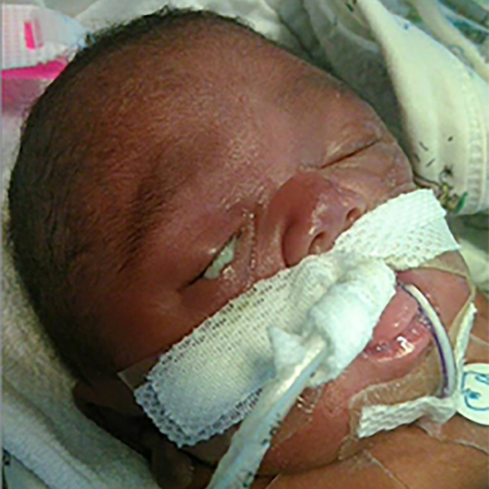 Figure 1. Photograph showing drainage spontaneously seeping from the right eye on day 48 of life.
 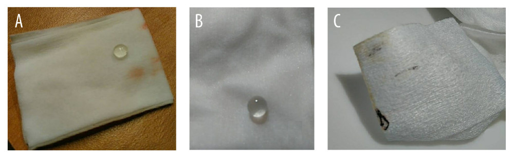 Figure 2. Photographs of collected contents expelled from the right eye. (A, B) Clear, firm tissue maintaining its shape after expulsion from the right eye, suspected to represent the crystalline lens. (C) Clear gel-like material and hyperpigmented tissue expelled from the right eye, suspected to represent vitreous humor and choroidal tissue.
 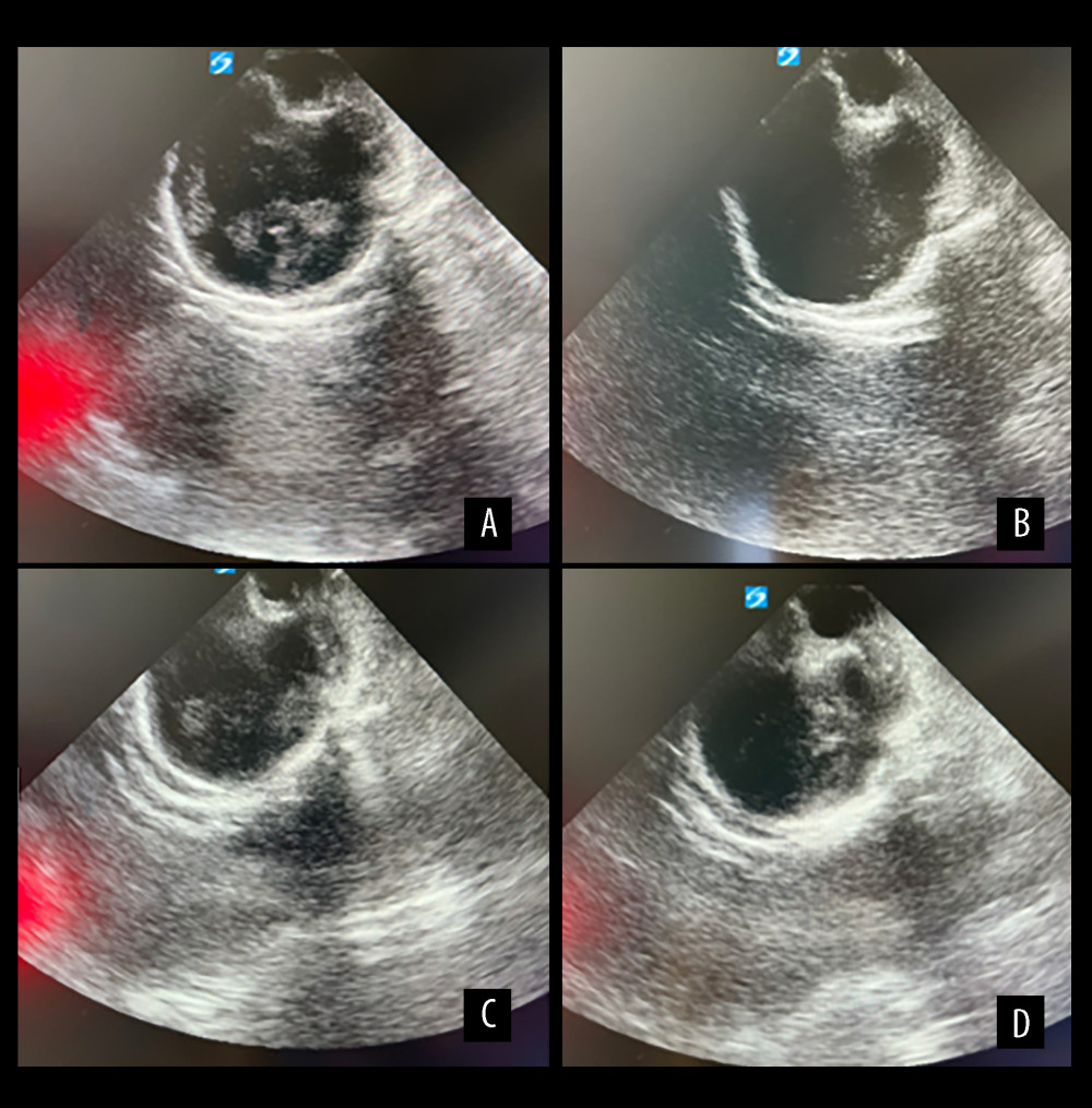 Figure 3. B-scan ultrasonography of the right eye demonstrating intravitreal condensations and distorted globe architecture (A, B), and of the left eye demonstrating intravitreal condensations (C, D).
 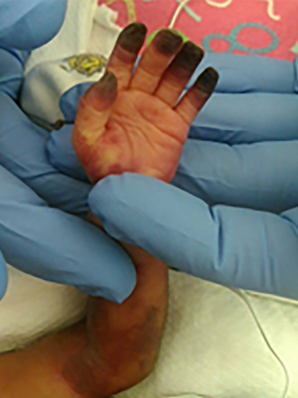 Figure 4. Photograph of the left upper extremity demonstrating pronounced bruising and necrosis of the fingertips.
 References
1. Li X, Zarbin MA, Bhagat N, Pediatric open globe injury: A review of the literature: J Emerg Trauma Shock, 2015; 8; 216-23
2. Skarbek-Borowska SE, Campbell KT, Globe rupture and nonaccidental trauma: Two case reports: Pediatr Emerg Care, 2011; 27; 544-46
3. Castellarin A, Lipskey S, Sternberg P, Iatrogenic open globe eye injury following sinus surgery: Am J Ophthalmol, 2004; 137; 175-76
4. Alallah J, Mohtisham F, Khan Z, Spontaneous bilateral corneal perforation in a neonate: A case report: Int J Pediatr Adolesc Med, 2020; 7; 53-55
5. Al-Khersan H, Pirakitikulr N, Kalavar M, Primary evisceration for neonatal endogenous endophthalmitis: A report of two cases: Am J Ophthalmol Case Rep, 2021; 22; 101081
6. Basu S, Kumar A, Kapoor K, Neonatal endogenous endophthalmitis: A report of six cases: Pediatrics, 2013; 131; e1292-97
7. Kernt M, Kampik A, Endophthalmitis: Pathogenesis, clinical presentation, management, and perspectives: Clin Ophthalmol, 2010; 4; 121-35
8. Kobayashi R, Hashida N, Overview of cytomegalovirus ocular diseases: Retinitis, corneal endotheliitis, and iridocyclitis: Viruses, 2024; 16; 1110
9. Kaur R, Singh H, Sehgal A, Singh J, Spontaneous globe rupture: unusual ophthalmic manifestation with dengue hemorrhagic shock syndrome: Am J Ophthalmol Case Rep, 2022; 25; 101297
10. Camacho-Gonzalez A, Spearman PW, Stoll BJ, Neonatal infectious diseases: Evaluation of neonatal sepsis: Pediatr Clin North Am, 2013; 60; 367-89
11. Jackson TL, Eykyn SJ, Graham EM, Stanford MR, Endogenous bacterial endophthalmitis: A 17-year prospective series and review of 267 reported cases: Surv Ophthalmol, 2003; 48; 403-23
12. Schuster K, Goelz R, Speckmann C, Henneke P, Symptomatic cytomegalovirus infections in the first year of life: When is antiviral therapy conceived to be justified?: Pediatr Infect Dis J, 2017; 36; 224-27
13. Schleiss MR, Cytomegalovirus in the neonate: Immune correlates of infection and protection: Clin Dev Immunol, 2013; 2013; 501801
14. Lavine JA, Mititelu M: J Ophthalmic Inflamm Infect, 2015; 5; 54
15. Duerksen K, Barlow WE, Stasior OG, Fused eyelids in premature infants: Ophthalmic Plast Reconstr Surg, 1994; 10; 234-40
16. Fierson WMAmerican Academy of Pediatrics Section on Ophthalmology; American Academy of Ophthalmology; American Association for Pediatric Ophthalmology and Strabismus; American Association of Certified Orthoptists, Screening examination of premature infants for retinopathy of prematurity: Pediatrics, 2018; 142(6); e20183061 [published erratum appears in Pediatrics. 2019;143(3):e20183810]
Figures
Figure 1. Photograph showing drainage spontaneously seeping from the right eye on day 48 of life.Figure 2. Photographs of collected contents expelled from the right eye. (A, B) Clear, firm tissue maintaining its shape after expulsion from the right eye, suspected to represent the crystalline lens. (C) Clear gel-like material and hyperpigmented tissue expelled from the right eye, suspected to represent vitreous humor and choroidal tissue.Figure 3. B-scan ultrasonography of the right eye demonstrating intravitreal condensations and distorted globe architecture (A, B), and of the left eye demonstrating intravitreal condensations (C, D).Figure 4. Photograph of the left upper extremity demonstrating pronounced bruising and necrosis of the fingertips. In Press
Case report 
Am J Case Rep In Press; DOI: 10.12659/AJCR.953555
Case report
Am J Case Rep In Press; DOI: 10.12659/AJCR.953169
Case report 
Am J Case Rep In Press; DOI: 10.12659/AJCR.954027
Case report
Am J Case Rep In Press; DOI: 10.12659/AJCR.954380
Most Viewed Current Articles
07 Dec 2021 : Case report
28,713,109
DOI :10.12659/AJCR.934347
Am J Case Rep 2021; 22:e934347
06 Dec 2021 : Case report  180,916
DOI :10.12659/AJCR.934406
Am J Case Rep 2021; 22:e934406
21 Jun 2024 : Case report
124,581
DOI :10.12659/AJCR.944371
Am J Case Rep 2024; 25:e944371
08 May 2026 : Case report
95,842
DOI :10.12659/AJCR.952158
Am J Case Rep 2026; 27:e952158