


15 August 2026: Articles 
Single-Site ALK-Positive Histiocytosis With TFG-ALK Fusion: Expanding the Clinical Spectrum of This Rare Entity
Rare disease
Mona Dasgupta BCDEF 1*, David Horvath BCDE 2, Benjamin Bevill A 1, Ashley Scheiderer BCDE 2, Sean Jordan AEF 3DOI: 10.12659/AJCR.952807
Am J Case Rep 2026; 27:e952807


Abstract
BACKGROUND: Anaplastic lymphoma kinase (ALK)-positive histiocytosis is a rare and recently described histiocytic neoplasm characterized by ALK gene rearrangements. Several ALK fusion partners have been reported, particularly in patients with multisystem disease. We report the first case of single-site ALK-positive histiocytosis involving a TFG-ALK fusion, a rearrangement previously linked to disseminated disease.
CASE REPORT: A man in his 50s with obesity, obstructive sleep apnea, diabetes mellitus, hypertension, coronary artery disease, and a 40-pack-year smoking history underwent computed tomography for lung cancer screening; the results showed a solitary 1.6-cm left lower lobe pulmonary nodule. Positron emission tomography demonstrated hypermetabolic activity confined to the lesion. He subsequently underwent video-assisted thoracoscopic wedge resection with mediastinal lymphadenectomy. Histopathologic evaluation revealed a well-circumscribed spindle cell neoplasm expressing histiocytic markers CD4, CD68, and CD163, with cytoplasmic ALK positivity. A TFG-ALK gene fusion was detected using Archer FusionPlex Pan Solid Tumor panel testing with anchored multiplex polymerase chain reaction via Illumina NextSeq 500. Immunohistochemical and flow cytometric studies excluded alternative ALK-rearranged neoplasms, including inflammatory myofibroblastic tumor, anaplastic large cell lymphoma, and ALK-positive diffuse large B-cell lymphoma. The patient received no systemic therapy and remains disease-free on radiographic surveillance.
CONCLUSIONS: To our knowledge, this case represents the first reported instance of single-site ALK-positive histiocytosis with a TFG-ALK fusion. It expands the molecular and clinical spectrum of this rare entity, suggesting that certain ALK fusion partners are associated with localized disease and an indolent clinical course. Recognition of such presentations is essential to avoid unnecessary systemic therapy.
Keywords: Anaplastic Lymphoma Kinase, Gene Fusion, Histiocytosis, Immunohistochemistry, Thoracic Surgery
Introduction
Anaplastic lymphoma kinase (ALK)-positive histiocytosis is a relatively recently described clonal neoplasm characterized by histiocytes expressing abnormal ALK fusion proteins capable of clonal proliferation and tissue infiltration. The largest published cohort study by Kemps et al included 39 pediatric and adult patients, highlighting the rarity of this disease [1,2]. In that study, KIF5B-ALK was the most commonly reported fusion partner, identified in 27 patients, followed by 5 patients with ALK fluorescence in situ hybridization positivity (ALK-FISH+). Additional ALK fusion partners included CLTC-ALK, TPM3-ALK, EML4-ALK, DCTN1-ALK, and TRK-fused gene (TFG)-ALK. Clinically, ALK-positive histiocytosis demonstrates a broad spectrum of presentations, ranging from isolated single-organ lesions to diffuse multisystem disease [1]. The nervous system, liver, lung, bone, and skin are the most commonly involved organs [1,2]. Among reported ALK rearrangements, TFG-ALK appears to be less frequently identified and has primarily been documented in cases of multisystem disease.
Here, we report a case of single-site ALK-positive histiocytosis harboring a TFG-ALK fusion in a man in his 50s. To our knowledge, this represents the first recorded case of single-site ALK-positive histiocytosis with this specific fusion. The benign clinical course and apparent cure after surgical resection in our patient suggest that not all TFG-ALK rearrangements result in disseminated or aggressive disease.
Case Report
A man in his 50s with obesity, obstructive sleep apnea, diabetes mellitus, hypertension, coronary artery disease, and a 40-pack-year smoking history was referred to the thoracic surgery clinic after a low-dose computed tomography (CT) scan performed for lung cancer screening revealed a 1.6-cm left lower lobe pulmonary nodule (Figure 1). CT-guided biopsy demonstrated a spindle cell neoplasm. A positron emission tomography scan performed before surgical evaluation showed hypermetabolic activity confined to the left lower lobe nodule. Laboratory findings were unremarkable.
The patient underwent video-assisted thoracoscopic surgery with left lower lobe wedge resection and mediastinal lymphadenectomy for diagnostic and curative purposes. Pathologic examination revealed a well-circumscribed lesion composed of uniform spindle cells expressing histiocytic markers CD4, CD68, and CD163 (Figure 2), with positive cytoplasmic staining for ALK (CD246). A TFG-ALK gene fusion was detected using the Archer FusionPlex Pan Solid Tumor panel, an anchored multiplex polymerase chain reaction method, on the Illumina NextSeq 500 platform. Immunohistochemistry findings for CD1a, a key marker of Langerhans cell differentiation (Figure 3), smooth muscle actin, and desmin were negative, thereby excluding Langerhans cell histiocytosis, smooth muscle neoplasms, and fibroblastic/myofibroblastic tumors such as inflammatory myofibroblastic tumor (IMT). Tumor cells also showed CD30, CD20, CD43, CD3, CD5, and CD7 negativity according to immunohistochemistry. Additionally, flow cytometry performed on a peripheral blood sample showed no evidence of hematolymphoid neoplasia, making anaplastic large cell lymphoma (ALCL) and ALK-positive diffuse large B-cell lymphoma (DLBCL) unlikely. Based on the combined morphologic and immunohistochemical profile, a diagnosis of single-system ALK-positive histiocytosis with TFG-ALK rearrangement was established.
The patient was referred to malignant hematology for further evaluation. He did not receive systemic therapy and remains under surveillance with non-contrast chest CT scans every 6 months for the first 2 years, followed by annual scans for a minimum of 5 years.
Discussion
ALK-positive histiocytosis is a rare histiocytic neoplasm. The disease was first reported in 2008 in 3 infants with multisystem disease involving the liver and hematopoietic system [2]. ALK-positive histiocytosis can be categorized into 3 clinical groups: infants with multisystem disease involving the liver and hematopoietic system (group 1A), noninfants with multisystem disease (group 1B), and patients with single-system disease (group 2) [2].
The diagnostic criteria for ALK-positive histiocytosis applied in previously published case reports include: 1) cytoplasmic or membranous ALK immunoreactivity and/or molecular confirmation of ALK rearrangement, and 2) confirmation of histiocytic differentiation through lesional cell expression of at least 2 histiocytic markers (CD163, CD68, CD14, or CD4) [2]. These criteria were met in the present case.
Given the rarity of this entity, exclusion of other ALK-rearranged neoplasms (eg, IMT, ALCL, and ALK-positive DLBCL) is essential when establishing the diagnosis. A summary of the key differential diagnoses and distinguishing features is provided in Table 1. Smooth muscle actin and desmin are present in a substantial proportion of IMTs; however, both markers were absent in the present case. Additionally, S100 immunoreactivity, which showed patchy positivity in this case (Figure 4), has rarely been reported in IMT [3]. Negative expression results concerning T-cell and B-cell markers, as well as CD30, strongly argue against ALCL and DLBCL.
TFG-ALK fusion has been identified in only 2 previously reported cases. The first involved a man in his 20s with multisystem disease affecting the liver, skin, and colorectum; he was treated with corticosteroids and achieved a partial response but subsequently relapsed and died of sepsis [2]. The second case involved a man in his 30s with involvement of the lumbar spine, pelvic bones, proximal femurs, and peritoneum [4]. This patient initially received pegylated interferon-α (PEG-IFN-α) for 2 months with limited response, followed by alectinib therapy with a favorable response, as demonstrated by a reduction in the number of hypermetabolic osteolytic lesions on positron emission tomography imaging. At the time of publication, the patient remained on alectinib therapy [4].
In contrast to these reports, our patient exhibited isolated single-site disease confined to the lung, representing the first documented case of localized ALK-positive histiocytosis harboring a TFG-ALK fusion. Our report is also the first to describe disease resolution after surgical resection. These findings suggest that TFG-ALK rearrangements are associated with a broader clinical spectrum than previously recognized, ranging from disseminated multisystem disease to localized lesions amenable to surgical cure. At present, it remains unclear whether specific ALK fusion partners influence disease distribution, clinical behavior, or prognosis in ALK-positive histiocytosis. Larger case series with molecular characterization will be necessary to determine whether genotype-phenotype correlations exist.
Pulmonary involvement in ALK-positive histiocytosis is rare but recognized. It more commonly presents as multiple pulmonary nodules rather than a solitary lesion. KIF5B-ALK remains the most common fusion identified in both single- and multiple-nodule presentations.
For patients with localized single-system disease, complete surgical resection is often curative [2]. Our patient has remained disease-free since wedge resection. In cases in which resection is not feasible, treatment with ALK inhibitors has shown efficacy. Management of multisystem disease may include surgery, corticosteroids, chemotherapy, intravenous immunoglobulin, and ALK-targeted therapy [2,5].
Conclusions
Although understanding of ALK-positive histiocytosis continues to evolve, its clinicopathologic and molecular spectrum remains incompletely characterized. Recognition of localized forms is crucial to avoid unnecessary systemic therapy. This case underscores that ALK-positive histiocytosis can manifest as single-site disease, even with rare fusion partners such as TFG-ALK, and may follow an indolent clinical course.
Figures
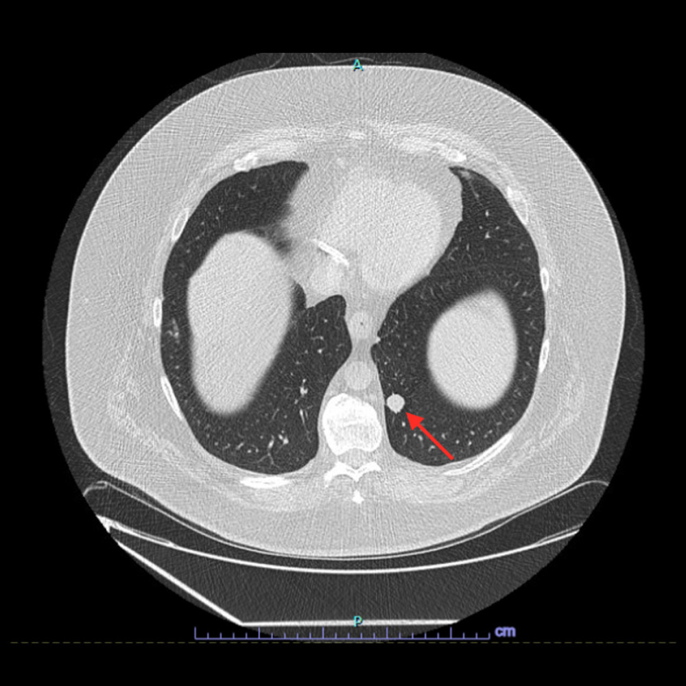 Figure 1. Low-dose computed tomography scan demonstrating a 1.6-cm left lower lobe pulmonary nodule (arrow).
 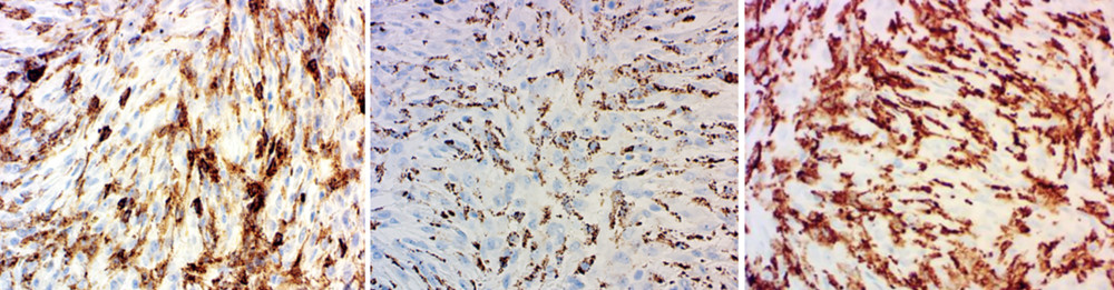 Figure 2. Immunohistochemical staining at 400× magnification showing positive expression of CD4, CD68, and CD163 from left to right, confirming histiocytic differentiation.
 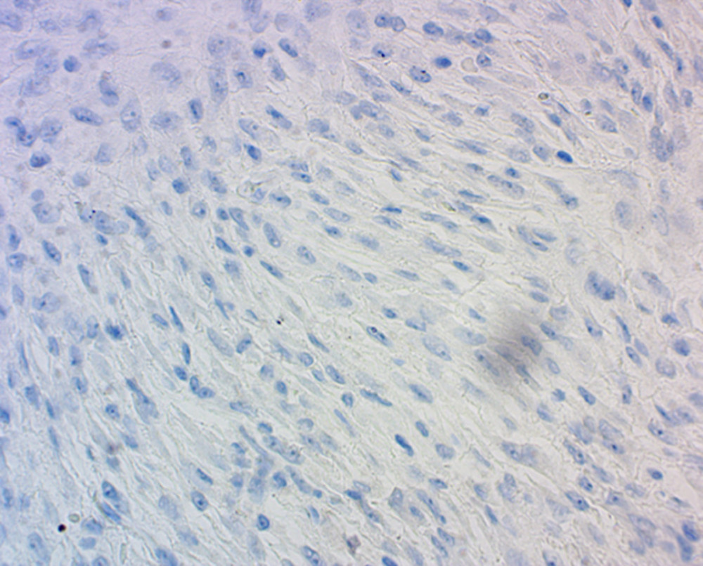 Figure 3. Immunohistochemical staining at 400× magnification showing absence of CD1a immunoreactivity, an important marker of Langerhans cell differentiation.
 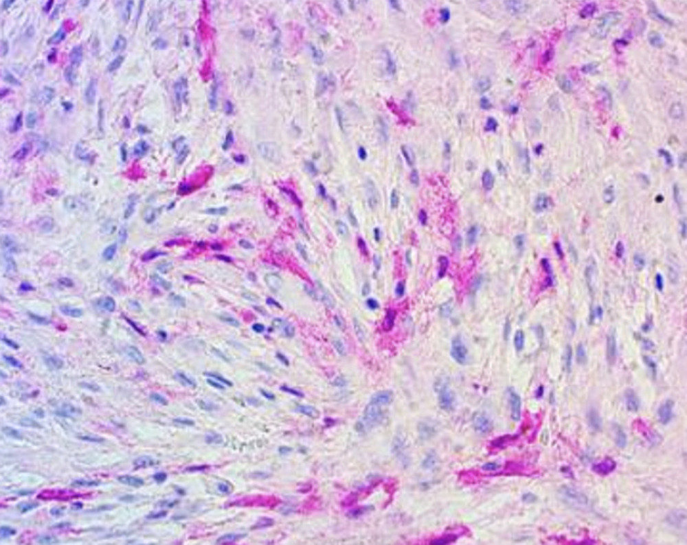 Figure 4. Immunohistochemical staining at 400× magnification demonstrating patchy cytoplasmic and nuclear S100 protein expression.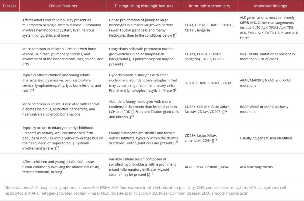
References
1. Breimer GE, Rozeman EA, Braunius WW, ALK-positive histiocytosis and a clonally related chronic myelomonocytic leukemia: Haematologica, 2026; 111(1); 423-26
2. Kemps PG, Picarsic J, Durham BH, ALK-positive histiocytosis: A new clinicopathologic spectrum highlighting neurologic involvement and responses to ALK inhibition: Blood, 2022; 139(2); 256-80
3. Wang QA, Wu RC, Lee CW, Clinicopathological features and immunochemical staining of inflammatory myofibroblastic tumor: A retrospective study of 48 cases: Anal Cell Pathol (Amst), 2025; 2025; 4948627
4. Lee JY, Lee KR, Na SY, ALK-positive adult histiocytosis with a TFG-ALK fusion gene: Oncologist, 2025; 30(9); oyaf221
5. Liu W, Liu HJ, Wang WY, Multisystem ALK-positive histiocytosis: A multi-case study and literature review: Orphanet J Rare Dis, 2023; 18; 53
6. McKinney RA, Wang G, Langerhans cell histiocytosis and other histiocytic lesions: Head Neck Pathol, 2025; 19(1); 26
7. Scurtu LG, Scurtu F, Simionescu O, Unraveling cutaneous histiocytosis: Insights into histology, pathogenesis, diagnosis, and treatment pitfalls: Front Med (Lausanne), 2025; 12; 1585815
8. Bruce-Brand C, Schneider JW, Schubert P, Rosai-Dorfman disease: An overview: J Clin Pathol, 2020; 73(11); 697-705
9. Abla O, Jacobsen E, Picarsic J, Consensus recommendations for the diagnosis and clinical management of Rosai-Dorfman-Destombes disease: Blood, 2018; 131(26); 2877-90
10. Goyal G, Young JR, Koster MJ, The Mayo Clinic Histiocytosis Working Group consensus statement for the diagnosis and evaluation of Erdheim-Chester disease: Mod Pathol, 2018; 31(4); 581-97
11. Volpicelli ER, Doyle L, Annes JP, Erdheim-Chester disease presenting with cutaneous involvement: A case report and literature review: J Cutan Pathol, 2011; 38(3); 280-85
12. Swerdlow SH, Campo E, Harris NL, The 2016 revision of the World Health Organization classification of lymphoid neoplasms: Blood, 2016; 127(20); 2375-90
13. Collie JS, Harper CD, Fillman EP, Juvenile xanthogranuloma: StatPearls [Internet], 2025, Treasure Island (FL), StatPearls Publishing Available from: https://www.ncbi.nlm.nih.gov/books/NBK526103/
14. Choi JH, Inflammatory myofibroblastic tumor: An updated review: Cancers (Basel), 2025; 17(8); 1327
15. Gleason BC, Hornick JL, Inflammatory myofibroblastic tumours: Where are we now?: J Clin Pathol, 2008; 61(4); 428-37
Figures
Figure 1. Low-dose computed tomography scan demonstrating a 1.6-cm left lower lobe pulmonary nodule (arrow).Figure 2. Immunohistochemical staining at 400× magnification showing positive expression of CD4, CD68, and CD163 from left to right, confirming histiocytic differentiation.Figure 3. Immunohistochemical staining at 400× magnification showing absence of CD1a immunoreactivity, an important marker of Langerhans cell differentiation.Figure 4. Immunohistochemical staining at 400× magnification demonstrating patchy cytoplasmic and nuclear S100 protein expression. In Press
Case report 
Am J Case Rep In Press; DOI: 10.12659/AJCR.953543
Case report 
Am J Case Rep In Press; DOI: 10.12659/AJCR.954216
Case report 
Am J Case Rep In Press; DOI: 10.12659/AJCR.954932
Case report
Am J Case Rep In Press; DOI: 10.12659/AJCR.954146
Most Viewed Current Articles
07 Dec 2021 : Case report
28,712,695
DOI :10.12659/AJCR.934347
Am J Case Rep 2021; 22:e934347
06 Dec 2021 : Case report  180,721
DOI :10.12659/AJCR.934406
Am J Case Rep 2021; 22:e934406
21 Jun 2024 : Case report
124,419
DOI :10.12659/AJCR.944371
Am J Case Rep 2024; 25:e944371
08 May 2026 : Case report
95,462
DOI :10.12659/AJCR.952158
Am J Case Rep 2026; 27:e952158