


14 August 2026: Articles 
A Rare Case of Takotsubo Cardiomyopathy Recurrence With Alternate Reverse Phenotypes
Challenging differential diagnosis, Diagnostic / therapeutic accidents, Unusual setting of medical care, Rare disease, Educational Purpose (only if useful for a systematic review or synthesis)
Joud Fahed AEF 1, Sara Samadani AEF 1, Abdul Mohammad AEF 2, Wongelawit F. Zerihun AEF 1*, Mohamed H. Abdalaziz E 3, Danhue Moodie AEF 1
DOI: 10.12659/AJCR.952861
Am J Case Rep 2026; 27:e952861


Abstract
BACKGROUND: Takotsubo cardiomyopathy (TCM) is a stress-induced cardiomyopathy characterized by transient left ventricular systolic dysfunction and reversible regional wall-motion abnormalities extending beyond the distribution of a single coronary artery. Atypical variants of TCM’s classic apical ballooning pattern include mid-ventricular, focal, and reverse TCM. Reverse TCM, defined by basal and mid-ventricular akinesis with relative preservation or hyperkinesis of the apical segments, is uncommon (~2%). Recurrent TCM is rare, and phenotypic switching between different variants in the same patient is exceedingly unusual, posing diagnostic and clinical management challenges.
CASE REPORT: A 61-year-old woman with a history of apical TCM presented with acute epigastric pain, severe nausea, and vomiting. Laboratory evaluation revealed leukocytosis, and mildly elevated troponin. Electrocardiography demonstrated normal sinus rhythm without acute ischemic changes. Echocardiogram revealed basal-to-mid left-ventricular hypokinesis with apical sparing and LV ejection fraction 40%. These findings were consistent with reverse TCM, likely precipitated by a physiologic stress response related to intractable gastrointestinal symptoms. During a similar hospitalization 1 year earlier, she had been diagnosed with apical TCM following coronary angiography. This is a rare example of recurrent TCM with phenotypic switching between the classic/apical and reverse variants.
CONCLUSIONS: This case underscores the heterogeneity of TCM presentations and highlights the importance of recognizing atypical variants, particularly in patients with recurrent disease. Phenotypic switching, while rare, should be considered in patients presenting with troponin elevation and non-coronary patterns of wall-motion abnormalities. Careful clinical assessment, awareness of characteristic features, and use of serial echocardiography or other cardiac imaging modalities are essential to establish diagnosis, guide management, and avoid unnecessary invasive testing.
Keywords: Cardiology, Case Reports, Echocardiography, Phenotype, Recurrence, takotsubo cardiomyopathy
Introduction
Takotsubo cardiomyopathy (TCM), also referred to as stress-induced cardiomyopathy or “broken heart syndrome,” is a transient disorder of left ventricular systolic function characterized by reversible regional wall-motion abnormalities extending beyond a single coronary artery distribution. The condition derives its name from the Japanese term
The underlying pathophysiology remains incompletely elucidated, though several mechanisms have been proposed, including catecholamine-mediated myocardial stunning, microvascular dysfunction, and epicardial coronary vasospasm. While the apical variant predominates, accounting for the majority of cases, atypical morphologic patterns have been described. While the classic phenotype—apical akinesis or dyskinesis with apical ballooning—is well recognized, rare variants such as reverse TCM exhibit basal akinesis or dyskinesis. This atypical presentation is uncommon, accounting for only approximately 2.2% of reported TCM cases [1]. The mechanism is usually the same, induced primarily by catecholamine surge and sympathetic nervous system hyperactivation, with higher incidence noted in acute neurological conditions [2]. We describe the clinical course of a rare case of a 61-year-old woman managed for recurrent stress-induced TCM recurring in a rare alternate phenotype, highlighting the heterogeneity of this syndrome and the diagnostic challenge it poses to clinicians.
Case Report
HISTORY OF PRESENTATION:
A 61-year-old woman with a history of prior apical TCM presented with 3 days of epigastric pain, nausea, and vomiting. She reported waking with sharp epigastric pain radiating to the left side, accompanied by persistent nausea that prevented oral intake. She initially sought care at an urgent care center, where she was found to have elevated troponins and was referred to the emergency department for further evaluation. At home, she only takes daily pantoprazole and as needed hydroxyzine.
PHYSICAL EXAMINATION:
On presentation, the patient was arousable but appeared lethargic and fatigued. Her temperature was 36.5 °C, heart rate 80 beats per minute, respiratory rate 17 breaths per minute, blood pressure 94/68 mm Hg, and oxygen saturation 99% on room air. Cardiac and pulmonary examinations were normal, with no added heart sounds and clear breath sounds. Her abdomen was soft and non-tender with normal bowel sounds. There was no peripheral or pedal edema. Neurological examination was unremarkable, and she was oriented and able to follow commands.
PAST MEDICAL HISTORY:
Her history was significant for prior apical variant TCM, hypertension, hyperlipidemia, and anxiety. During a similar presentation 1 year earlier, cardiac catheterization had demonstrated non-obstructive coronary artery disease. Echocardiography at that time showed apical hypokinesis with preserved basal function, consistent with classic apical TCM, with an ejection fraction of approximately 40% that was presumed to be brought on by an acute respiratory illness.
LABORATORY INVESTIGATIONS:
Laboratory evaluation revealed leukocytosis of 14 800 cells per microliter without bandemia, sodium 129 mEq/L, potassium 3.0 mEq/L, and an elevated troponin T of 0.36 ng/mL that decreased to 0.18 ng/mL. Chest radiography showed no acute abnormalities. Electrocardiography demonstrated normal sinus rhythm with non-specific ST/T-wave changes (Figures 1, 2).
Transthoracic echocardiography with contrast showed severe hypokinesis/akinesis of the basal-to-mid segments with normal-to-hyperdynamic function in the distal and apical segments, with wall-motion score index of 1.4. Regional wall-motion abnormalities that do not fit a coronary distribution were seen. Systolic function was mildly to moderately reduced, at 40% by the biplane method of disks with end-diastolic dimension (EDD) 4.0 cm and end-systolic dimension (ESD) of 3.7 cm. Due to facility capabilities, strain analysis was not performed. Normal left ventricular outflow tract gradient and overall normal valves were noted. These findings were consistent with reverse TCM (Figures 3, 4).
Prior echocardiograms from the previous admission documented the opposite pattern, with apical hypokinesis and preserved basal contractility (Figure 5).
DIFFERENTIAL DIAGNOSIS:
The patient’s presentation of epigastric pain, nausea, elevated troponin levels, and new regional wall-motion abnormalities prompted consideration of several diagnostic possibilities. Acute coronary syndrome remained an initial concern given the biomarker elevation, although the absence of ischemic changes on electrocardiography and the non-coronary distribution of dysfunction made this less likely. Recurrent TCM, including both the classic apical variant and the reverse phenotype, was strongly considered based on the patient’s prior history and the echocardiographic pattern. Myocarditis was another potential etiology, particularly in the setting of leukocytosis and systolic dysfunction. Other considerations included stress-related cardiomyopathy due to catecholamine surge, metabolic disturbances related to significant hyponatremia and hypokalemia, and noncardiac causes of epigastric pain. Ultimately, the characteristic echocardiographic imaging findings, preserved coronary anatomy, elevated cardiac biomarkers including troponins, and significant brain natriuretic peptide elevations in a postmenopausal woman, favored a diagnosis of reverse TCM triggered by physiologic stress in the setting of intractable nausea and vomiting as per InterTak criteria with an InterTak score of 50.
MANAGEMENT:
The patient was admitted to the telemetry unit for continuous cardiac monitoring. She remained hemodynamically stable and did not experience any arrhythmias during hospitalization. Her symptoms improved shortly after admission with supportive care. Given that the pattern of regional wall-motion abnormalities did not correspond to a single coronary artery distribution and was characteristic of reverse TCM, with prior normal coronary arteries on cardiac catheterization, further ischemic evaluation was deferred. She was continued on guideline-directed medical therapy for cardiomyopathy, including her home medications, and her electrolytes were repleted.
The patient was counseled on the benign and self-limited nature of the condition, the potential for recurrence, and the importance of close outpatient follow-up. She was discharged in stable condition with instructions to follow up with her outpatient cardiologist and obtain repeat imaging to assess recovery of left ventricular function. She was unfortunately lost to follow-up so repeat echocardiogram could not be obtained, which is a significant limitation.
FOLLOW-UP:
At discharge, the patient was hemodynamically stable and reported complete resolution of her epigastric pain, nausea, and vomiting. She was instructed to follow up with her outpatient cardiologist within 1 to 2 weeks for reassessment and medication review. A repeat transthoracic echocardiogram was recommended to evaluate improvement in left ventricular systolic function and resolution of the previously noted basal-to-mid regional wall-motion abnormalities. Given her prior history of apical TCM, she was counseled on the possibility of recurrence and the importance of seeking prompt evaluation for any return of chest discomfort, dyspnea, or gastrointestinal symptoms. The patient expressed understanding of the diagnosis, the need for continued surveillance, and the importance of adherence to guideline-directed medical therapy.
Discussion
PATHOPHYSIOLOGIC MECHANISMS:
A study revealed that TCM patients usually have multiple predisposing risk factors such as hypertension, dyslipidemia, diabetes, or chronic kidney disease, with frequencies similar to those in acute coronary syndrome [4]. Several pathogenic mechanisms have been suggested, including endothelial dysfunction, coronary vasospasm, and catecholamine-mediated myocardial stunning [5].
This case is particularly noteworthy due to the recurrence of TCM in a different anatomic pattern. While the recurrence rate of TCM is estimated at 1.2% at 6 months and up to 5% at 6 years [6], cases involving alternating phenotypes are exceedingly rare. Data from the InterTAK registry, which studied over 1400 patients, report a recurrence rate of 4.7%, with inverted TCM occurring in only 0.3% [7]. Similarly, the GErman Italian STress cardiomyopathy (GEIST) registry noted a 4% recurrence rate, with very few patients demonstrating changes in the pattern of ventricular involvement [8].
This case also demonstrates the value of serial cardiac imaging, including transthoracic echocardiography with tissue Doppler, to evaluate for regional wall-motion abnormalities and left ventricular outflow tract obstruction [9]. The significant decline in ejection fraction and the development of basal-predominant dysfunction highlight the dynamic course of TCM and the importance of ongoing assessment.
CLINICAL DECISION-MAKING:
The patient’s elevated troponin levels, leukocytosis, electrolyte abnormalities, and gastrointestinal symptoms initially raised concern for acute coronary syndrome. However, the absence of ischemic electrocardiographic changes and the regional wall-motion abnormalities not fitting a single coronary distribution supported a diagnosis of reverse TCM. The prior cardiac catheterization demonstrating non-obstructive coronary disease further reduced suspicion for ischemia.
The patient was monitored on telemetry without arrhythmias and improved clinically with supportive therapy. Given the characteristic imaging findings and the mismatch between dysfunction and coronary anatomy, additional ischemic evaluation was deferred.
LIMITATIONS:
Coronary angiography was not performed in this case as the pattern of wall-motion abnormalities did not correspond to a single coronary artery distribution and a fairly recent cardiac catheterization had shown normal coronary arteries. Cardiac magnetic resonance imaging (CMRI) was not performed during hospitalization due to the unavailability of inpatient CMRI in our community hospital.
Conclusions
This case offers several important insights for clinicians. Reverse TCM remains an uncommon variant that may mimic acute coronary syndrome and requires careful imaging interpretation. The recurrence of TCM with an alternate phenotype within a relatively short period of time (a year) illustrates the heterogeneity of this syndrome and the need for clinical vigilance. Serial echocardiography is essential, as evolving regional wall-motion abnormalities may not be evident initially. Recognizing atypical patterns helps avoid unnecessary invasive evaluation and guides appropriate management.
Figures
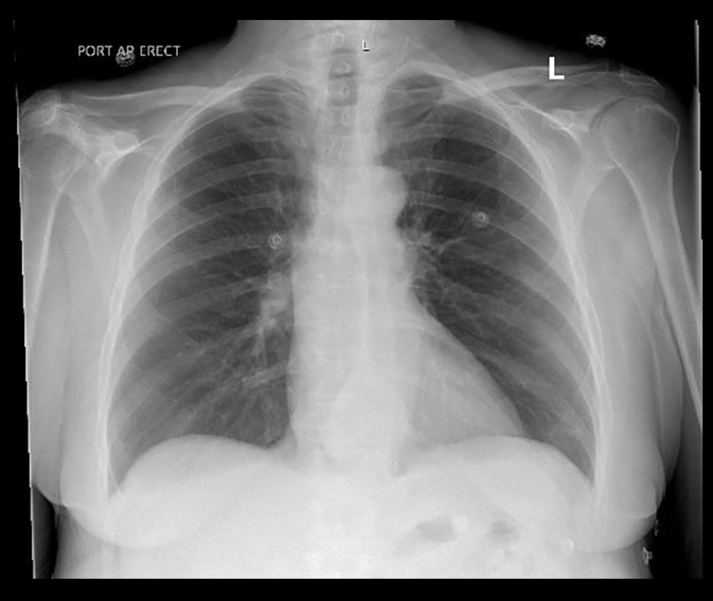 Figure 1. Chest radiograph. Chest X-ray demonstrating no acute cardiopulmonary abnormalities.
 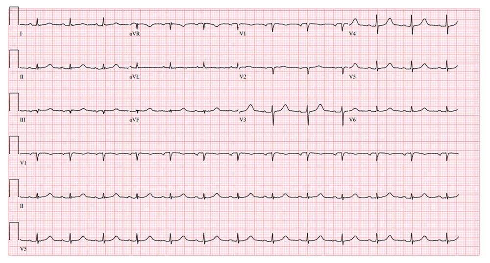 Figure 2. Electrocardiogram. Twelve-lead electrocardiogram showing normal sinus rhythm with no specific ischemic changes.
 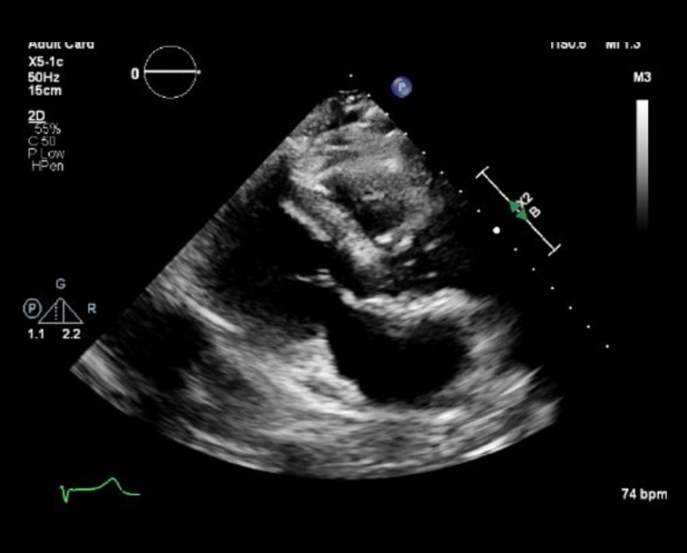 Figure 3. Contrast-enhanced echocardiogram. Contrast-enhanced transthoracic echocardiogram showing severe hypokinesis and akinesis of the basal-to-mid left ventricular segments with preserved apical contraction, consistent with reverse takotsubo cardiomyopathy.
 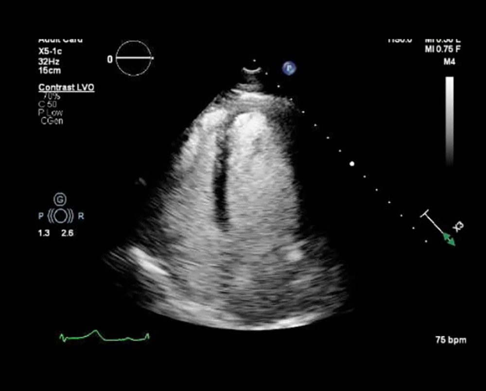 Figure 4. Echocardiogram, parasternal and short-axis views. Standard transthoracic echocardiogram demonstrating basal-predominant dysfunction with apical sparing.
 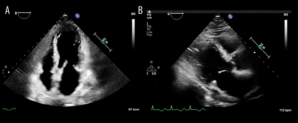 Figure 5. (A) Prior echocardiogram, 4-chamber view. Echocardiogram from the prior admission showing apical hypokinesis with preserved basal function, consistent with classic apical takotsubo cardiomyopathy. (B) Prior echocardiogram, parasternal long-axis view. Parasternal long-axis view from the previous episode demonstrating apical dysfunction with preserved basal contractility.
 References
1. Carey C, Nucifora G, Macnab A, A case report of recurrent takotsubo cardiomyopathy including the rare inverted form: Eur Heart J Case Rep, 2022; 6(6); ytac207
2. Ives CW, Smith PA, Atchley TJ, Pogwizd SM, A novel case of reverse takotsubo cardiomyopathy following brain biopsy: Echocardiography, 2020; 37(7); 1080-83
3. Lyon AR, Bossone E, Schneider B, Current state of knowledge on takotsubo syndrome: A position statement from the Taskforce on Takotsubo Syndrome of the Heart Failure Association of the European Society of Cardiology: Eur J Heart Fail, 2016; 18(1); 8-27
4. Summers MR, Lennon RJ, Prasad A, Pre-morbid psychiatric and cardiovascular diseases in apical ballooning syndrome (tako-tsubo/stress-induced cardiomyopathy): Potential pre-disposing factors: J Am Coll Cardiol, 2010; 55(7); 700-1
5. Sethi Y, Murli H, Kaiwan O, Broken heart syndrome: Evolving molecular mechanisms and principles of management: J Clin Med, 2023; 12(1); 125
6. Singh K, Carson K, Usmani Z, Systematic review and meta-analysis of incidence and correlates of recurrence of takotsubo cardiomyopathy: Int J Cardiol, 2014; 174(3); 696-701
7. Templin C, Ghadri JR, Diekmann J, Clinical features and outcomes of takotsubo (stress) cardiomyopathy: N Engl J Med, 2015; 373(10); 929-38
8. El-Battrawy I, Santoro F, Stiermaier T, Incidence and clinical impact of recurrent takotsubo syndrome: Results from the GEIST registry: J Am Heart Assoc, 2019; 8(9); e010753
9. Medina de Chazal H, Del Buono MG, Keyser-Marcus L, Stress cardiomyopathy diagnosis and treatment: JACC state-of-the-art review: J Am Coll Cardiol, 2018; 72(16); 1955-71
Figures
Figure 1. Chest radiograph. Chest X-ray demonstrating no acute cardiopulmonary abnormalities.Figure 2. Electrocardiogram. Twelve-lead electrocardiogram showing normal sinus rhythm with no specific ischemic changes.Figure 3. Contrast-enhanced echocardiogram. Contrast-enhanced transthoracic echocardiogram showing severe hypokinesis and akinesis of the basal-to-mid left ventricular segments with preserved apical contraction, consistent with reverse takotsubo cardiomyopathy.Figure 4. Echocardiogram, parasternal and short-axis views. Standard transthoracic echocardiogram demonstrating basal-predominant dysfunction with apical sparing.Figure 5. (A) Prior echocardiogram, 4-chamber view. Echocardiogram from the prior admission showing apical hypokinesis with preserved basal function, consistent with classic apical takotsubo cardiomyopathy. (B) Prior echocardiogram, parasternal long-axis view. Parasternal long-axis view from the previous episode demonstrating apical dysfunction with preserved basal contractility. In Press
Case report 
Am J Case Rep In Press; DOI: 10.12659/AJCR.953543
Case report 
Am J Case Rep In Press; DOI: 10.12659/AJCR.954216
Case report 
Am J Case Rep In Press; DOI: 10.12659/AJCR.954932
Case report
Am J Case Rep In Press; DOI: 10.12659/AJCR.954146
Most Viewed Current Articles
07 Dec 2021 : Case report
28,712,695
DOI :10.12659/AJCR.934347
Am J Case Rep 2021; 22:e934347
06 Dec 2021 : Case report  180,721
DOI :10.12659/AJCR.934406
Am J Case Rep 2021; 22:e934406
21 Jun 2024 : Case report
124,419
DOI :10.12659/AJCR.944371
Am J Case Rep 2024; 25:e944371
08 May 2026 : Case report
95,462
DOI :10.12659/AJCR.952158
Am J Case Rep 2026; 27:e952158