


10 June 2026: Articles 
Thoracoscopic Primary Repair for Spontaneous Esophageal Rupture in a Patient With Acute Irreducible Inguinal Hernia: A Case Report
Unusual clinical course, Management of emergency care
Masahiro Takahashi ABCDEF 1*, Ken Ishikawa A 1, Keisuke Tomoda A 1, Yuki Inagaki B 1, Satoru Takayama A 1
A 1
DOI: 10.12659/AJCR.953095
Am J Case Rep 2026; 27:e953095


Abstract
BACKGROUND: Spontaneous esophageal rupture is a life-threatening emergency caused by a rapid increase in intraesophageal pressure. Timely diagnosis and surgical intervention are crucial for survival. Typical cases involve vomiting-related rupture of the left distal esophagus with left pleural effusion. However, atypical presentations, including right-sided rupture, have been reported and can contribute to delayed diagnosis. Cases occurring in association with acute irreducible inguinal hernia are extremely rare; their clinical significance and optimal management remain unclear. Such atypical presentations may hinder accurate diagnosis.
CASE REPORT: A 70-year-old Japanese woman presented with acute-onset epigastric pain after experiencing nausea and retching without vomiting. Imaging revealed pneumomediastinum, right pleural effusion, right pneumothorax, and a right inguinal hernia containing small bowel. Based on the clinical presentation and imaging findings, spontaneous esophageal rupture associated with retching in the context of an acute irreducible inguinal hernia was diagnosed. Given the atypical right-sided presentation and absence of definitive bowel obstruction, careful diagnostic evaluation was required to avoid misdiagnosis. Thoracoscopic primary repair and chest drainage were performed approximately 3 hours after symptom onset. The hernia was manually reduced and subsequently managed with elective laparoscopic repair. The postoperative course was uneventful, without major complications or recurrence.
CONCLUSIONS: This case suggests an association between acute irreducible inguinal hernia and spontaneous esophageal rupture through retching-related increases in intraesophageal pressure, although a direct causal relationship cannot be definitively established. Prompt imaging, timely thoracoscopic repair, and staged management of coexisting abdominal pathology may contribute to favorable outcomes, even in cases of atypical right-sided perforation.
Keywords: Esophageal Perforation, Hernia, Inguinal, Pneumothorax, Thoracic Surgery
Introduction
Spontaneous esophageal rupture is a life-threatening condition caused by a rapid increase in intraesophageal pressure, resulting in full-thickness disruption of the esophageal wall. Most cases are associated with vomiting and involve the left wall of the distal esophagus [1,2]. However, atypical mechanisms of onset and right-sided perforations have been reported; they may contribute to delayed diagnosis [3]. Spontaneous esophageal rupture occurring in association with acute irreducible inguinal hernia is extremely rare, and only a limited number of cases have been described in the literature [4].
Management strategies should be tailored according to the patient’s clinical status, perforation site, degree of contamination, and time from onset. In selected patients, minimally invasive thoracoscopic repair has been reported as a feasible option [5]; however, evidence regarding right-sided rupture combined with abdominal pathology remains scarce. We report a case of spontaneous esophageal rupture after retching in the context of an acute, initially irreducible inguinal hernia; the case was successfully managed with thoracoscopic primary repair and staged laparoscopic hernia repair.
Case Report
A 70-year-old Japanese woman presented to the emergency department with acute-onset epigastric pain that developed immediately after nausea and retching without vomiting. She reported no history of smoking or alcohol consumption. Her medical history included hypertension, osteoporosis, and sustained virologic response following hepatitis C treatment. Her surgical history included bilateral hip arthroplasty. Current medications included losartan and alendronate.
On admission, vital signs were stable. Physical examination revealed mild abdominal distension, severe epigastric tenderness, and a right inguinal bulge. However, medical records did not clearly document the presence or absence of tenderness associated with the inguinal mass, which represents a limitation of the clinical assessment. Laboratory testing demonstrated leukocytosis (13 300/μL) and a slightly elevated C-reactive protein level (0.02 mg/dL).
Chest radiography revealed a right pneumothorax with increased opacity in the right lower lung zone. Contrast-enhanced computed tomography (CT) demonstrated pneumomediastinum with periesophageal air and fluid, right pleural effusion containing high-attenuation material, right pneumothorax (Figure 1), and a right inguinal hernia containing small bowel (Figure 2). Based on its anatomical location, the hernia was considered an indirect inguinal hernia. Mild small-bowel dilatation with minimal air-fluid levels was noted; however, these findings were insufficient to establish a definitive diagnosis of bowel obstruction. CT showed no obvious abnormal esophageal course, diaphragmatic hernia, large hiatal hernia, distal obstruction, tumor, or diverticulum. The hernia was initially irreducible and required physician-assisted manual reduction in the emergency department.
Although a direct causal association could not be definitively established, the inguinal hernia was considered a possible contributing factor to the patient’s nausea and retching. Based on the clinical presentation and imaging findings, spontaneous esophageal rupture after retching in the context of an acute, initially irreducible inguinal hernia was diagnosed. Given that no signs of strangulation or bowel necrosis were observed, management of the esophageal perforation was prioritized.
Surgery was initiated approximately 3 hours after symptom onset. With the patient in the prone position, thoracoscopic repair was performed using 3 ports: a 5-mm port in the fourth intercostal space at the anterior axillary line, a 12-mm port in the seventh intercostal space at the midaxillary line, and a 12-mm port in the ninth intercostal space at the posterior axillary line. A large amount of dark pleural fluid containing food debris was evacuated. An approximately 35-mm longitudinal tear was identified on the right wall of the lower thoracic esophagus (Figure 3A). Although the defect was sizable, the surrounding tissue appeared fresh, well perfused, and minimally edematous, without gross necrosis or severe contusion.
Using a 3-0 absorbable barbed monofilament suture (V-Loc™, Medtronic, Minneapolis, MN, USA), full-thickness closure was performed in a continuous fashion (Figure 3B). Suture tension was carefully controlled to prevent tissue strangulation. The adjacent visceral pleura was also repaired. The thoracic cavity was thoroughly irrigated, and a chest drain was placed near the repair site. The operative time was 116 minutes, with minimal blood loss.
The patient was extubated and transferred out of the intensive care unit on postoperative days 1 and 2, respectively. On postoperative day 5, contrast esophagography followed by CT revealed no evidence of leakage (Figure 4), allowing resumption of oral intake. Because no clinical or radiological evidence of leakage was observed, postoperative endoscopy was not performed. Abdominal CT also demonstrated slight protrusion of the small bowel through the inguinal orifice without evidence of incarceration (Figure 5). The chest drain was removed on postoperative day 6, and the patient was discharged on postoperative day 18 without major complications.
Two months later, elective laparoscopic transabdominal preperitoneal repair was performed for the right inguinal hernia (Figure 6). Although imaging revealed no clinically significant enlargement of the internal inguinal ring, intraoperative findings confirmed an indirect inguinal hernia, corresponding to a small defect consistent with European Hernia Society classification L1. After more than 2.5 years of follow-up, the patient remains in good health without recurrence.
Discussion
This case illustrates several clinically significant features of spontaneous esophageal rupture. First, although vomiting is the most common precipitating factor [6], our patient developed esophageal rupture after retching without vomiting. CT findings suggested localized bowel involvement associated with the acute, initially irreducible inguinal hernia; however, definitive features of complete bowel obstruction were not identified. Even in the absence of definitive bowel obstruction, localized irritation or transient functional disturbance may have contributed to nausea and retching. Therefore, the hernia was considered a possible contributing factor associated with retching and transient increases in intraesophageal pressure.
Second, spontaneous esophageal rupture most frequently occurs on the left wall of the distal esophagus [7]; however, right-sided perforations have been reported [3,8–12]. In the present case, imaging revealed no predisposing factors, including prior thoracic or upper abdominal surgery, anatomic abnormalities, diaphragmatic or hiatal hernia, distal obstruction, tumor, or diverticulum; therefore, no clear structural predisposition was identified. Right-sided pleural contamination can mimic primary pulmonary disease and contribute to diagnostic delay. However, early CT facilitated prompt recognition of the esophageal source despite the atypical laterality.
Third, surgical timing and tissue viability greatly influenced operative decision-making. The short interval between symptom onset and surgery (approximately 3 hours) may have limited inflammatory progression. Although the tear measured 35 mm, the tissue edges remained viable with minimal edema or necrosis, supporting primary repair rather than esophageal resection [12–14]. A thoracoscopic approach was selected over open surgery due to the early presentation, stable clinical condition, and need for prompt management in the context of suspected pleural contamination. Early intervention is a major prognostic factor in spontaneous esophageal rupture, and the present case highlights the benefits of rapid surgical treatment.
Management of the acute irreducible inguinal hernia was staged. Following successful manual reduction and confirmation of the absence of strangulation, infection control and patient stabilization were prioritized. Current international guidelines support elective mesh repair several weeks after successful reduction [15], and delayed laparoscopic repair in the present case was consistent with recommended practice.
This report is limited by its single-case design, and a causal association between hernia incarceration and esophageal rupture cannot be definitively established. Nevertheless, it contributes to the literature by describing an uncommon clinical presentation, right-sided perforation, and staged minimally invasive management.
Conclusions
This case suggests that an acute, initially irreducible inguinal hernia may have been associated with spontaneous esophageal rupture through retching-related increases in intraesophageal pressure. Prompt imaging, timely surgical intervention, and tissue-based operative decision-making may facilitate successful thoracoscopic primary repair in selected cases, even in atypical right-sided perforations. In carefully selected patients, staged management of associated abdominal pathology may represent a safe treatment strategy.
Figures
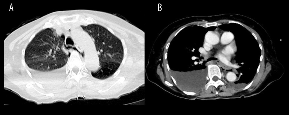 Figure 1. (A, B) Contrast-enhanced computed tomography revealing pneumomediastinum with periesophageal air, right pneumothorax, and right pleural effusion containing a high-density component.
 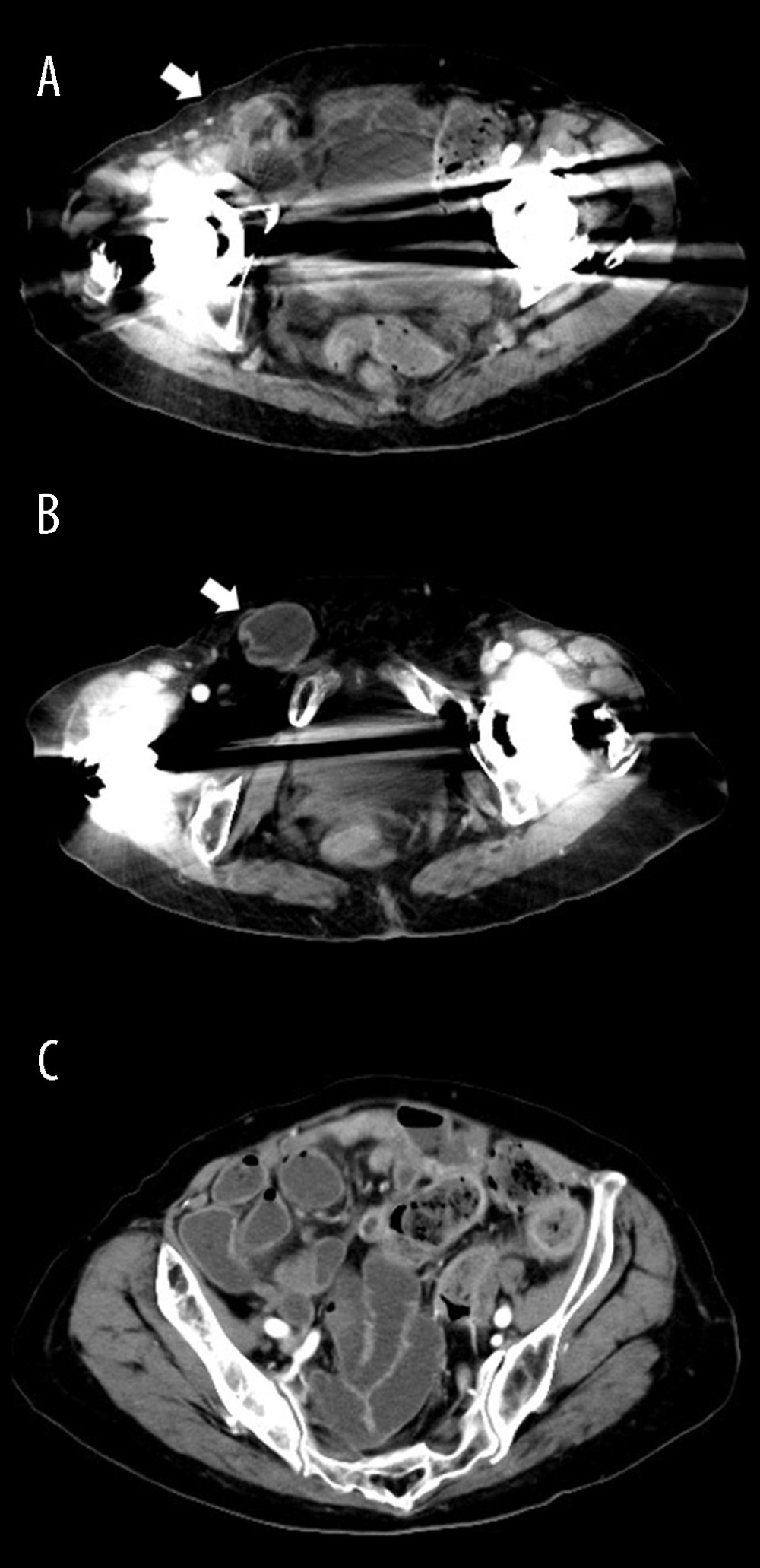 Figure 2. Contrast-enhanced axial computed tomography findings of the right inguinal hernia. (A) Small-bowel herniation at the internal inguinal ring (arrow). (B) Potentially incarcerated small bowel within the inguinal canal (arrow). (C) Mild localized proximal small-bowel dilatation.
 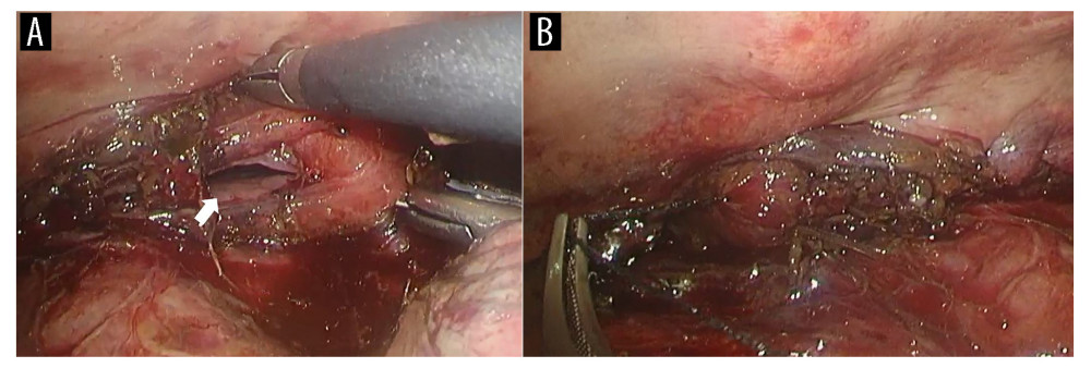 Figure 3. Thoracoscopic findings during surgical repair. (A) An approximately 35-mm longitudinal esophageal tear on the right wall of the lower thoracic esophagus (arrow). (B) The esophageal defect after closure with a continuous full-thickness suture using a 3-0 absorbable barbed monofilament.
 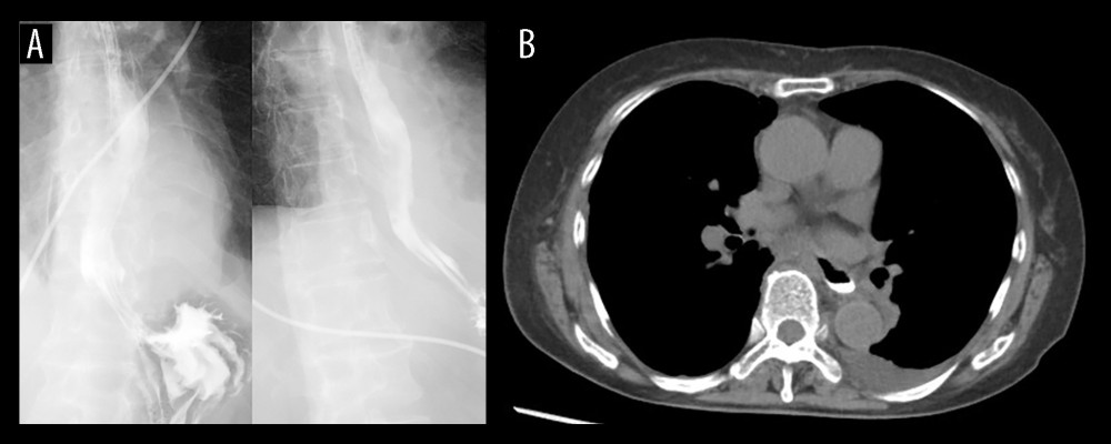 Figure 4. Postoperative imaging findings. (A) Contrast esophagography using Gastrografin demonstrating no evidence of leakage. (B) Computed tomography performed immediately after esophagography confirming the absence of leakage.
 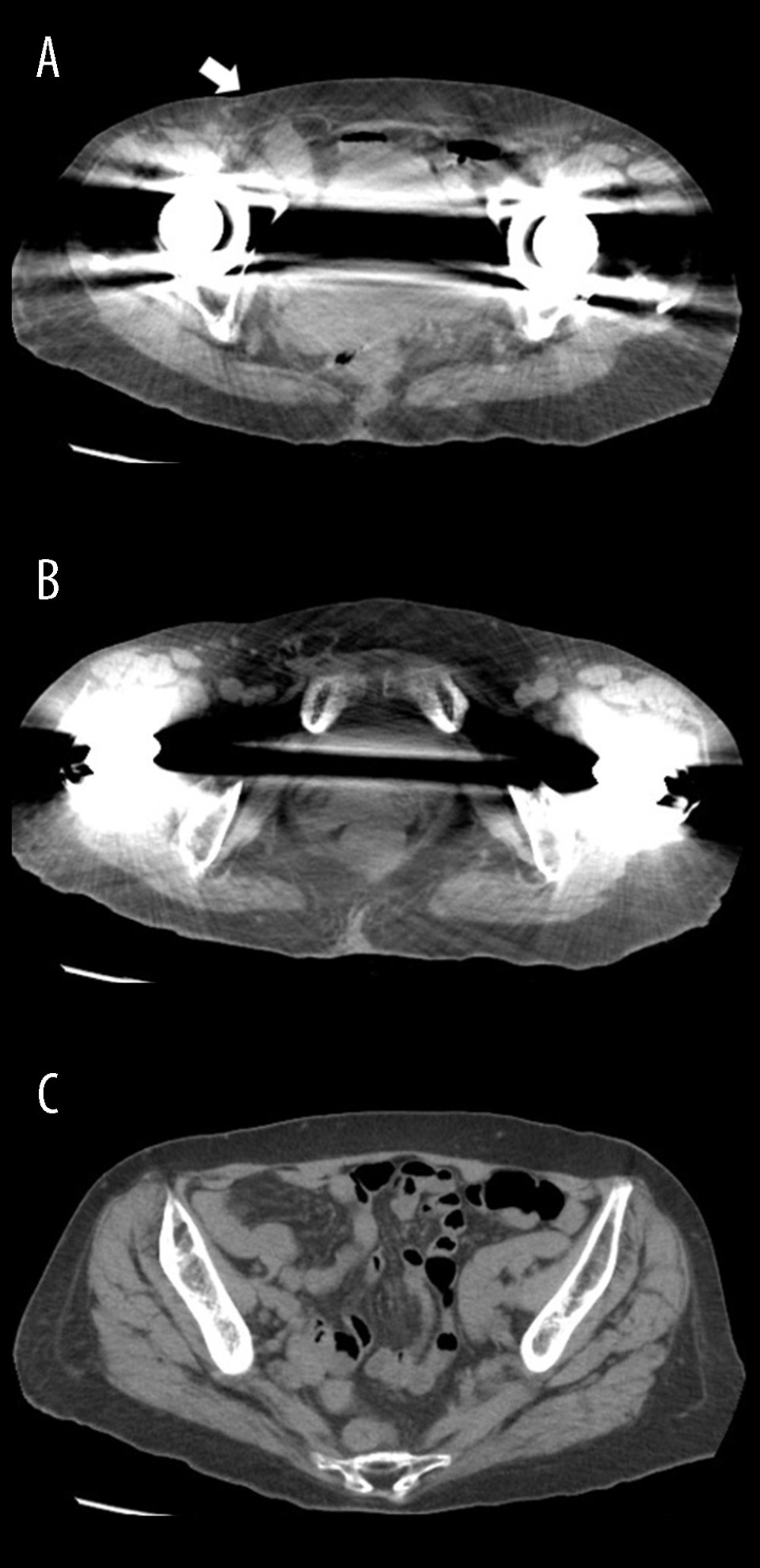 Figure 5. (A–C) Abdominal computed tomography findings. Axial contrast-enhanced computed tomography image demonstrating slight small-bowel protrusion through the inguinal orifice (arrow) without evidence of incarceration.
 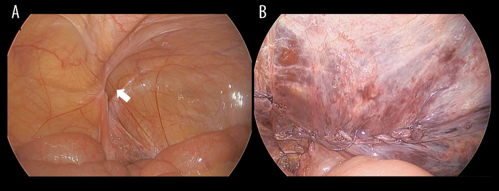 Figure 6. Laparoscopic findings during elective transabdominal preperitoneal repair. (A) Laparoscopic view demonstrating a right inguinal hernia with a small defect (arrow). (B) Laparoscopic view after mesh placement and peritoneal closure.
 References
1. Pan J, Ge Y, Feng T, Outcome of treatment modalities for spontaneous esophageal rupture: A meta-analysis and case series: Int J Surg, 2024; 111; 1135-43
2. Korn O, Oñate JC, López R, Anatomy of the Boerhaave syndrome: Surgery, 2007; 141; 222-28
3. Reardon ES, Martin LW, Boerhaave’s syndrome presenting as a mid-esophageal perforation associated with a right-sided pleural effusion: J Surg Case Rep, 2015; 2015; rjv142
4. Ashrafi AS, Horkoff MJ, Mohammad WM, Boerhaave’s syndrome secondary to an incarcerated inguinal hernia: A case report: Int J Surg Case Rep, 2016; 28; 234-36
5. Pickering O, Pucher PH, De’Ath H, Minimally invasive approach in Boerhaave’s syndrome: Case series and systematic review: J Laparoendosc Adv Surg Tech A, 2021; 31; 1254-61
6. Derbes VJ, Mitchell RE, Hermann Boerhaave’s Atrocis, nec descripti prius, morbi historia: The first translation of the classic case report of rupture of the esophagus, with annotations: Bull Med Libr Assoc, 1955; 43; 217-40
7. Brinster CJ, Singhal S, Lee L, Evolving options in the management of esophageal perforation: Ann Thorac Surg, 2004; 77; 1475-83
8. Yang ST, Devanand A, Tan KL, Eng PC, Boerhaave’s syndrome presenting as a right-sided pleural effusion: Ann Acad Med Singap, 2003; 32; 415-17
9. Chang YC, Liu HC, Huang CJ, Wu CL, Right-sided pleural effusion in spontaneous esophageal perforation: Ann Thorac Cardiovasc Surg, 2000; 6; 73-76
10. Razi E, Davoodabadi A, Razi A, Spontaneous esophageal perforation presenting as a right-sided pleural effusion: A case report: Tanaffos, 2013; 12; 53-57
11. Jagminas L, Silverman RA, Boerhaave’s syndrome presenting with abdominal pain and right hydropneumothorax: Am J Emerg Med, 1996; 14; 53-56
12. Tsalis K, Vasiliadis K, Tsachalis T, Management of Boerhaave’s syndrome: Report of three cases: J Gastrointestin Liver Dis, 2008; 17; 81-85
13. Kollmar O, Lindemann W, Richter S, Boerhaave’s syndrome: Primary repair vs. esophageal resection—Case reports and meta-analysis of the literature: J Gastrointest Surg, 2003; 7; 726-34
14. García-Moreno V, Maiocchi K, Gómez-Quiles L, Treatment of esophageal perforation: A review of our experience at a tertiary referral hospital spanning the past 19 years: Rev Gastroenterol Mex, 2022; 87; 405-10
15. Stabilini C, van Veenendaal N, Aasvang E, Update of the international HerniaSurge guidelines for groin hernia management: BJS Open, 2023; 7; zrad080
Figures
Figure 1. (A, B) Contrast-enhanced computed tomography revealing pneumomediastinum with periesophageal air, right pneumothorax, and right pleural effusion containing a high-density component.Figure 2. Contrast-enhanced axial computed tomography findings of the right inguinal hernia. (A) Small-bowel herniation at the internal inguinal ring (arrow). (B) Potentially incarcerated small bowel within the inguinal canal (arrow). (C) Mild localized proximal small-bowel dilatation.Figure 3. Thoracoscopic findings during surgical repair. (A) An approximately 35-mm longitudinal esophageal tear on the right wall of the lower thoracic esophagus (arrow). (B) The esophageal defect after closure with a continuous full-thickness suture using a 3-0 absorbable barbed monofilament.Figure 4. Postoperative imaging findings. (A) Contrast esophagography using Gastrografin demonstrating no evidence of leakage. (B) Computed tomography performed immediately after esophagography confirming the absence of leakage.Figure 5. (A–C) Abdominal computed tomography findings. Axial contrast-enhanced computed tomography image demonstrating slight small-bowel protrusion through the inguinal orifice (arrow) without evidence of incarceration.Figure 6. Laparoscopic findings during elective transabdominal preperitoneal repair. (A) Laparoscopic view demonstrating a right inguinal hernia with a small defect (arrow). (B) Laparoscopic view after mesh placement and peritoneal closure. In Press
Case report 
Am J Case Rep In Press; DOI: 10.12659/AJCR.953543
Case report
Am J Case Rep In Press; DOI: 10.12659/AJCR.954216
Case report 
Am J Case Rep In Press; DOI: 10.12659/AJCR.954932
Case report 
Am J Case Rep In Press; DOI: 10.12659/AJCR.954146
Most Viewed Current Articles
07 Dec 2021 : Case report
28,712,695
DOI :10.12659/AJCR.934347
Am J Case Rep 2021; 22:e934347
06 Dec 2021 : Case report  180,721
DOI :10.12659/AJCR.934406
Am J Case Rep 2021; 22:e934406
21 Jun 2024 : Case report
124,419
DOI :10.12659/AJCR.944371
Am J Case Rep 2024; 25:e944371
08 May 2026 : Case report
95,462
DOI :10.12659/AJCR.952158
Am J Case Rep 2026; 27:e952158