


03 September 2026: Articles 
A Near-Fatal Leak: Acute Cardiac Tamponade From Contrast Extravasation via a Thrombosed Port-a-Cath
Rare disease
Hamza Janjua ABEF 1, Monica Wilbekin E 2, Hira Janjua E 1, Kushal Das E 1,3, Zulfiqar Qutrio Baloch E 4, Rehan Ishaque E 4*
DOI: 10.12659/AJCR.953197
Am J Case Rep 2026; 27:e953197


Abstract
BACKGROUND: Cardiac tamponade caused by intrapericardial contrast extravasation is a rare but life-threatening iatrogenic complication associated with central venous access devices (CVADs) during power injection. CVADs, including implanted ports (port-a-caths), are susceptible to fibrin sheath formation and mural thrombosis over time, which can tether the catheter tip against the vessel wall and predispose it to erosion or perforation. When high-pressure contrast is injected through a compromised device, contrast may bypass the vessel lumen entirely and accumulate within the pericardial sac, producing acute obstructive shock.
CASE REPORT: We report a 61-year-old woman with a history of diabetes mellitus, cerebrovascular accident, and an indwelling port-a-cath who underwent computed tomography (CT) angiography because of difficult peripheral venous access. Immediately after contrast administration, she developed sudden cardiovascular collapse and cardiac arrest. CT imaging demonstrated a large hyperdense pericardial effusion with superior vena cava thrombosis adjacent to the catheter tip, suggesting catheter-related vessel wall injury and direct contrast extravasation into the pericardial sac. Bedside echocardiography confirmed tamponade physiology with right ventricular diastolic collapse and chamber compression. Emergent pericardiocentesis drained 1 L of contrast-containing serosanguinous fluid and resulted in immediate hemodynamic recovery after cardiac arrest.
CONCLUSIONS: Contrast extravasation through a long-term indwelling port-a-cath is a rare but catastrophic cause of acute cardiac tamponade. Sudden cardiovascular collapse during or after contrast-enhanced CT in a patient with a long-term central venous device should prompt immediate suspicion for this diagnosis. Additionally, echocardiography enables rapid confirmation, and emergent pericardiocentesis is life-saving.
Keywords: Cardiology, Case Reports, central venous catheters, Contrast Media, Iatrogenic Disease, Radiology, Cardiac Tamponade, Pericardial Effusion
Introduction
Cardiac tamponade resulting from intrapericardial contrast extravasation is an exceedingly rare and life-threatening iatrogenic complication [1]. Most documented cases involve central venous access devices, including peripherally inserted central catheters, tunneled lines, and implanted ports (port-a-caths) [2]. These devices are typically composed of biocompatible materials such as silicone or polyurethane. While polyurethane catheters tolerate higher injection pressures, their relative stiffness compared with silicone may increase long-term mechanical contact with the vessel wall. Over time, these catheters are prone to fibrin sheath formation and mural thrombosis, which can tether the catheter tip against the vein wall or superior vena cava, predisposing it to erosion or perforation [3].
The pathophysiology of this event involves a high-pressure contrast bolus bypassing the vessel lumen through a perforation or erosion. This results in contrast extravasation, the direct leakage of radiopaque media into the pericardial sac, which is often accompanied by secondary hemopericardium if the vascular injury is significant. The rapid accumulation of these fluids within the non-compliant pericardial space increases intrapericardial pressure above the intracardiac diastolic pressure. This leads to right-sided chamber collapse, impaired filling, and acute obstructive shock [4].
While the mechanism is well understood, the clinical manifestation is often abrupt, occurring during or immediately following contrast administration. Because tamponade physiology can precipitate cardiovascular collapse within minutes, the window for diagnosis and intervention is critically narrow. The rising use of automated power injectors in computed tomography (CT) angiography, combined with an increasing population of patients with long-term indwelling devices, necessitates heightened vigilance [3]. Long-term complications such as tip migration, vessel wall irritation, and tip-associated thrombus create a “fixed” point of contact that can erode adjacent structures, forming a direct conduit for extravasated contrast.
Early recognition is paramount. A new pericardial effusion immediately after contrast infusion should raise suspicion for contrast extravasation until proven otherwise. Additionally, transthoracic echocardiography remains the fastest method for confirming tamponade physiology, and emergent pericardiocentesis remains the definitive life-saving intervention [5]. This report emphasizes the necessity of verifying catheter patency and tip position before high-pressure injection and underscores the importance of prompt clinical suspicion in cases of unexplained hemodynamic instability during imaging.
Case Report
A 61-year-old woman with a history of diabetes mellitus, gastroparesis, and a prior cerebrovascular accident (with residual right-sided weakness and blindness) presented to the emergency department with abdominal pain, lower back pain, nausea, and vomiting. On arrival, her initial vital signs were stable, with a blood pressure of 138/84 mm Hg and a heart rate of 88 beats/min; however, she exhibited altered mental status. She was initially treated with intravenous (IV) fluids and empiric antibiotics for a presumed severe urinary tract infection.
Due to extremely difficult peripheral venous access and multiple unsuccessful attempts at new IV placement, the patient’s existing indwelling port-a-cath was utilized for a CT angiography of the abdomen and pelvis. The standard institutional power-injection protocol for CT angiography was used, with 100 mL of Isovue-370 administered at 325 psi and an injection rate of 1.5 mL/s. Shortly after returning from the CT suite, the patient developed acute respiratory distress followed by rapid hemodynamic decompensation. Laboratory studies at the time were notable for a new-onset metabolic acidosis (pH 7.18), while hematocrit remained stable at 34%, suggesting the acute collapse was not due to massive internal hemorrhage. The patient subsequently had a sudden cardiovascular collapse and a 2-minute episode of cardiac arrest. Advanced cardiac life support was initiated immediately. During resuscitation, emergent alternative access was established via intraosseous placement and a right internal jugular central venous catheter; the port-a-cath was strictly avoided once iatrogenic injury was suspected. Return of spontaneous circulation was achieved, and she was stabilized with IV fluids and vasopressors (vasopressin and norepinephrine) administered exclusively through the newly established intraosseous and internal jugular access to avoid further pericardial infusion via the compromised port-a-cath.
Subsequent review of the CT imaging revealed a large, hyperdense pericardial effusion (Figures 1, 2), consistent with direct contrast extravasation into the pericardial sac. In addition, a focal thrombus was identified in the superior vena cava adjacent to the distal tip of the indwelling port-a-cath (Figure 3), suggesting catheter-associated venous thrombosis that may have contributed to impaired contrast flow and increased intraluminal pressure during power injection. An emergent bedside echocardiogram confirmed tamponade physiology, showing a large effusion with right ventricular diastolic collapse (Figure 4) and preserved left ventricular systolic function, with an ejection fraction of 55%. Emergent pericardiocentesis was performed in the emergency department, followed by the placement of a pericardial drain, which yielded a total of 1 L of serosanguinous fluid and contrast. No blood transfusions were required during the hospital course. The patient’s hemodynamics improved immediately after drainage. She was successfully weaned from vasopressors within 24 hours, the drain was removed on day 3, and she was discharged on day 5. A follow-up echocardiogram showed complete resolution of the effusion (Figure 5).
Discussion
PATHOPHYSIOLOGY OF CATHETER-RELATED INJURY:
The risk of vessel wall injury is influenced by the physical properties of the catheter and its hemodynamic environment. Catheter length and internal diameter (French size) significantly affect flow dynamics during power injection. According to Poiseuille’s law, smaller diameters increase resistance, requiring higher pressures to achieve the necessary flow rates for CT angiography. This increased pressure, especially when the catheter tip is fixed by a fibrin sheath or mural thrombus, can create a jet effect against the vessel wall.
In our case, the thrombus likely contributed to catheter fixation and chronic endothelial irritation, while the high-pressure contrast bolus probably served as the immediate trigger for extravasation. A localized thrombus at the superior vena cava–atrial junction can tether the catheter tip, causing chronic mechanical friction and weakening of the vessel wall [7]. When the high-pressure contrast bolus was introduced, the weakened wall could no longer withstand the shear stress, resulting in perforation or erosion. The presence of air at the catheter tip on CT further supports a breach in the integrity of the vessel-catheter interface, creating a direct conduit into the pericardial space.
CLINICAL RECOGNITION AND MANAGEMENT:
Catheter-linked tamponade occurs in approximately 0.0001% to 1.4% of cases, but mortality can exceed 60% if diagnosis is delayed [8]. Diagnosis is often difficult because symptoms may mimic the patient’s primary illness, such as sepsis or metabolic acidosis. This case underscores the necessity of early bedside transthoracic echocardiography, which, in the hands of trained healthcare professionals, is the gold standard for rapid identification of right ventricular diastolic collapse and tamponade physiology.
Once iatrogenic injury is suspected, a critical management step is the immediate cessation of use of the implicated line. Using the same malpositioned or perforated line for fluid resuscitation would inadvertently worsen the pericardial effusion. Alternative access (eg, intraosseous or a new central line in a different vessel) must be established immediately for hemodynamic support.
THE ROLE OF SURGICAL INTERVENTION:
Although our patient improved with pericardiocentesis alone, cardiothoracic surgical evaluation remains important in cases in which persistent leakage or recurrent effusion is suspected. In iatrogenic vascular perforation, the pericardial window can be considered when persistent leakage is suspected or when recurrent effusion cannot be adequately managed by percutaneous drainage [9].
PREVENTION STRATEGIES:
This complication is preventable through meticulous vigilance. Catheter patency and tip position must be verified, ideally with recent imaging or aspiration of free-flowing blood before high-pressure infusions are administered. Clinicians should maintain a high index of suspicion for tamponade in any patient who experiences sudden, unexplained cardiovascular collapse during or shortly after imaging [10].
This case illustrates the importance of considering tamponade in any patient with indwelling central venous access who develops unexplained hypotension, altered mental status, or rapid hemodynamic decompensation. The most rapid and certain form of making a diagnosis is bedside transthoracic echocardiography. The echocardiogram of this case revealed the right ventricular diastolic collapse and massive effusion, which is a sign of acute tamponade. CT imaging was useful in this case, revealing the presence of a large, contrast-filled pericardial effusion, as illustrated on coronal reconstruction.
Conclusions
Contrast extravasation through an indwelling port-a-cath is a rare but catastrophic cause of acute cardiac tamponade. In this case, long-term catheter-associated thrombosis likely predisposed to vessel wall injury, while power injection precipitated intrapericardial contrast leakage and sudden cardiovascular collapse. Immediate bedside echocardiography enabled diagnosis, and emergent pericardiocentesis was life-saving. Verification of catheter patency and tip position before power injection is essential, particularly in long-standing central venous devices.
Figures
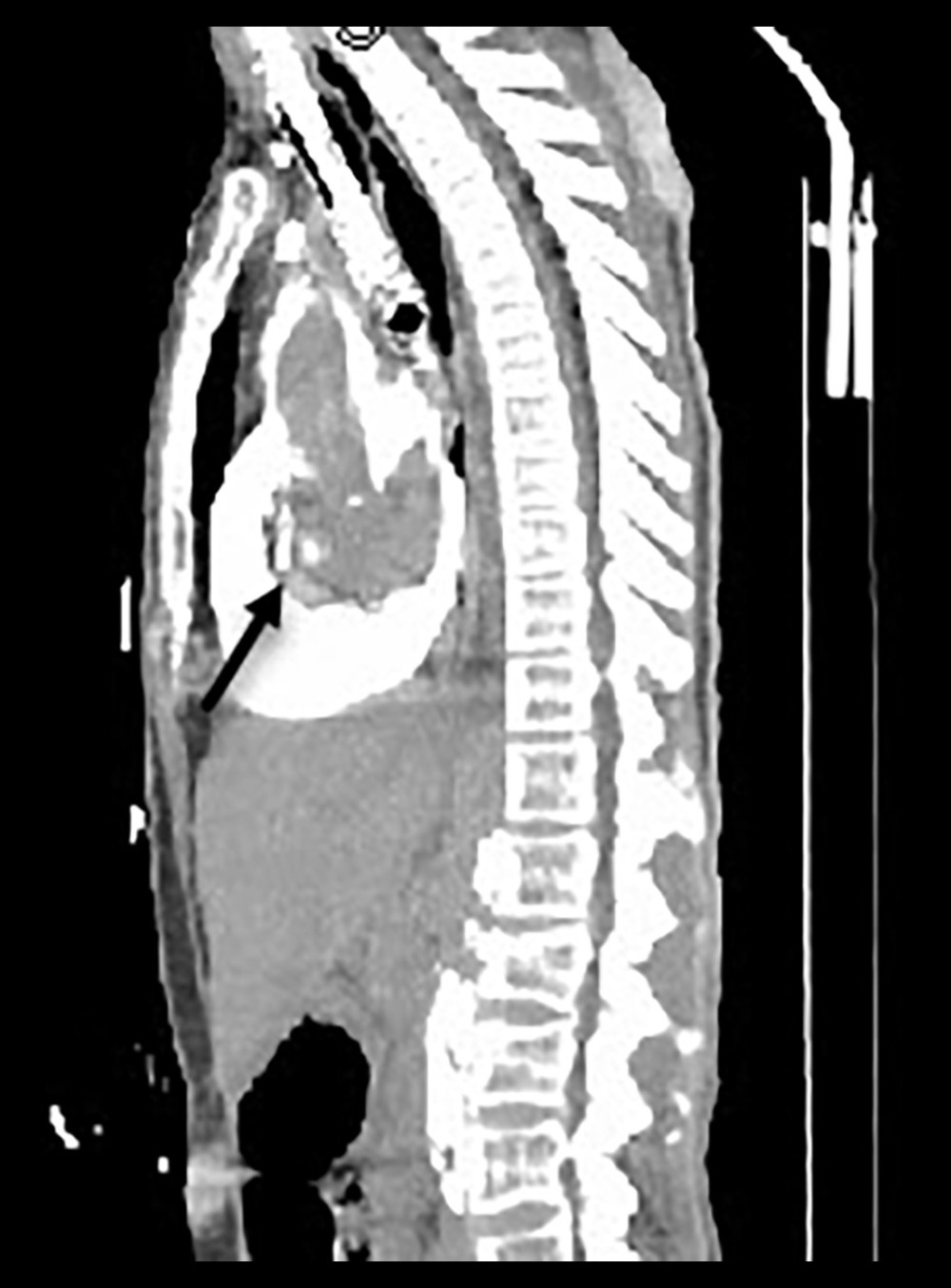 Figure 1. Sagittal contrast-enhanced computed tomography reconstruction demonstrating dense contrast accumulation within the pericardial sac, consistent with intrapericardial contrast extravasation resulting in cardiac tamponade. Arrow indicates the hyperdense pericardial collection.
 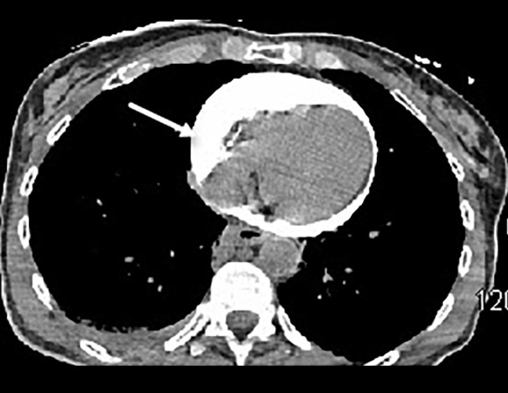 Figure 2. Axial contrast-enhanced computed tomography image showing circumferential hyperdense pericardial effusion compressing the cardiac chambers, consistent with contrast extravasation into the pericardial space. Arrow indicates the hyperdense effusion surrounding the heart.
 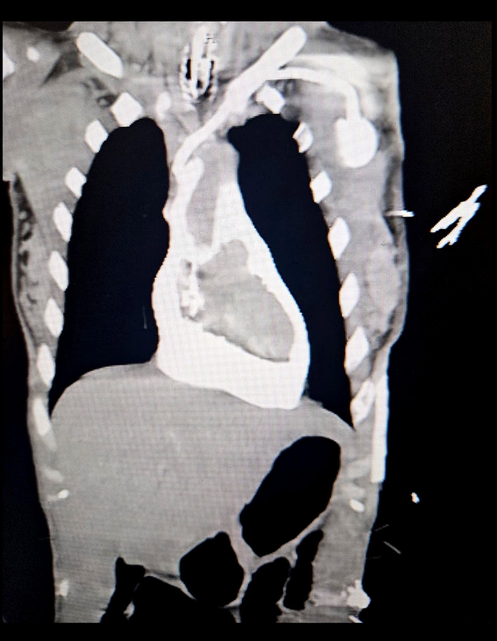 Figure 3. Coronal computed tomography image demonstrating a focal filling defect within the superior vena cava, adjacent to the distal tip of the indwelling port-a-cath, consistent with catheter-associated thrombus.
 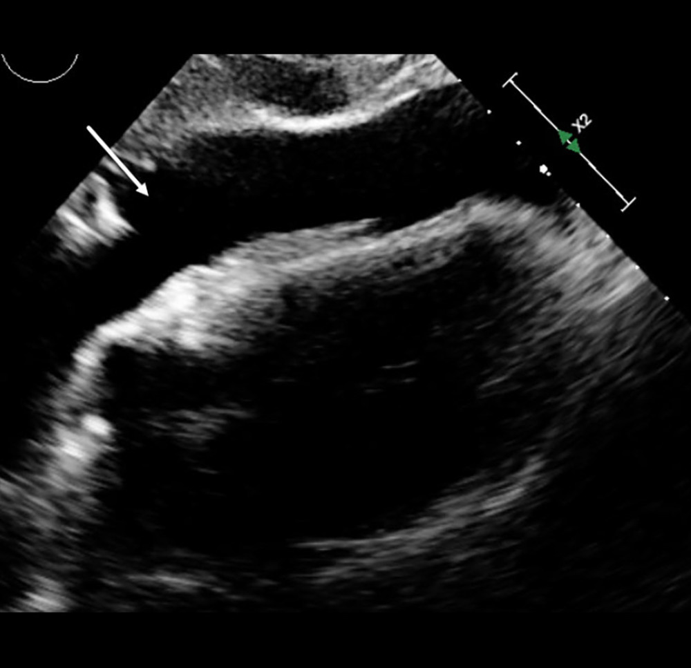 Figure 4. Bedside transthoracic echocardiography demonstrating a large pericardial effusion with right ventricular diastolic collapse, consistent with acute cardiac tamponade. Arrow indicates the effusion compressing the right ventricle.
 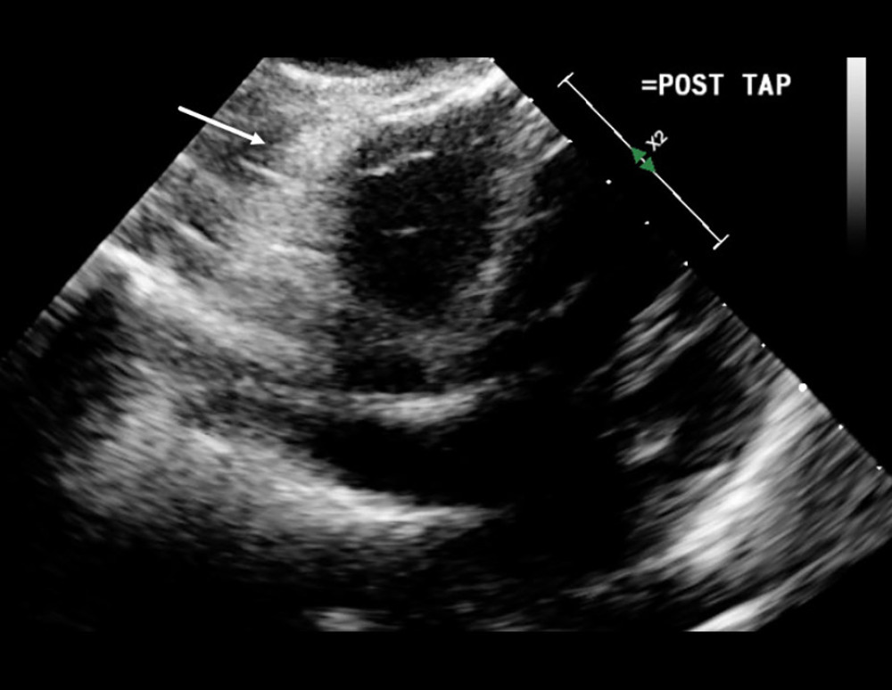 Figure 5. Follow-up transthoracic echocardiography after pericardiocentesis demonstrating resolution of the pericardial effusion and normalization of cardiac chamber filling. Arrow indicates the now-decompressed pericardial space.
 References
1. Liang CD, Ko SF, Huang CF, Catheter-malposition-induced cardiac tamponade via contrast media leakage during computed tomography study: Cardiovasc Intervent Radiol, 2005; 28(6); 822-24
2. Schummer C, Sakr Y, Steenbeck J, Risk of extravasation after power injection of contrast media via the proximal port of multilumen central venous catheters: Case report and review of the literature: ROFO Fortschr Geb Rontgenstr Nuklearmed, 2010; 182(1); 14-19
3. Machat S, Eisenhuber E, Pfarl G, Complications of central venous port systems: A pictorial review: Insights Imaging, 2019; 10(1); 86
4. Collier PE, Goodman GB, Cardiac tamponade caused by central venous catheter perforation of the heart: A preventable complication: J Am Coll Surg, 1995; 181(5); 459-63
5. Takamatsu J, Pericardial injury with cardiac tamponade and bleeding from the pericardium confirmed using contrast-enhanced computed tomography: A case report: Surg Case Rep, 2019; 5(1); 32
6. Kalen V, Medige TA, Rinsky LA, Pericardial tamponade secondary to perforation by central venous catheters in orthopaedic patients: J Bone Joint Surg Am, 1991; 73(10); 1503-6
7. Premuzic V, Katalinic L, Pasalic M, Jurin H, Nonfatal cardiac perforation after central venous catheter insertion: Saudi J Anaesth, 2018; 12(1); 118-20
8. Messina Alvarez AA, Bilal MA, Manasrah N, Chaudhary A, Iatrogenic cardiac tamponade secondary to central venous catheter placement: A literature review: Cureus, 2023; 15(4); e37695
9. Azevedo AC, Flor de Lima I, Brito VCardiac tamponade: A rare complication of central venous catheter – a clinical case report: Braz J Anesthesiol Elsevier, 2018; 68(1); 104-8 [in Portuguese]
10. Shields LBE, Hunsaker DM, Hunsaker JC, Iatrogenic catheter-related cardiac tamponade: A case report of fatal hydropericardium following subcutaneous implantation of a chemotherapeutic injection port: J Forensic Sci, 2003; 48(2); 414-18
Figures
Figure 1. Sagittal contrast-enhanced computed tomography reconstruction demonstrating dense contrast accumulation within the pericardial sac, consistent with intrapericardial contrast extravasation resulting in cardiac tamponade. Arrow indicates the hyperdense pericardial collection.Figure 2. Axial contrast-enhanced computed tomography image showing circumferential hyperdense pericardial effusion compressing the cardiac chambers, consistent with contrast extravasation into the pericardial space. Arrow indicates the hyperdense effusion surrounding the heart.Figure 3. Coronal computed tomography image demonstrating a focal filling defect within the superior vena cava, adjacent to the distal tip of the indwelling port-a-cath, consistent with catheter-associated thrombus.Figure 4. Bedside transthoracic echocardiography demonstrating a large pericardial effusion with right ventricular diastolic collapse, consistent with acute cardiac tamponade. Arrow indicates the effusion compressing the right ventricle.Figure 5. Follow-up transthoracic echocardiography after pericardiocentesis demonstrating resolution of the pericardial effusion and normalization of cardiac chamber filling. Arrow indicates the now-decompressed pericardial space. In Press
Case report 
Am J Case Rep In Press; DOI: 10.12659/AJCR.953543
Case report 
Am J Case Rep In Press; DOI: 10.12659/AJCR.954216
Case report 
Am J Case Rep In Press; DOI: 10.12659/AJCR.954932
Case report
Am J Case Rep In Press; DOI: 10.12659/AJCR.954146
Most Viewed Current Articles
07 Dec 2021 : Case report
28,712,695
DOI :10.12659/AJCR.934347
Am J Case Rep 2021; 22:e934347
06 Dec 2021 : Case report  180,721
DOI :10.12659/AJCR.934406
Am J Case Rep 2021; 22:e934406
21 Jun 2024 : Case report
124,419
DOI :10.12659/AJCR.944371
Am J Case Rep 2024; 25:e944371
08 May 2026 : Case report
95,462
DOI :10.12659/AJCR.952158
Am J Case Rep 2026; 27:e952158