


04 September 2026: Articles 
CytoSorb Application for Successful Control of Life-Threatening Gastrointestinal Bleeding Following Rivaroxaban Administration
Management of emergency care
Liliya Raeva ABCDEFG 1*, Miroslav Petrov CD 1, Mihail Cholakov AF 1, Aya Taha B 2, Asen Petrov CF 3, Stoyan Handzhiev BC 3DOI: 10.12659/AJCR.953201
Am J Case Rep 2026; 27:e953201


Abstract
BACKGROUND: Direct oral anticoagulants (DOACs) are increasingly being prescribed to prevent thromboembolic events. They are preferred over warfarin due to their rapid action and because they do not require routine blood monitoring. However, they are associated with a risk of severe bleeding, particularly in elderly patients with multiple comorbidities. Managing major gastrointestinal hemorrhage in such patients remains challenging, especially when standard reversal agents are unavailable (due to high costs) or contraindicated based on hypersensitivity reactions.
CASE REPORT: We present the case of a 68-year-old man with an extensive cardiovascular and vascular surgical history who developed severe lower gastrointestinal bleeding while receiving rivaroxaban therapy (20 mg daily). He had a history of colonic diverticular disease. Conventional resuscitative and endoscopic measures were insufficient to control the hemorrhage, and emergency surgery was deemed extremely high risk. Our center did not have the specific reversal agent (andexanet alfa) for rivaroxaban on site due to its high cost, and the patient’s condition was deteriorating rapidly, with evidence of ongoing bleeding. Hemoperfusion therapy using CytoSorb in combination with continuous renal replacement therapy was initiated as a rescue strategy. The patient achieved rapid hemodynamic stabilization and cessation of bleeding within hours, thus avoiding emergent colectomy.
CONCLUSIONS: This case study illustrates the potential of extracorporeal hemoperfusion as an adjunctive or alternative treatment for patients experiencing life-threatening bleeding associated with direct oral anticoagulants, particularly when reversal agents are unavailable. It provides rapid action and reduces the need for blood transfusions, potentially leading to a significantly shorter hospital stay for patients.
Keywords: Case Reports, Gastroenterology, Hemostasis, Rivaroxaban
Introduction
Direct oral anticoagulants (DOACs) have transformed antithrombotic therapy, offering predictable pharmacokinetics, fixed dosing, and reduced monitoring requirements compared with vitamin K antagonists. DOACs, such as rivaroxaban, a factor Xa inhibitor, are widely prescribed for patients with atrial fibrillation, venous thromboembolism, or peripheral vascular disease [1]. Despite these advantages, severe bleeding events remain a significant clinical concern [2]. Gastrointestinal bleeding is one of the most common and potentially fatal complications associated with DOAC therapy, especially in older patients with underlying diverticular disease or renal or hepatic impairment [3,4].
Strategies for reversing DOAC-associated bleeding include supportive resuscitation, prothrombin complex concentrates, antifibrinolytics and, where available, specific antidotes [3,5]. However, limitations such as cost, availability, and incomplete efficacy can complicate decision-making processes. Extracorporeal blood purification techniques, including hemadsorption devices such as CytoSorb (CytoSorbents Inc., Princeton, NJ, USA), have emerged as potential adjuncts capable of removing certain circulating drugs and inflammatory mediators [6]. Current evidence also suggests that CytoSorb can be used for the removal of tricagrelor or DOACs (eg apixaban or rivaroxaban) from blood, thereby mitigating the risk of severe bleeding events. In general, CytoSorb is a Conformité Européenne (CE)-marked hemoadsorption device developed for the extracorporeal removal of hydrophobic substances in the molecular weight range up to 60 kDa. The device contains a highly porous polymer bead matrix composed of polystyrene-divinylbenzene. Each polymer bead is between 300 and 800 μm in size and has pores and channels, resulting in an effective surface area of approximately 40 000 m2 for binding hydrophobic small- and medium-sized molecules. The beads are coated with a biocompatible layer to minimize immunogenicity and are housed in a cartridge that can be integrated into various extracorporeal platforms. The device has been officially approved for the removal of excessive inflammatory mediators (cytokines), bilirubin, myoglobin, and specific anti-thrombotic drugs (ticagrelor, rivaroxaban).
This report describes the successful use of CytoSorb hemoperfusion in a critically ill patient with massive lower gastrointestinal hemorrhage associated with rivaroxaban therapy, after standard measures had failed and surgical intervention would have carried a prohibitive risk.
Case Report
On 15 August 2025, a 68-year-old man with an extensive history of cardiovascular and peripheral vascular disease presented with acute rectal bleeding. His past medical history included the implantation of an abdominal aortic aneurysm stent graft (Endurant II, Medtronic, MN, USA) and prior coronary angioplasty of the left anterior descending coronary artery, while the right coronary artery showed subtotal occlusion. Furthermore, he had undergone multiple vascular surgical interventions, including thromboendarterectomy and peripheral bypass procedures involving the lower extremities. Given this medical history, he was taking rivaroxaban (20 mg daily, at 7: 00 AM). This was long-term rivaroxaban treatment that he had been undergoing for many years, in addition to other cardiovascular medications, as part of his long-term blood-thinning regimen.
The patient had a known diagnosis of colonic diverticular disease, established several years earlier following evaluation for intermittent rectal bleeding, and had been receiving treatment since July 2021. His renal function had been mildly impaired, with estimated glomerular filtration rates typically indicating stage 3a chronic kidney disease (an estimated glomerular filtration rate of 49 mL/min/1.73 m2, indicating mild to moderate decreased function). These comorbidities collectively increased his risk of thrombosis and hemorrhage.
On the day of admission, after taking his morning dose of 20 mg rivaroxaban (at 7: 00 AM), the patient experienced an onset of profuse rectal bleeding (at 11: 00 AM) while performing routine daily activities. Emergency medical services transported him to the hospital, where he was initially admitted to the gastroenterology department and subsequently transferred to the intensive care unit (ICU) due to clinical deterioration. Upon arrival in the ICU (at 1: 00 PM), he was conscious but lethargic, pale, sweaty, and hypotensive, with borderline blood pressure and tachycardia (initial blood pressure: 105/70 mm Hg; heart rate: 95 bpm). Two large-bore peripheral intravenous lines were established and aggressive fluid resuscitation with crystalloid solutions was initiated (3 liters of crystalloid fluids).
Laboratory investigations revealed hemoglobin values initially within near-normal limits (13.1 g/dL), leukocytosis (12 500/μL), slightly elevated C-reactive protein levels (12.1 mg/L) and prolonged coagulation parameters, including an international normalized ratio (INR) of 1.66 and an activated partial thromboplastin time (aPTT) of 31.1 s, consistent with an anticoagulant effect. Due to the ongoing pharmacological activity of rivaroxaban, we also decided to measure the anti-factor Xa levels. Interestingly, the anti-factor Xa level was within the normal range (0.15 IU/mL; normal range: 0.2–0.4 IU/mL).
An urgent contrast-enhanced abdominal computed tomography (CT) scan was performed to potentially localize the source of the bleeding and to assess for a potential surgical pathology. Imaging revealed extensive diverticulosis of the colon with features of chronic diverticulitis, but no definitive signs of active contrast extravasation were evident. The previously implanted aortic stent graft appeared stable, with no evidence of endoleak or rupture. Minimal pelvic ascites and gallbladder sludge were incidentally noted. Despite the absence of clear CT evidence of active bleeding, the patient’s clinical condition continued to deteriorate. Shortly after the CT-scan, the patient experienced another episode of massive rectal hemorrhage followed by hemodynamic collapse (blood pressure 55/30 mm Hg). Rapid transfusion of blood products was initiated, including 4 units each of fresh frozen plasma and packed red blood cells, as well as antifibrinolytic agents (tranexamic acid) and colloid solutions (500 mL of Gelafundin).
Temporary stabilization was achieved, but bleeding persisted. An urgent, unprepared colonoscopy was performed after multidisciplinary consultation between gastroenterology, anesthesia, and the critical care teams. Visualization was limited by the absence of bowel preparation and the presence of large volumes of fresh and altered blood. The endoscope was advanced cautiously to approximately 45 cm from the anal verge. Severe, deforming diverticulosis of the left colon was observed, with multiple sites of active diffuse bleeding. Due to poor visibility and high risk of iatrogenic perforation, further advancement was not performed. The rectum and anal canal showed inflammatory and thrombotic changes but no single dominant bleeding lesion amenable to endoscopic hemostasis. The conclusion from the endoscopy was active, multifocal hemorrhage from advanced diverticular disease rather than a solitary treatable source (Figures 1–4).
Although the patient’s condition briefly stabilized following the procedure (blood pressure 115/75 mm Hg), subsequent laboratory tests revealed a dramatic fall in hemoglobin levels, confirming ongoing blood loss (hemoglobin 8.5 g/dL, hematocrit 24.5%). Although surgical consultation considered a subtotal colectomy, the patient’s ongoing coagulopathy, hemodynamic instability, comorbid vascular disease, and high perioperative mortality risk made emergent surgery extremely risky. Additionally, our center did not have any specific reversal agents for rivaroxaban on site. Meanwhile, his hemoglobin levels continued to drop despite ongoing transfusions of red blood cells and prothrombin complex concentrates (hemoglobin 6.1 g/dL, hematocrit 18.1%).
Given the failure of conservative and endoscopic measures, and prohibitive surgical risk, the multidisciplinary team decided to proceed with extracorporeal hemoperfusion therapy using CytoSorb integrated into a continuous renal replacement therapy (CRRT) platform as a rescue intervention. Hemoadsorption therapy started at 7: 00 PM on the day of admission. The rationale was twofold: the potential reduction of circulating rivaroxaban levels (despite normal anti-factor Xa activity), and the modulation of both the systemic inflammatory and coagulopathic responses associated with massive hemorrhage. Despite the elevated bleeding risk, a hemodialysis catheter was subsequently placed into the right internal jugular vein under ultrasound guidance, reflecting the life-threatening nature of the situation. CytoSorb hemoperfusion was initiated in conjunction with a Prismaflex CRRT system (Baxter Inc., Deerfield, Illinois, USA). The CRRT machine was set to Continuous Veno-Venous Hemodiafiltration mode, a conventional filter (Prismaflex ST150 Set; Baxter, Inc., Deerfield, Illinois, USA) was integrated, and the blood flow was maintained at approximately 180 mL/min. No systemic anticoagulation was used due to the patient’s impaired coagulation. The adsorber cartridge was positioned post-dialysis filter. A notable clinical improvement was observed within approximately 2 hours of therapy initiation. Active rectal bleeding diminished and then ceased entirely, and the patient’s hemodynamic parameters stabilized without the need for further escalation of vasopressor or transfusion requirements. The session was discontinued after 6 hours due to filter clotting. No additional adsorber was used thereafter since hemoglobin levels had stabilized at 8.1 g/dL and further transfusions were not necessary. Over the following days, the patient demonstrated progressive recovery. He remained hemodynamically stable with no recurrence of gastrointestinal bleeding. Renal function improved modestly and inflammatory markers declined. Four days after stabilization, the patient was transferred from the ICU to the gastroenterology ward for continued monitoring and supportive care. Three days later, he was discharged home in good general condition, with plans for outpatient follow-up and a reassessment of his long-term antithrombotic strategy.
Discussion
In the present case, we report on the emergency treatment of a patient with severe gastrointestinal bleeding who was undergoing chronic rivaroxaban therapy. Combined treatment with CRRT and CytoSorb hemoperfusion therapy was associated with rapid and effective clinical stabilization. Diverticular disease is a recognized cause of lower gastrointestinal hemorrhage, and DOAC therapy can exacerbate the severity and persistence of bleeding [7]. Furthermore, gastrointestinal bleeding in such cases is often diffuse, with no definitive bleeding source. In such situations, prothrombin complex concentrates and antifibrinolytics can be beneficial; however, their effectiveness varies and, most importantly, they do not directly eliminate circulating anticoagulant drugs. Although specific reversal agents for factor Xa inhibitors exist, these may be limited by cost or availability in many institutions, including ours [2,8]. CytoSorb has been shown to remove DOACs during cardiac surgery and has CE mark approval for the removal of rivaroxaban during cardiopulmonary bypass. Multiple studies have demonstrated that removing antithrombotic drugs improves clinical bleeding outcomes in cardiac surgery [9–12]. Recently, a multicenter randomized controlled trial showed a significant reduction in severe bleeding events in patients undergoing isolated coronary artery bypass graft surgery under ticagrelor [13]. In our case, we decided to use hemoperfusion off-label in a conventional dialysis machine as a last resort to remove rivaroxaban. This decision was made despite normal anti-factor Xa plasma levels. It should be noted, however, that DOACs are extensively distributed into the tissues, and a potential rebound effect may occur over time [14]. In this case, the rapid cessation of bleeding following the initiation of hemoperfusion therapy, after conventional measures had failed, supports the hypothesis that hemoperfusion contributed meaningfully to the clinical improvement, and that the underlying cause was still-active but undetectable drug concentrations of rivaroxaban.
However, because inflammatory cytokines may stabilize the endothelium and improve coagulation balance in severe inflammation, in the absence of abnormal anti-factor Xa plasma levels, modulation of the inflammatory response or the coagulopathic response also needs to be considered as a potential underlying mechanism in the current case, since inflammation itself can cause coagulopathy. Recently, antithrombotic removal in cardiac surgery has been included into several guidelines and was also proposed as a new pillar in the treatment of blood-thinner–related bleeding events [2].
We must acknowledge that, in certain situations involving suspected localized bleeding, digital subtraction angiography may be useful [15]. Since we were unable to localize the source of bleeding during the initial contrast-enhanced abdominal CT, we suspected, based on the clinical situation, diffuse and generalized bleeding due to recent rivaroxaban treatment. Furthermore, digital subtraction angiography is currently unavailable at our hospital, and we decided against transferring the hemodynamically unstable patient to another hospital in our group.
The overall treatment time in this case was 6 hours, after which the adsorber clotted. This could be taken as an indirect sign that almost all of the antithrombotic drugs had been removed, since no other anticoagulation for the extracorporeal circuit had been administered. Furthermore, as there were no further signs of bleeding and no additional transfusions were required, we decided against prolonging hemoadsorption, although it could be useful in certain cases.
Another 2 case reports have analyzed the off-label use of CytoSorb (outside of cardiopulmonary bypass) in a patient undergoing DOAC therapy (apixaban) who urgently required a nephrostomy [16,17]. In both cases the authors were also able to demonstrate direct apixaban concentration measurements. Currently, direct drug measurements alongside anti-factor Xa level evaluations are unavailable at our center.
To the best of our knowledge, this is the second published case outside the cardio-surgical indication that did not use a heart-lung machine to evaluate antithrombotic removal with hemoperfusion. Furthermore, it is the first case in Bulgaria.
Conclusions
Massive gastrointestinal bleeding in patients receiving DOAC therapy poses a significant therapeutic challenge, particularly when standard reversal strategies are ineffective or unavailable and surgical options are risky. This case study shows that combining CytoSorb hemoperfusion with CRRT can effectively stabilize hemodynamics and control hemorrhage, and might be a potential treatment option in emergency patients on DOACs presenting with bleeding who cannot wait for natural drug washout. Of note, the clinical improvement in the current case occurred despite the absence of laboratory evidence for elevated rivaroxaban levels. Further prospective studies outside cardio-surgical indications are needed. Recently, a protocol for analyzing antithrombotic removal in orthopedic patients was published [18].
Figures
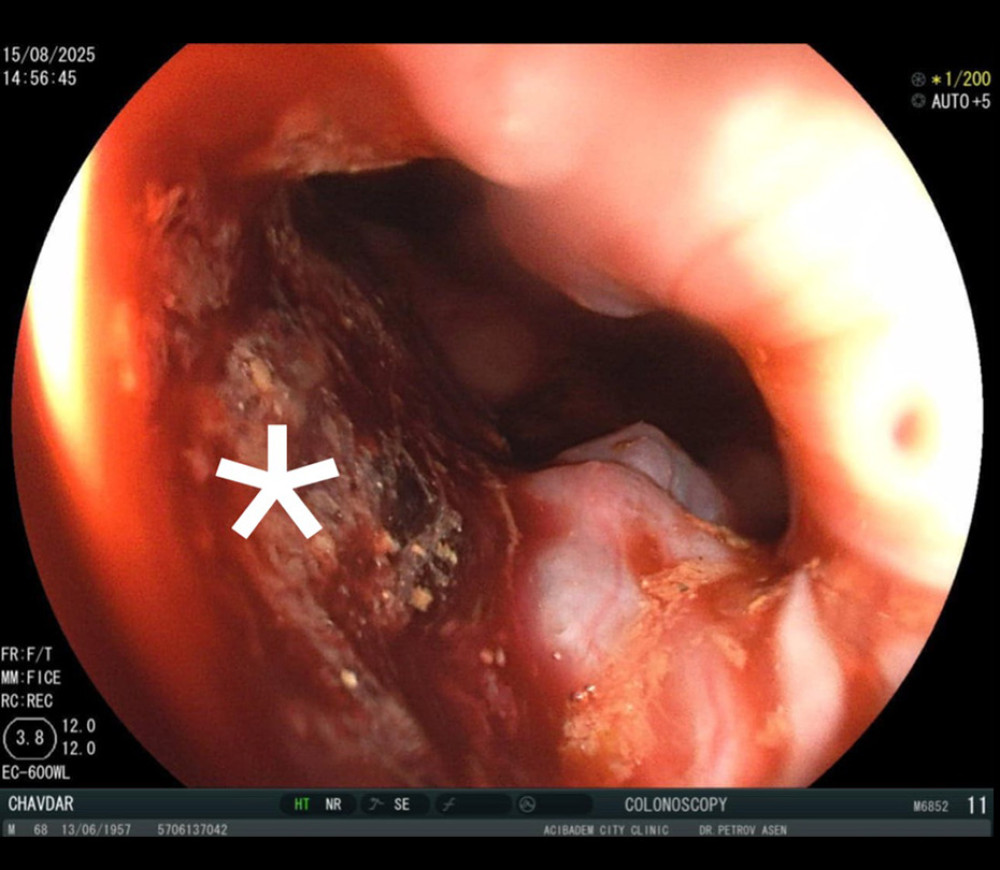 Figure 1. Colonoscopic view. Multifocal and diffuse hemorrhage was observed (*).
 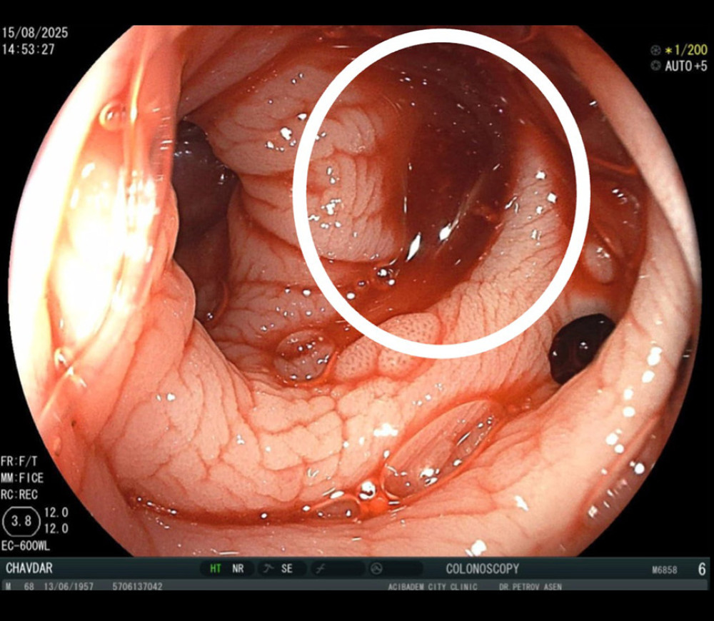 Figure 2. Colonoscopic view. Diffuse bleeding of the colon haustra (white circle). No blood clots were visible.
 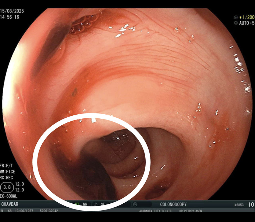 Figure 3. Colonoscopic view. Blood clots within the colon haustra (white circle).
 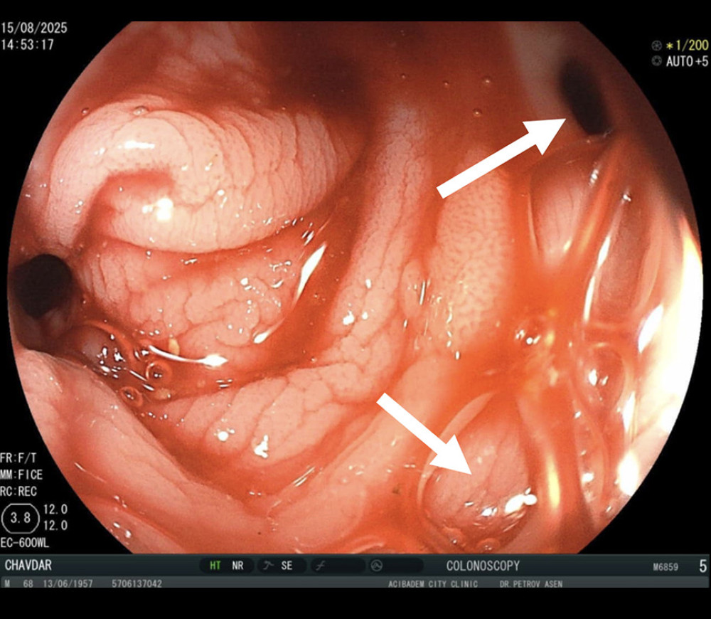 Figure 4. Colonoscopic view. Diverticulosis of the left colon (white arrows).
 References
1. Hassan K, Thielmann M, Easo J, Removal of apixaban during emergency cardiac surgery using hemoadsorption with a porous polymer bead sorbent: J Clin Med, 2022; 11(19); 5889
2. Cao D, Amabile N, Chiarito M, Reversal and removal of oral antithrombotic drugs in patients with active or perceived imminent bleeding: Eur Heart J, 2023; 44(20); 1780-94
3. Cormier S, Siegal DM, A framework for managing severe DOAC-related bleeding: Recognize, reverse, resume, reduce: Thrombosis Update, 2024; 15; 100165
4. Milling TJ, Refaai MA, Sengupta N, Anticoagulant reversal in gastrointestinal bleeding: Review of treatment guidelines: Dig Dis Sci, 2021; 66(11); 3698-714
5. Milling TJ, Ziebell CM, A review of oral anticoagulants, old and new, in major bleeding and the need for urgent surgery: Trends Cardiovasc Med, 2020; 30(2); 86-90
6. Tripathi R, Morales J, Lee V, Antithrombotic drug removal from whole blood using haemoadsorption with a porous polymer bead sorbent: Eur Heart J Cardiovasc Pharmacother, 2022; 8(8); 847-56
7. Turpin M, Gregory P, Direct oral anticoagulant use and risk of diverticular hemorrhage: A systematic review of the literature: Can J Gastroenterol Hepatol, 2019; 2019; 9851307
8. Douketis JD, Spyropoulos AC, Perioperative management of patients taking direct oral anticoagulants: A review: JAMA, 2024; 332(10); 825-34
9. Schmitto J, De Somer F, Thielmann M, Intraoperative antithrombotic drug removal during heart transplantation: A case series from the International Safe and Timely Antithrombotic Removal (STAR) registry: JHLT Open, 2025; 10; 100369
10. Schmoeckel M, Thielmann M, Hassan K, Intraoperative haemoadsorption for antithrombotic drug removal during cardiac surgery: Initial report of the international safe and timely antithrombotic removal (STAR) registry: J Thromb Thrombolysis, 2024; 57(6); 888-97
11. Storey RF, Hassan K, Meyer AL, Early CABG with intraoperative hemoadsorption in patients on ticagrelor: Real-world data from the international Safe and Timely Antithrombotic Removal (STAR) registry: Cardiovasc Revasc Med, 2026; 82; 50-56
12. Schmoeckel M, Thielmann M, Vitanova K, Direct-acting oral anticoagulant removal by intraoperative hemoadsorption in CABG and/or single valve surgery: Interim analysis of the International Safe and Timely Antithrombotic Removal (STAR) registry: J Cardiothorac Surg, 2025; 20(1); 74
13. Mack MJ, Whitlock R, Chu MWA, Randomized, sham-controlled trial of intraoperative ticagrelor removal to reduce perioperative bleeding: J Thorac Cardiovasc Surg, 2026; 172(1); 128-37e4
14. Budd AN, Kertai MD, Wyler von Ballmoos MC, Management of direct oral anticoagulants in adult patients undergoing cardiac surgery: A joint consensus statement by the Society of Cardiovascular Anesthesiologists and the Society of Thoracic Surgeons: Anesth Analg, 2026; 142(4); 668-81
15. Sinduja R, Vijayakumar C, Sudharshan M, Pseudoaneurysm of the gastroduodenal artery secondary to diverticulitis of the first part of duodenum: A rare presentation of upper gastrointestinal bleed: BMJ Case Rep, 2021; 14(1); e238232
16. Dalmastri V, Angelini A, Minerva V, Extracorporeal hemoadsorption therapy as a potential therapeutic option for rapid removal of Apixaban in high risk-surgical patients: A case report: J Med Case Rep, 2023; 17(1); 283
17. Hudowenz O, Nitschke M, Lapshyn H, Muck P, Successful kidney transplantation despite therapeutic anticoagulation-effective apixaban elimination by hemoadsorption: Clin Transplant, 2024; 38(10); e15463
18. Melegari G, Gazzotti F, Arturi F, Potential clinical use of CytoSorb((R)) for ticagrelor and rivaroxaban elimination prior to emergency orthopedic surgery in trauma patients: Life (Basel), 2025; 15(7); 1065
Figures
Figure 1. Colonoscopic view. Multifocal and diffuse hemorrhage was observed (*).Figure 2. Colonoscopic view. Diffuse bleeding of the colon haustra (white circle). No blood clots were visible.Figure 3. Colonoscopic view. Blood clots within the colon haustra (white circle).Figure 4. Colonoscopic view. Diverticulosis of the left colon (white arrows). In Press
Case report 
Am J Case Rep In Press; DOI: 10.12659/AJCR.953555
Case report
Am J Case Rep In Press; DOI: 10.12659/AJCR.953169
Case report 
Am J Case Rep In Press; DOI: 10.12659/AJCR.954027
Case report
Am J Case Rep In Press; DOI: 10.12659/AJCR.954380
Most Viewed Current Articles
07 Dec 2021 : Case report  28,713,109
DOI :10.12659/AJCR.934347
Am J Case Rep 2021; 22:e934347
06 Dec 2021 : Case report  180,916
DOI :10.12659/AJCR.934406
Am J Case Rep 2021; 22:e934406
21 Jun 2024 : Case report
124,581
DOI :10.12659/AJCR.944371
Am J Case Rep 2024; 25:e944371
08 May 2026 : Case report
95,842
DOI :10.12659/AJCR.952158
Am J Case Rep 2026; 27:e952158