


25 August 2026: Articles 
Pilomatricoma in an Octogenarian: A Case Report of an Exceptional Late-Life Presentation
Unusual clinical course, Challenging differential diagnosis
Oliwia Gunia BCEF 1*, Maëlys Bresson BCEF 1, Claude Depardieu BCD 1, Gilles Dolivet AB 1,2, Giacomo Gravante CD 3, Romina Mastronicola ABCDEFG 1,2DOI: 10.12659/AJCR.953320
Am J Case Rep 2026; 27:e953320


Abstract
BACKGROUND: Pilomatricoma, also known as Malherbe’s calcifying epithelioma, is a benign adnexal tumor originating from hair follicle matrix cells and occurs predominantly in children and young adults. Its presentation in an older adult is exceptionally rare and can pose diagnostic challenges. We report a rare case of pilomatricoma in an 86-year-old patient.
CASE REPORT: We report the case of an 86-year-old man who presented with a tender, subcutaneous cyst on the right scalp. The lesion had been evolving for several months, enlarging from approximately 5 cm to 8 cm. His medical history included multiple comorbidities: coronary artery disease, insulin-requiring type 2 diabetes mellitus, advanced chronic kidney disease, hypertension, dyslipidemia, and chronic obstructive pulmonary disease. Initial biopsy suggested pilomatricoma without malignant features; however, clinicopathological discordance and tumor progression raised suspicion of malignancy. Owing to significant anesthetic risk, initial management consisted of close surveillance. Following the onset of bleeding, surgical excision was performed. Histopathological analysis confirmed a benign pilomatricoma with clear margins. Postoperative recovery was uneventful, and follow-up was scheduled.
CONCLUSIONS: Giant pilomatricoma can present atypically in older patients, mimicking malignancy and leading to diagnostic uncertainty. This case underscores the importance of considering this diagnosis despite age and rapid growth, and supports complete surgical excision for definitive diagnosis and management.
Keywords: Pilomatrixoma, Case Reports, Geriatrics, surgical oncology, Ablation Techniques
Introduction
Pilomatricoma, formerly referred to as calcifying epithelioma of Malherbe, is a benign adnexal cutaneous tumor arising from hair follicle matrix cells. First described at the end of the nineteenth century, it corresponds to a tumor proliferation showing differentiation toward hair matrix structures and is histologically characterized by the association of basophilic cells and keratinized ghost/shadow cells [1].
It is a relatively rare tumor, accounting for approximately 0.1% of all cutaneous tumors in large histopathological series [1]. However, its true incidence is probably underestimated, mainly due to frequent preoperative misdiagnoses [2].
From an epidemiological standpoint, pilomatricoma predominantly affects the pediatric population and young adults. Most cases occur before the age of 20 years, with nearly 40% diagnosed before the age of 10 years [2,3]. Several studies report a mean age at diagnosis of 16.5 years, with an age range spanning from 5 months to 97 years [3].
The age distribution is classically described as bimodal, with a major peak during childhood (5 to 15 years) and a second much smaller peak observed in adulthood between 50 and 65 years [3]. Nevertheless, cases occurring in older patients remain exceptional, with some series reporting only rare occurrences beyond the age of 70 years [4].
Clinically, pilomatricoma usually presents as a firm, well-circumscribed, slow-growing subcutaneous nodule, most often located in the head and neck region, which is rich in hair follicles [2]. Its clinical diagnosis remains challenging because of its variable presentation and the wide range of differential diagnoses, making histopathological examination the gold standard for definitive diagnosis [2,3].
The occurrence of pilomatricoma in elderly patients therefore is an unusual presentation. This observation raises several pathophysiological hypotheses, including late tumorigenesis related to the accumulation of somatic mutations or, more likely, delayed diagnosis of a long-standing lesion that remained paucisymptomatic for years [3,5].
Through this case of an 86-year-old patient, we report an atypical presentation of pilomatricoma, highlighting the diagnostic particularities and the clinical implications related to its occurrence in the geriatric cohort. We aim to demonstrate that pilomatricoma can mimic aggressive scalp lesions in older adults, leading to a significant clinicopathological discrepancy between suspicious clinical features and benign histology. Additionally, we discuss appropriate management options for this high-surgical-risk population.
Case Report
An 85-year-old male patient, with no relevant ethnic background, presented in March 2025 with a lesion of the right scalp that had been evolving for approximately 1 month at the time of initial clinical evaluation. The lesion appeared as a subcutaneous mass of cystic appearance, tender on palpation, and without associated inflammatory signs or systemic symptoms. The patient denied any history of local trauma. He was a non-smoker, reported no alcohol consumption, and denied illicit drug use. No relevant occupational exposure was identified, as he was retired.
The patient’s medical history was significant for multivessel coronary artery disease requiring multiple percutaneous coronary interventions with stent placement. He also had insulin-dependent type 2 diabetes mellitus, advanced chronic kidney disease, hypertension, and dyslipidemia. Benign prostatic hyperplasia was suspected. In addition, his history included recurrent pulmonary infections requiring several hospitalizations over the previous 2 years in the setting of chronic obstructive pulmonary disease. He had no personal history of cutaneous or extracutaneous malignancy.
The patient was receiving treatment adapted to these comorbidities. Diabetes mellitus was managed with insulin glargine. Cardiovascular prevention related to coronary artery disease included acetylsalicylic acid combined with bisoprolol. Hypertension was treated with amlodipine. Dyslipidemia was managed with a combination of ezetimibe and atorvastatin. Chronic kidney disease was treated with furosemide. Lower urinary tract symptoms suggestive of benign prostatic hyperplasia were managed with tamsulosin. Chronic obstructive pulmonary disease was treated with an inhaled combination of fluticasone and vilanterol.
The scalp lesion had in fact been known since February 2025. In July 2025, its initial estimated size was approximately 2 × 2 cm in diameter. A skin biopsy performed at this time revealed histological features consistent with pilomatricoma without evidence of malignancy. Ultrasound examination revealed no suspicious lymphadenopathy at the occipital and cervical levels. However, due to clinicopathological discordance—particularly given the tumor size and its clinical evolution—the differential diagnosis of a malignant cutaneous tumor, especially squamous cell carcinoma or basal cell carcinoma, remained under consideration. Additional sampling was therefore discussed to rule out a possible collision tumor.
Nevertheless, in the absence of histological malignancy and given the patient’s overall condition and significant cardiovascular history—including a prior cardiorespiratory arrest during previous general anesthesia—an initial conservative approach with clinical surveillance was favored. In this context, radiotherapy was excluded.
The patient re-presented in December 2025 because of bleeding from the lesion that had been evolving for approximately 2 weeks. Clinical examination also revealed an increase in tumor volume to 5 cm in greatest dimension. The nodule was subcutaneous and freely mobile relative to both the overlying skin and deep structures. The lesion remained stable in appearance across consultations, characterized by an erythematous and ulcerated surface, without clinical signs of calcification upon palpation (Figure 1). Given this unfavorable progression, radiotherapy was reconsidered. However, histological confirmation was mandatory prior to treatment initiation. Following a thorough preoperative assessment, an excisional biopsy under general anesthesia was indicated.
At the time of surgery, performed in February 2026, the lesion measured 8 cm in greatest dimension, reflecting significant growth compared with its initial size. Complete excision was achieved with 5-mm safety margins. The resulting defect was managed using bolster dressings with secondary intention healing.
Definitive histopathological examination of the surgical specimen confirmed the diagnosis of giant pilomatricoma without malignant features. Microscopic analysis revealed a well-circumscribed dermal tumor composed of islands of epithelial cells showing a dual population: peripheral basaloid cells and central anucleated ghost cells, with areas of keratinization and foreign-body giant cell reaction (Figure 2). Given the absence of invasiveness, the fairly numerous and non-atypical mitoses, and the presence of ghost cells, carcinoma could be ruled out. The lesion was removed with complete excision and clear margins. No postoperative complications were observed at 1-week follow-up. Long-term clinical surveillance was scheduled to monitor for potential local recurrence.
Follow-up visits were planned to evaluate wound healing at 1 week and 1 month after excision. For long-term monitoring and at least 1 yearly consultation, the patient was referred back to his dermatologist. A re-evaluation by the surgeon will be scheduled if there are any concerns about the patient’s condition or the development of scars.
Discussion
Pilomatricoma is a benign cutaneous tumor originating from the uncontrolled proliferation of pluripotent precursors of hair matrix cells. Despite being well-characterized histologically, it is often misdiagnosed clinically [6]. Cases are often preceded by a history of trauma, bite, or antecedent surgery [7]. It commonly develops in the first 2 decades of life in the head and neck area and is rare in older adults. Previous studies on over 2000 cases of pilomatricoma reported a mean age at excision of 16 years 7 months, with a range from 5 months to 97 years [8,9].
This case illustrates several critical aspects of a fast-growing giant pilomatricoma in an 85-year-old man, which brings up several points of interrogation compared to the existing literature. The difference between the initial biopsy results and clinical evolution underscores the diagnostic hurdles of pilomatricomas. This led us to include squamous cell carcinoma and basal cell carcinoma in the differential diagnosis.
While sources report a diagnostic accuracy of 16% to 34% for pilomatricoma, our case demonstrates that diagnostic uncertainty can persist even after initial histological analysis when the tumor size exceeds typical dimensions [9]. The threshold definition for a giant pilomatricoma varies in the literature, fluctuating between diameters greater than 4 cm and 5 cm [10]. A giant pilomatricoma measuring 6 × 4 cm was reported in the preauricular region of a 65-year-old woman, with a clinical history evolving over 1 year [6]. Another case involved a 38-year-old woman with an exceptionally large 11 × 10 × 5 cm pilomatricoma located on the truncal dorsal midline, which rapidly evolved over a 3-month period [10]. In comparison, our patient’s 8-cm lesion clearly qualifies as a giant variant and underscores the possibility of a giant pilomatricoma presentation at an advanced age. Such large tumors can clinically imitate malignancy, as described in several previous reports. Our case ruled out the initial clinical suspicion of malignancy.
In the present case, preoperative imaging could have been valuable in refining the initial diagnosis. Recent data support the utility of high-frequency ultrasound, the commonest imaging modality used in this diagnosis [9]. Typical ultrasonographic features often include heterogeneous hypoechoic masses with internal echogenic foci (calcifications), a hypoechoic halo, and posterior acoustic shadowing. The presence of these signs could have confirmed the diagnosis and potentially influenced initial management. Other imaging modalities, such as computed tomography and magnetic resonance imaging, could also have been used; these highlight calcifications and heterogeneous T2 hyperintensity with enhancement on magnetic resonance imaging, correlating with histopathological findings.
The initial conservative approach with monitoring was justified by the patient›s severe cardiovascular comorbidities, notably a history of cardiorespiratory arrest under general anesthesia, and remained reasonable given the benign histological report from the initial biopsy. However, this case highlights several limitations of conservative management for giant pilomatricoma. The progression of 3 cm in 12 months (from 5 cm to 8 cm), coupled with bleeding episodes, ultimately necessitated the initially deferred intervention. It is essential to address the question of the optimal timing for surgical intervention in patients with multimorbidity and polypharmacy.
Surgical excision remains the gold standard treatment for pilomatricoma [7,9]. Although there is limited literature regarding the malignant transformation of pilomatricoma, the clinical behavior of giant variants can be unpredictable. Three cases of giant pilomatricoma have been reported, suggesting clinical malignancy, including 1 case complicated by hypercalcemia due to parathyroid hormone-related protein production [11]. The rapid growth and bleeding observed in our case—contrasting with the final histology that excluded malignancy—demonstrate that clinical behavior cannot always be predicted by the initial biopsy, especially when the sample is not representative of the entire tumor.
Conclusions
Although pilomatricoma is well-known, it is frequently misdiagnosed. These findings suggest that pilomatricoma should be considered in the differential diagnosis of masses with a large diameter in the head and neck area, especially among older individuals showing rapid erythematous progression, where this diagnosis may be prematurely ruled out based on known epidemiological data [8]. Histological confirmation is essential, as benign pilomatricoma can clinically imitate malignant variants [12]. While complete surgical excision remains the definitive treatment, therapeutic decisions in older adults must carefully balance the benefits, medical burden, and potential anesthetic risks.
Figures
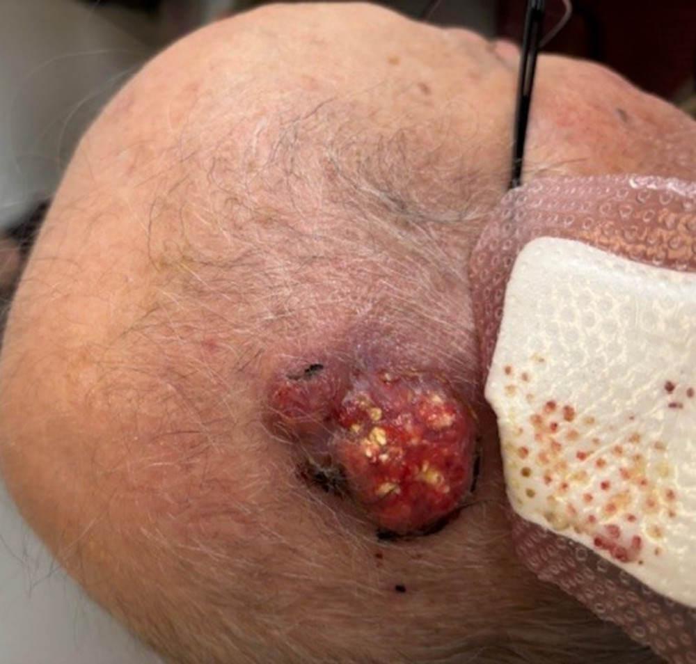 Figure 1. Clinical photography of lesion 2 months prior to surgical excision. Freely mobile, ulcerated, and erythematous lesion on the scalp, measuring 5 cm in its largest diameter, with no bone involvement.
 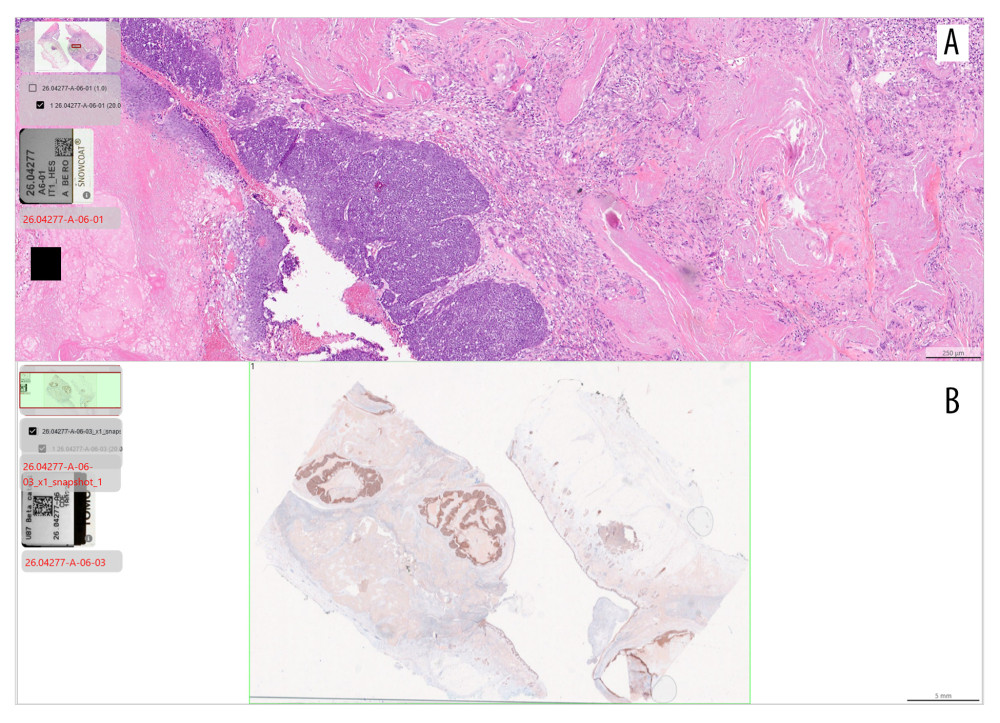 Figure 2. Histopathological section: (A) H&E: Well-circumscribed, lobulated dermal neoplasm. At the cellular level, it shows islands of basaloid cells with mitotic activity, exhibiting abrupt keratinization into anucleated ghost cells. The tumor keratin has elicited a foreign-body giant cells granulomatous inflammatory response. No signs of invasion or atypical features are present. (B) Immunohistochemistry (beta-catenin): Low-power view showing nuclear and membranous positivity. The areas of shadow cells and keratins remain negative.
 References
1. Moehlenbeck FW, Pilomatrixoma (calcifying epithelioma): A statistical study: Arch Dermatol, 1973; 108(4); 532-34
2. Lan MY, Lan MC, Ho CY, Pilomatricoma of the head and neck: A retrospective review of 179 cases: Arch Otolaryngol Head Neck Surg, 2003; 129(12); 1327-30
3. Rao J, Arshad H, Aulakh A, Elston DM, Pilomatrixoma (pilomatricoma): Background, pathophysiology, epidemiology: Medscape, 2024 https://emedicine.medscape.com/article/1058965-overview
4. Lin SF, Xu SH, Xie ZL, Calcifying epithelioma of Malherbe (pilomatrixoma): Clinical and sonographic features: J Clin Ultrasound, 2018; 46(1); 3-7
5. Wang J, Cobb CJ, Martin SE, Pilomatrixoma: Clinicopathologic study of 51 cases with emphasis on cytologic features: Diagn Cytopathol, 2002; 27(3); 167-72
6. Yuca K, Kutluhan A, Çankaya H, Akman E, Giant pilomatrixoma arising in the preauricular region: A case report: Kulak Burun Bogaz Ihtis Derg, 2004; 12(5); 147-49
7. Guinot-Moya R, Valmaseda-Castellon E, Berini-Aytes L, Gay-Escoda C, Pilomatrixoma: Review of 205 cases: Med Oral Patol Oral Cir Bucal, 2011; 16(4); e552-e55
8. Behnke N, Schulte K, Ruzicka T, Megahed M, Pilomatricoma in elderly individuals: Dermatology, 1998; 197(4); 391-93
9. Jones CD, Ho W, Robertson BF, Gunn E, Morley S, Pilomatrixoma: A comprehensive review of the literature: Dermatol Surg, 2018; 40(9); 631-41
10. Göktay F, Mansur AT, Aydingöz IE, A rare presentation of giant pilomatricoma Located on the back: Dermatol Surg, 2007; 33(5); 596-600
11. Yamauchi M, Yotsuyanagi T, Saito T, Three cases of giant pilomatrixoma – considerations for diagnosis and treatment of giant skin tumours with abundant inner calcification present on the upper body: J Plast Reconstr Aesthet Surg, 2010; 63(6); e519-e24
12. Blegen K, Samaniego M, Stetson C, Sturgeon A, A rapidly growing forearm pilomatricoma in an elderly patient: Cureus, 2023; 15(5); e39043
Figures
Figure 1. Clinical photography of lesion 2 months prior to surgical excision. Freely mobile, ulcerated, and erythematous lesion on the scalp, measuring 5 cm in its largest diameter, with no bone involvement.Figure 2. Histopathological section: (A) H&E: Well-circumscribed, lobulated dermal neoplasm. At the cellular level, it shows islands of basaloid cells with mitotic activity, exhibiting abrupt keratinization into anucleated ghost cells. The tumor keratin has elicited a foreign-body giant cells granulomatous inflammatory response. No signs of invasion or atypical features are present. (B) Immunohistochemistry (beta-catenin): Low-power view showing nuclear and membranous positivity. The areas of shadow cells and keratins remain negative. In Press
Case report 
Am J Case Rep In Press; DOI: 10.12659/AJCR.953543
Case report 
Am J Case Rep In Press; DOI: 10.12659/AJCR.954216
Case report 
Am J Case Rep In Press; DOI: 10.12659/AJCR.954932
Case report 
Am J Case Rep In Press; DOI: 10.12659/AJCR.954146
Most Viewed Current Articles
07 Dec 2021 : Case report
28,712,695
DOI :10.12659/AJCR.934347
Am J Case Rep 2021; 22:e934347
06 Dec 2021 : Case report  180,721
DOI :10.12659/AJCR.934406
Am J Case Rep 2021; 22:e934406
21 Jun 2024 : Case report
124,419
DOI :10.12659/AJCR.944371
Am J Case Rep 2024; 25:e944371
08 May 2026 : Case report
95,462
DOI :10.12659/AJCR.952158
Am J Case Rep 2026; 27:e952158