


08 September 2026: Articles 
Suspected Donor-Site Implantation of Floor of Mouth Squamous Cell Carcinoma After Free Flap Reconstruction: A Case Report
Unusual clinical course, Unusual or unexpected effect of treatment, Diagnostic / therapeutic accidents
TianWen Zhang BCEF 1, Bo Lin ACF 1, HuiJun Yang ADE 1, HongYu Yang ADG 1*
DOI: 10.12659/AJCR.953340
Am J Case Rep 2026; 27:e953340


Abstract
BACKGROUND: Oral squamous cell carcinoma (OSCC) is the most common malignant tumor in the head and neck. A free flap is often used to repair tissue defects left after surgery. Due to the existence of 2 surgical sites—the donor site and recipient site—cross-contamination should be prevented during the operation. This report presents a case of donor tumor implantation suspected to be caused by cross-contamination.
CASE REPORT: A 57-year-old Chinese man was admitted with a mass in the left floor of the mouth, first noticed 2 months before. The biopsy confirmed squamous cell carcinoma (SCC). Under general anesthesia, extended resection of the left floor of the mouth SCC and repair with a left anterolateral thigh free flap were performed. Five months after surgery, a mass was found in the scar on the left thigh. Extensive resection of the left thigh mass was performed, and postoperative pathology revealed metastatic SCC. At the same time, a tumor was found on the right back and another on the chest wall. These 2 tumors were resected at the same time and were also confirmed as SCC. Seven months after surgery, the patient gradually developed local recurrence and systemic multiple metastases, and he died 10 months after surgery.
CONCLUSIONS: The “no tumor left behind” principle should be strictly followed in malignant tumor surgery to avoid malignant tumor implantation. Oral and maxillofacial surgeons should be aware of this and use appropriate techniques to avoid such incidents.
Keywords: Carcinoma, Squamous Cell, Implants, Experimental, metastasectomy, Surgery, Oral
Introduction
Oral squamous cell carcinoma (OSCC) is the most common malignant tumor of the head and neck [1,2]. Surgical resection is the main treatment [3,4]. A free flap is often used to repair tissue defects left after surgery [5,6]. Due to the existence of 2 surgical sites—the donor site and recipient site—cross-contamination should be prevented during the operation. Advanced head and neck malignancies often undergo systemic metastasis [7], but tumor implantation has been rarely reported in the literature. This report presents a case of donor tumor implantation suspected to be caused by cross-contamination. The tumor happened to appear at the scar site of the donor area.
Case Report
A 57-year-old Chinese man was admitted with a mass in the left floor of the mouth, first noticed 2 months before. Since first discovery, the mass had grown slowly and no numbness of the tongue had occurred. His general condition was good, with no previous history of oral or maxillofacial malignancy.
Clinical examination showed no obvious abnormality in facial appearance. A cauliflower mass could be seen at the left floor of mouth, involving the tongue and abdomen, ulcerative, about 4 × 3 cm in size, hard, and immobile, and swollen lymph nodes were found in the left neck (Figure 1).
Imaging examination revealed an irregular, lobulated, soft-tissue mass on the left tongue and the floor of mouth, with unclear boundary. It was up to the abdomen of the tongue, down to the floor of mouth, with a range of about 32 × 15 × 20 mm. The plain scan density was uneven and showed obvious uneven enhancement. The left sublingual gland, submandibular gland, and tonsil were enlarged, the local boundary was unclear, and the enhancement was significantly enhanced compared with the contralateral side. The left salivary gland duct was dilated, and the adjacent hyoid bone and mandible had no obvious bone destruction. Multiple enlarged lymph nodes were observed in the first to fourth region of the left neck, and the largest was about 15 × 9 mm (Figure 2).
After admission, an examination was completed, and the biopsy was confirmed as squamous cell carcinoma. Under general anesthesia, we performed extended resection of left floor of the mouth squamous cell carcinoma (SCC), partial resection of left mandibular and bilateral lymph nodes of the neck, dissection and repair of left anterolateral thigh free flap, and tracheotomy (Figure 3). Two separate surgical fields were used with separate teams and instruments for each field. Head and neck tumor resection and cervical lymph node dissection were performed concurrently with flap harvesting. A sterile drape was placed at the level of the abdomen as an isolation barrier, establishing a strict protocol requiring surgical personnel from both teams to place instruments within their respective areas. After tumor resection and cervical lymph node dissection were completed, the recipient-site surgical team changed surgical gowns and gloves, and simultaneously replaced surgical instruments and sterile drapes. The donor-site surgical team irrigated the operative field with copious amounts of normal saline before wound closure. Postoperative pathology showed SCC T3N3bMx, stage IV, without invasion of bone tissue (Figure 4).
Adjuvant radiotherapy was administered 6 weeks after surgery. The clinical target volume (CTV) radiotherapy doses were 60 Gy (tumor area) and CTV 54Gy (lymph node area). Chemotherapy was also administered, with cisplatin 50 mg d1–3/21d.
Five months after surgery, a mass was found in the scar on the left thigh. Clinical examination showed that the 3 × 2 cm mass was located under the scar. It was soft and mobile (Figure 5). B-ultrasound showed a 30 × 23 × 18 mm hypoechoic mass located 3 mm from the skin surface in the subcutaneous soft tissue of the left thigh, which was elliptical, with a clear margin, internal echo not uniform, and the upper and lower had “rat tail sign”, the distance (Figure 6). A computed tomography (CT) scan showed a subcutaneous, round, slightly low-density shadow in front of the thigh of the left side, about 21 × 22 mm in size, with blurred edges, unclear boundary with the rectus femoris, and increased density of the surrounding subcutaneous fat space (Figure 7). Extensive resection of the left thigh mass was performed (Figure 8), and postoperative pathology revealed metastatic squamous cell carcinoma (SCC) (Figure 9). At the same time, a tumor was found on the right back and another in the chest wall, each about 2 × 2 cm, and both were tough and indistinct from the surrounding tissue. These 2 tumors were resected at the same time and it were also confirmed as SCC (Figure 10). Seven months after surgery, the patient gradually developed local recurrence and systemic multiple metastases, and he died 10 months after surgery.
Discussion
In recent years, surgery and adjuvant chemoradiotherapy for OSCC have improved the overall survival rate and control rate of local lesions. However, the distant metastasis rate has an increasing trend. OSCC often metastases to regional lymph nodes, and distant metastasis is rare [8], but when it happens, it worsens patients’ quality of life and prognosis [9]. Distant metastases are more common in the lungs than elsewhere. The presence of primary mandibular intraosseous carcinoma, grade IV and grade V cervical lymph node metastasis, local recurrence, and presence of pathological extrinsic extension are important risk factors for distant metastasis [10–12]. In addition, non-surgical treatment of local lesions is also a risk factor for distant metastases [13]. There are many studies on distant metastasis of oral cancer, especially in-depth studies on its risk factors, but there have been few reports on implantation metastasis in donor sites during repair and reconstruction.
In this case, the tumor was at the donor-site incision, raising the strong suspicion that the “no tumor left behind” principle was not strictly observed during the operation, resulting in implantation of malignant tumors. Multiple intraoperative mechanisms can promote the dissemination or implantation of tumor cells [14–16]: glove contamination [17], contaminated surgical instruments [18,19], aerosolized tumor cells or dispersed blood and tissue fluids [20], and mechanical manipulation of the tumor that induces cellular shedding [21]. Kuhar et al confirmed that gloves used in head and neck malignant tumor surgery can be tested for malignant tumor cells, as these cells can cause tumor implantation and metastasis [22]. Although great care was taken to avoid cross-contamination, the limited surgical operating space prevents isolation zones established using sterile drapes from being an absolute physical barrier [23]. Instruments used in different surgical areas can inadvertently be transferred across the isolation zone due to operator error, leading to instrument contamination [24]. Furthermore, garment contact between surgeons from different teams can also occur.
Preventive measures should be strictly implemented to minimize such risks. These include: (1) using separate instrument trays and designated surgical teams for the primary tumor resection and the donor site harvesting; (2) changing gloves and gowns after completing tumor resection and before accessing the donor site [25,26]; (3) adopting a “no-touch” technique during tumor handling and thoroughly irrigating the surgical site with sterile water before wound closure [27–29]; (4) applying physical barriers between the 2 surgical fields; (5) minimizing tumor manipulation [30,31]; and (6) performing donor-site procedures sequentially (eg, harvesting the flap before tumor resection when anatomically feasible) to preclude cross-contamination [32].
Tumor metastasis also appeared in the soft tissues of our patient’s chest and back, indicating that the mass in the donor site can also be a source of hematologic metastasis of malignant tumors. Wescott et al reported the case of a patient with advanced tongue SCC who underwent gastrostomy before surgery due to dysphagia, but the tumor still metastasized to the fistula [33], which was apparently independent of whether the “no tumor left behind” principle was followed during the operation. The same situation was reported by Sharma et al [34]. There is a theory that the blood or lymphatic spread of cancer cells from peripheral blood to the gastrostomy site is related to surgical stress during PEG tube placement. The same may be true in our case.
To further differentiate implantation metastasis from hematogenous metastasis, several criteria should be considered. Implantation metastasis is characterized by (1) tumor localization at the surgical incision or wound site rather than in parenchymal organs or randomly distributed soft tissues; (2) a close temporal relationship to the surgical procedure, often manifesting earlier than widespread systemic dissemination; (3) histopathological continuity between the implanted tumor and the primary lesion, with similar morphological features; and (4) the absence of other distant metastases at the time of donor-site lesion detection, or a metastatic pattern anatomically restricted to the operative field [35]. In contrast, hematogenous metastasis typically presents with multiple lesions in distant organs (eg, lungs, liver, bone) or soft tissues, often synchronous or metachronous with the primary tumor, and is associated with vascular invasion or large numbers of circulating tumor cells [36,37]. In our case, the simultaneous presence of chest and back soft-tissue metastases raised the possibility of a hematogenous component. Nevertheless, the location of the donor-site mass precisely at the surgical incision, its occurrence before widespread systemic spread, and the lack of other pulmonary metastases at initial detection argue in favor of implantation metastasis. However, the possibility that the donor-site lesion represents the first clinical manifestation of hematogenous dissemination cannot be entirely excluded. Therefore, we present donor-site implantation as the most plausible mechanism, rather than a proven cause. Future studies using molecular clonality analysis (eg, comparative genomic hybridization or next-generation sequencing) could help distinguish between these 2 mechanisms by comparing the genetic profiles of the primary tumor, donor-site mass, and distant soft-tissue metastases [38,39].
Regardless of the exact mechanism, adherence to the “no tumor left behind” principle should be strictly followed during malignant tumor surgery to avoid malignant tumor implantation. Oral and maxillofacial surgeons should be aware of this and use appropriate techniques to avoid such incidents. Understanding the mechanisms of intraoperative tumor cell dissemination, adopting preventive measures, and meticulously adhering to the “no tumor left behind” principle are essential to improve surgical outcome and prognosis [40,41].
Conclusions
In our case, the tumor was at the incision of the donor site, which leads to the strong suspicion that the “no tumor left behind” principle was not strictly observed during the operation, potentially resulting in implantation of malignant tumors. Nevertheless, hematogenous dissemination cannot be definitively excluded, particularly given the concurrent soft-tissue metastases on the chest and back. Therefore, donor-site implantation is presented as the most likely, but not proven, mechanism. A key limitation is that we did not perform molecular clonality analysis or genomic comparison between the primary tumor and the metastatic lesion, which precludes definitive distinction between implantation and hematogenous dissemination. Future studies incorporating such methods are warranted. The “no tumor left behind” principle should be strictly followed in the malignant tumor surgery to avoid malignant tumor implantation.
Figures
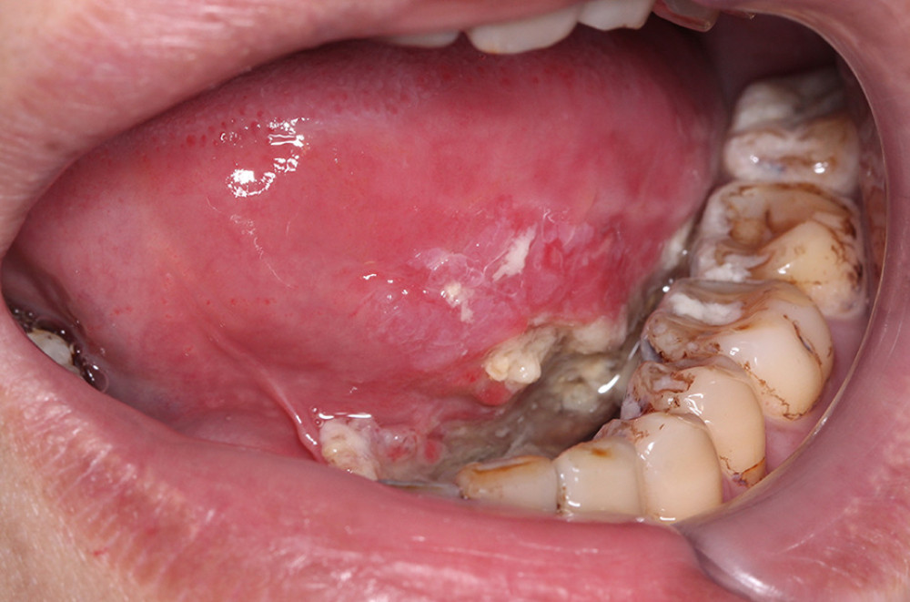 Figure 1. A mass located on the left floor of the mouth.
 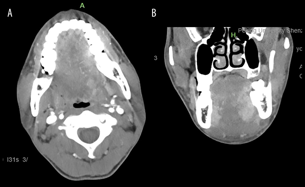 Figure 2. Computed tomography images of the mass. (A) Axial plane of the mass. (B) Coronal plane of the mass.
 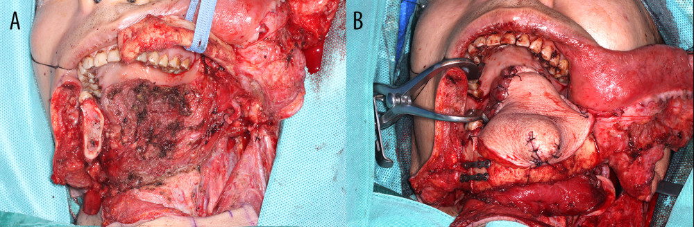 Figure 3. Intraoperative photos. (A) Postresection. (B) Free flap repair.
 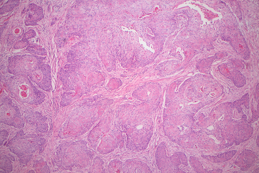 Figure 4. Pathology section of the tumor (H&E staining, magnification 50 ×).
 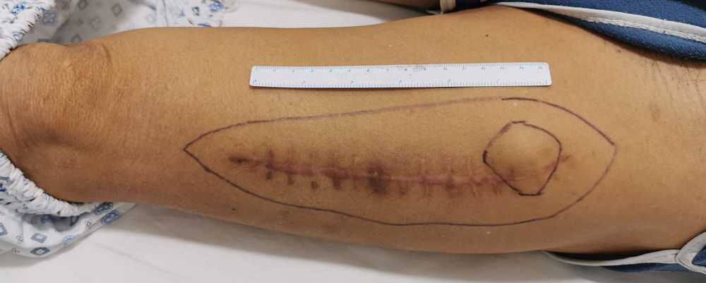 Figure 5. The mass in the left thigh.
 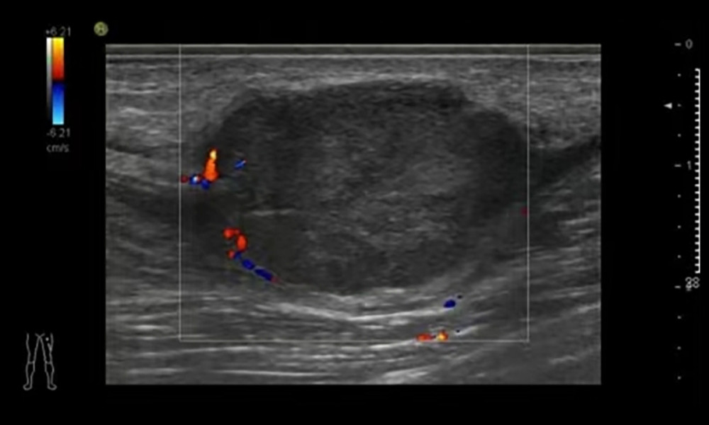 Figure 6. Ultrasound image of the mass in the left thigh.
 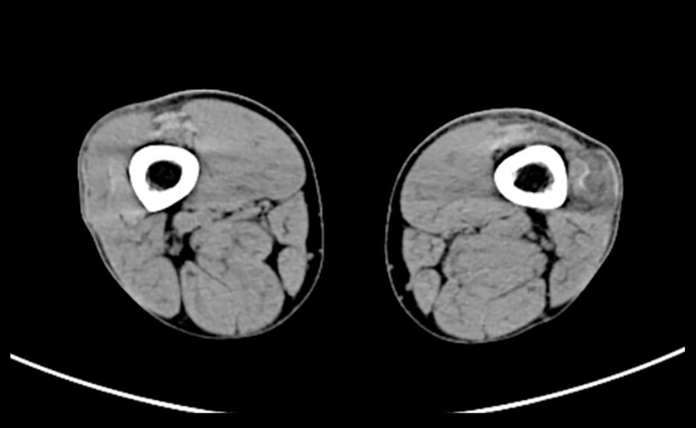 Figure 7. Computed tomography scan of the left thigh.
 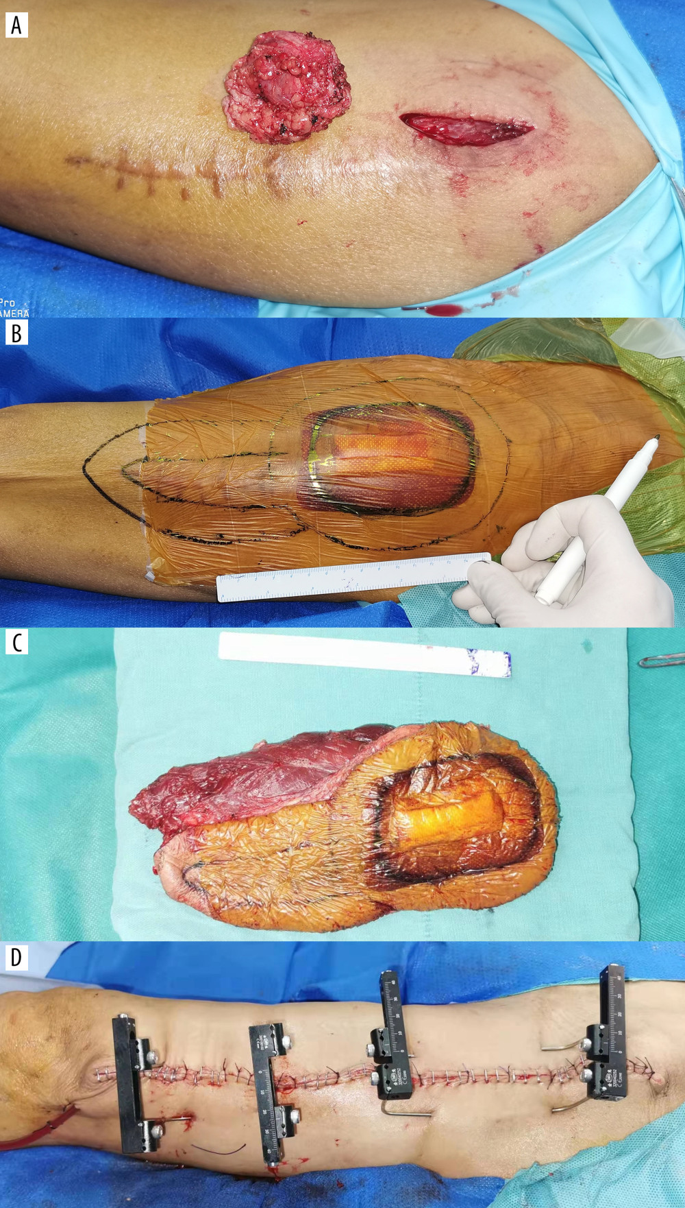 Figure 8. Intraoperative excision of the mass in the left thigh. (A) Local resection of the thigh mass. (B) After pathological confirmation of malignant tumor, the tumor was enlarged and resected. (C) Excision specimen. (D) Closure of the wound.
 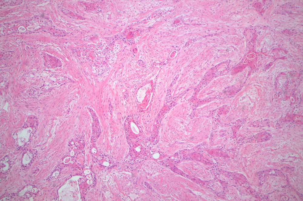 Figure 9. Pathology of the left thigh (H&E staining, magnification 50 ×).
 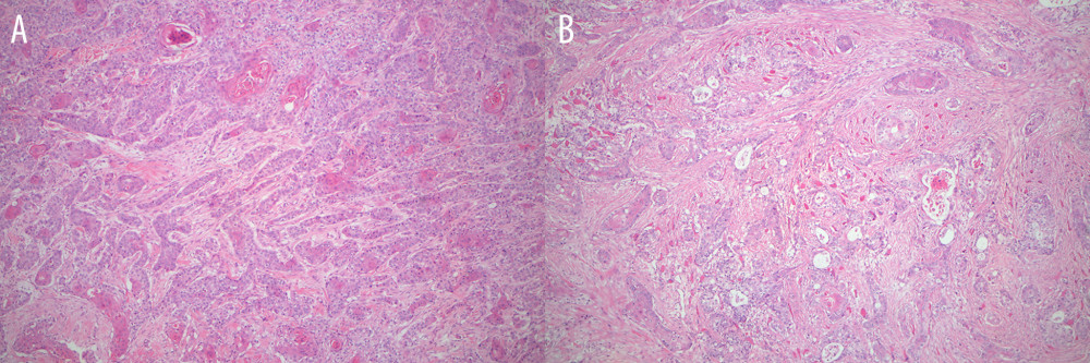 Figure 10. Pathological sections of the masses on the back and chest. (A) The back mass. (B) The chest mass (H&E staining, magnification 25 ×).
 References
1. Vitório JG, Duarte-Andrade FF, Dos Santos Fontes Pereira T, Metabolic landscape of oral squamous cell carcinoma: Metabolomics, 2020; 16(10); 105
2. Bugshan A, Farooq I, Oral squamous cell carcinoma: Metastasis, potentially associated malignant disorders, etiology and recent advancements in diagnosis: F1000Res, 2020; 9; 229
3. Sunga GM, Cobos Perez A, Molina AH, Translationally-relevant tumor resection model for murine preclinical models of oral squamous cell carcinoma: J Vis Exp, 2026(230), doi: 10.3791/68621
4. Xue N, Wang Y, Wang Z, Trends in immunotherapy for oral squamous cell carcinoma: Cell Oncol (Dordr), 2025; 48(5); 1159-79
5. Lovětínská V, Sukop A, Klein L, Free-flap monitoring: review and clinical approach: Acta Chir Plast, 2020; 61(1–4); 16-23
6. Othman S, Robinson E, Kamdar D, Microvascular free-flap head and neck reconstruction: The utility of the modified frailty five-item index: J Reconstr Microsurg, 2024; 41(3); 270-76
7. Hsieh RW, Borson S, Tsagianni A, Immunotherapy in recurrent/metastatic squamous cell carcinoma of the head and neck: Front Oncol, 2021; 11; 705614
8. Liu JC, Bhayani M, Kuchta K, Patterns of distant metastasis in head and neck cancer at presentation: Implications for initial evaluation: Oral Oncology, 2019; 88; 131-36
9. Aires Felipe T, Lin Chin S, Matos Leandro L, Risk factors for distant metastasis in patients with oral cavity squamous cell carcinoma undergoing surgical treatment: ORL J Otorhinolaryngol Relat Spec, 2017; 79(6); 347-55
10. Tomioka H, Yamagata Y, Oikawa Y, Risk factors for distant metastasis in locoregionally controlled oral squamous cell carcinoma: A retrospective study: Scientific Reports, 2021; 11(1); 5213
11. Sakamoto Y, Matsushita Y, Yamada S-i, Risk factors of distant metastasis in patients with squamous cell carcinoma of the oral cavity: Oral Surg Oral Med Oral Pathol Oral Radiol, 2016; 121(5); 474-80
12. Hasegawa T, Tanakura M, Takeda D, Risk factors associated with distant metastasis in patients with oral squamous cell carcinoma: Otolaryngol Head Neck Surg, 2015; 152(6); 1053-60
13. Sumioka S, Sawai NY, Kishino M, Risk factors for distant metastasis in squamous cell carcinoma of the oral cavity: J Oral Maxillofac Surg, 2013; 71(7); 1291-97
14. Matsunaga K, Sasaki K, Nozawa H, Clinicopathological characteristics of anastomotic recurrence after curative resection for colorectal cancer: Comparison with nonanastomotic local recurrences: Dis Colon Rectum, 2022; 66(10); e1014-e22
15. Nakaoka M, Nemoto T, Chiba H, Recurrence of gastric cancer caused by implantation of tumor cells after curative endoscopic submucosal dissection: Gastric Cancer, 2021; 24(5); 1160-66
16. Javed S, Khan A, Khalid A, Scalp metastasis from atypical meningioma: A case report and literature review: Clin Case Rep, 2024; 12(5); e8789
17. Gresham E, Don Parsa F, Iatrogenic implantation of cancer cells during surgery: Hawaii J Health Soc Welf, 2020; 79(1); 4-6
18. Link T, Guidelines in practice: Instrument cleaning: AORN J, 2021; 114(3); 241-51
19. Oya A, Matsumura N, Fujie A, Investigation of intraoperative bacterial contamination in surgical instruments used for total hip arthroplasty: Arthroplast Today, 2026; 39; 102027
20. Chen JX, Workman AD, Chari DA, Demonstration and mitigation of aerosol and particle dispersion during mastoidectomy relevant to the COVID-19 era: Otol Neurotol, 2020; 41(9); 1230-39
21. Moghassemi S, Dadashzadeh A, de Azevedo RB, Secure transplantation by tissue purging using photodynamic therapy to eradicate malignant cells: J Photochem Photobiol B, 2022; 234; 112546
22. Kuhar HN, Shay A, Revenaugh PC, Cytopathologic assessment of gloves and instruments after major head and neck surgery: Am J Otolaryngol, 2021; 42(3); 102876
23. Qvistgaard M, Almerud-Österberg S, Lovebo J, Covering surgical instruments with single- or double-layer drape pending surgery: An experimental study in a perioperative setting: J Infect Prev, 2020; 22(3); 126-31
24. Nofal MR, Tesfaye A, Gebeyehu N, Global scaling of a surgical infection prevention program to 5 low-resource countries: JAMA Surg, 2025; 160(9); 1004-11
25. Sagers K, Shields JS, Pollock DC, Characterizing particle evacuation in the setting of positive-pressure body exhaust suit using a novel gloving technique: J Surg Orthop Adv, 2021; 30(3); 166-69
26. Hegde S, Which type of personal protective equipment (PPE) and which method of donning or doffing PPE carries the least risk of infection for healthcare workers?: Evid Based Dent, 2020; 21(2); 74-76
27. Gao Q, Guo L, Wang B, The pathogenesis and prevention of port-site metastasis in gynecologic oncology: Cancer Manag Res, 2020; 12; 9655-63
28. Kanao H, Aoki Y, Fusegi A, Feasibility and outcomes of “no-look no-touch” laparoscopic radical trachelectomy for early-stage cervical cancer: J Clin Med, 2021; 10(18); 4154
29. Chen S, Zheng Y, Tong L, Laparoendoscopic single-site radical hysterectomy with vaginal closure and without uterine manipulator for FIGO IB1 cervical cancer: J Minim Invasive Gynecol, 2020; 27(7); 1471-72
30. Yasukawa M, Sawabata N, Kawaguchi T, Wedge resection of tumor before lobectomy for lung cancer could be a no-touch isolation technique: In Vivo, 2020; 34(2); 779-85
31. Dietl A, Aumann K, Beckmann MW, Tumor handling of early-stage cervical cancer: A literature analysis of villoglandular adenocarcinoma of the cervix: Anticancer Res, 2020; 40(6); 3049-53
32. Schmidt M, Zaussinger M, Duscher D, Preauricular pull through flap for reconstruction of the auricle: J Plast Reconstr Aesthet Surg, 2020; 74(1); 130-34
33. Wescott B, Seegmiller S, Mohamed Elfadil O, Seeding of gastrostomy tube site in patient with squamous cell carcinoma of the tongue: A case report: Nutrition in Clinical Practice, 2020; 36(3); 648-53
34. Sharma P, Berry SM, Wilson K, Metastatic implantation of an oral squamous-cell carcinoma at a percutaneous endoscopic gastrostomy site: Surg Endosc, 1994; 8(10); 1232-35
35. West JD, Martin AN, Kim J, Squamous cell carcinoma metastasis to the supraclavicular artery island flap donor site: Laryngoscope, 2020; 131(2); E440-E42
36. Li F, Liang H, You H, Targeting HECTD3-IKKα axis inhibits inflammation-related metastasis: Signal Transduct Target Ther, 2022; 7(1); 264
37. Orrapin S, Udomruk S, Lapisatepun W, Clinical implication of circulating tumor cells expressing Epithelial Mesenchymal Transition (EMT) and Cancer Stem Cell (CSC) markers and their perspective in HCC: A systematic review: Cancers (Basel), 2022; 14(14); 3373
38. Zheng C, Wang Y, Luo Y, Synchronous lung and multiple soft tissue metastases developed from osteosarcoma of tibia: A rare case report and genetic profile analysis: BMC Musculoskelet Disord, 2022; 23(1); 74
39. Song E, Song DE, Ahn J, Genetic profile of advanced thyroid cancers in relation to distant metastasis: Endocr Relat Cancer, 2020; 27(5); 285-93
40. Adachi H, Ito H, Sawabata N, Circulating tumor cells and the non-touch isolation technique in surgery for non-small-cell lung cancer: Cancers (Basel), 2022; 14(6); 1448
41. Kawase A, Sekihara K, Matsutani N, Circulating tumor cells from surgical manipulation predict recurrence and poor prognosis in non-small cell lung cancer: J Clin Med, 2025; 14(6); 2070
Figures
Figure 1. A mass located on the left floor of the mouth.Figure 2. Computed tomography images of the mass. (A) Axial plane of the mass. (B) Coronal plane of the mass.Figure 3. Intraoperative photos. (A) Postresection. (B) Free flap repair.Figure 4. Pathology section of the tumor (H&E staining, magnification 50 ×).Figure 5. The mass in the left thigh.Figure 6. Ultrasound image of the mass in the left thigh.Figure 7. Computed tomography scan of the left thigh.Figure 8. Intraoperative excision of the mass in the left thigh. (A) Local resection of the thigh mass. (B) After pathological confirmation of malignant tumor, the tumor was enlarged and resected. (C) Excision specimen. (D) Closure of the wound.Figure 9. Pathology of the left thigh (H&E staining, magnification 50 ×).Figure 10. Pathological sections of the masses on the back and chest. (A) The back mass. (B) The chest mass (H&E staining, magnification 25 ×). In Press
Case report 
Am J Case Rep In Press; DOI: 10.12659/AJCR.954216
Case report
Am J Case Rep In Press; DOI: 10.12659/AJCR.954932
Case report 
Am J Case Rep In Press; DOI: 10.12659/AJCR.954146
Case report
Am J Case Rep In Press; DOI: 10.12659/AJCR.953555
Most Viewed Current Articles
07 Dec 2021 : Case report
28,712,748
DOI :10.12659/AJCR.934347
Am J Case Rep 2021; 22:e934347
06 Dec 2021 : Case report  180,779
DOI :10.12659/AJCR.934406
Am J Case Rep 2021; 22:e934406
21 Jun 2024 : Case report
124,457
DOI :10.12659/AJCR.944371
Am J Case Rep 2024; 25:e944371
08 May 2026 : Case report
95,565
DOI :10.12659/AJCR.952158
Am J Case Rep 2026; 27:e952158