


24 September 2026: Articles 
Atypical Foster Kennedy Syndrome With Orbital Apex Involvement Secondary to Intracranial Meningiomatosis: A Case Report
Rare disease
Hatim Bazhar ABCDEFG 1*, Loubna Moulahid ACDEF 1, Zakaria Azemour ABDEF 1, Nabil Bouslous ABDEF 2, Moulay Omar Moustaine ACDEF 1, Louaya Shamil ABDE 3
DOI: 10.12659/AJCR.953495
Am J Case Rep 2026; 27:e953495


Abstract
BACKGROUND: Foster Kennedy syndrome is a rare neuro-ophthalmologic entity classically associated with anterior cranial fossa tumors, particularly meningiomas. Its slow evolution often leads to insidious, initially asymptomatic visual loss, resulting in delayed diagnosis. We describe an atypical presentation with unusual clinical features and a complex pathophysiological mechanism.
CASE REPORT: A 43-year-old man presented with left conjunctival chemosis as the initial clinical manifestation. His history was notable for long-standing endocrine abnormalities without prior specialized evaluation. Ophthalmologic examination additionally revealed bilateral proptosis, left complete ophthalmoplegia with ipsilateral optic atrophy, and contralateral optic disc edema. Neuroimaging demonstrated intracranial meningiomatosis; all of these findings were consistent with type 1 Foster Kennedy syndrome. The chemosis was attributed to meningioma extension into the left cavernous sinus and orbital apex, resulting in orbital apex syndrome. Contralateral papilledema reflected intracranial hypertension due to impaired dural venous outflow via superior sagittal sinus obstruction. A conservative approach was used, including corticosteroids to reduce mass effect and acetazolamide to decrease intracranial pressure, aiming to preserve optic nerve function. The clinical evolution was favorable.
CONCLUSIONS: This case illustrates an atypical form of Foster Kennedy syndrome with distinctive etiopathogenic and pathophysiological features. Hormonal disturbances may have contributed to meningiomatosis development and growth, while orbital apex involvement suggests a complex mechanism beyond isolated optic nerve compression. Conservative management remains appropriate first-line therapy in multifocal disease with intracranial hypertension, with the goal of preserving visual function.
Keywords: Cranial Nerve Diseases, Exophthalmos, Magnetic Resonance Imaging, Meningioma, Orbit, papilledema
Introduction
Foster Kennedy syndrome, named after Robert Foster Kennedy, is characterized by ipsilateral optic disc atrophy and contralateral papilledema. In type 1 cases, optic atrophy results from direct compression of the optic nerve by an anterior cranial fossa mass, while papilledema develops due to increased intracranial pressure. Patients can present with symptoms and signs of elevated intracranial pressure, including headache, nausea, vomiting, and transient visual disturbances. Anosmia or hyposmia can also occur, although inconsistently [1,2]. The lesions most commonly responsible are meningiomas located in the frontal lobe, falx cerebri, olfactory groove, sphenoid wing, or subfrontal region [2]. Here, we report an unusual case of type 1 Foster Kennedy syndrome in a man with hormonal disturbances, in whom intracranial meningiomatosis caused a left orbital apex syndrome. Direct invasion by a sphenoid wing meningioma led to optic disc pallor, ophthalmoplegia, and chemosis, while papilledema in the contralateral eye was secondary to increased intracranial pressure from impaired dural venous sinus outflow.
Case Report
A 43-year-old North African man presented for evaluation of a recently noted conjunctival swelling of the left eye. His medical history was notable for childhood growth retardation associated with psychomotor delay. According to his family, he was born with ambiguous external genitalia and was initially assigned female gender at birth. Because no birth records or neonatal examination reports were available, the exact genital phenotype could not be confirmed. Progressive virilization during childhood prompted clinical reassessment, after which he was reassigned and raised as male. During puberty, he experienced gynecomastia and erectile dysfunction; these manifestations were never investigated and no endocrine diagnosis had been established despite repeated recommendations for specialist evaluation. In addition, he described progressive bilateral proptosis beginning at age 33, for which he had not previously sought evaluation due to minimal symptoms. He denied associated neurologic concerns, including headache and anosmia.
On examination, best-corrected visual acuity was 20/20 in the right eye and light perception with accurate projection in the left eye. Bilateral proptosis measured 26 mm OD (oculus dexter [right eye]) and 29 mm OS (oculus sinister [left eye]) using a Hertel exophthalmometer and was nonreducible, nonpulsatile, and painless (Figure 1A). The left eye demonstrated a sluggish pupillary reflex and marked limitation of ocular motility in all directions of gaze. Slit-lamp examination revealed a mild inferior exposure keratopathy in the right eye (grade 1 lagophthalmos) with grade 2 lagophthalmos-related exposure keratopathy in the left eye (Figure 1B), and corneal sensitivity was reduced in the left eye. Intraocular pressures were 20 mm Hg OD and 19 mm Hg OS. Fundus examination showed grade 2 papilledema OD (Figure 2A) and optic disc pallor OS (Figure 2B). Cranio-orbital magnetic resonance imaging demonstrated multiple intracranial lesions isointense on T1-weighted sequences with marked gadolinium enhancement, involving the superior sagittal sinus (Figure 3A), left frontal lobe (Figure 3B), left cavernous sinus (Figure 3C) and bone window computed tomography (CT) demonstrated bilateral sphenoid wing hyperostosis (Figure 3D), consistent with cerebral meningiomatosis. The combination of left optic disc pallor and contralateral papilledema, along with radiologic evidence of meningiomatosis, supported the diagnosis of type 1 Foster Kennedy syndrome. Additional findings in the left eye, including chemosis, ophthalmoplegia, and decreased corneal sensitivity, suggest an associated left orbital apex syndrome.
The patient received systemic corticosteroids (1 mg/kg/day, tapered) and oral acetazolamide 3 g/day. Ocular surface therapy included topical terramycin 4 times daily in both eyes and nighttime eyelid taping of the right eye. Because of severe lagophthalmos with advanced exposure keratopathy in the left eye, a tarsorrhaphy was performed. Serial visual field monitoring was recommended for the right eye. At 1-year follow-up, visual acuity in the right eye, and magnetic resonance imaging remained stable. The patient currently continues treatment with acetazolamide at a maintenance dose of 1 g/day, preservative-free lubricating ointment 4 times daily, together with bilateral nighttime eyelid taping.
Discussion
Although variants of Foster Kennedy syndrome have been described, their classification remains controversial. Type 2 is defined as bilateral papilledema with unilateral optic atrophy, while type 3 involves bilateral papilledema progressing to bilateral optic atrophy. A similar clinical picture is observed in pseudo-Foster Kennedy syndrome, which differs in etiology, as it results from sequential anterior ischemic optic neuropathy rather than an intracranial mass [3]. Awareness of these variants is important to differentiate true Foster Kennedy syndrome from mimicking conditions and to guide appropriate evaluation and management.
Type 1 Foster Kennedy syndrome is most commonly caused by meningiomas, which are typically slow-growing tumors that may reach a large size before becoming symptomatic. At this stage, they can directly compress the ipsilateral optic nerve, leading to optic disc pallor, while causing contralateral papilledema through increased intracranial pressure. In the present case, a venous outflow disturbance likely contributed to the development of intracranial hypertension, as compression of the superior sagittal sinus can impair cerebral venous drainage, thereby increasing intracranial pressure. This mechanism may help explain the occurrence of papilledema even in the absence of direct optic nerve compression on the contralateral side. In this condition, patients can present with cognitive deterioration but often report little or no headache, likely owing to the gradual onset of this process [4,5].
Meningiomas are commonly located along the convexities, parasagittal region, sphenoid ridge, olfactory groove, and suprasellar or parasellar areas. They can present either as solitary lesions or as multiple tumors, the latter defining meningiomatosis, which is frequently associated with neurofibromatosis type 2. However, in patients without clinical or radiological features suggestive of this condition, alternative contributing mechanisms have been proposed. Among these, hormonal factors, particularly progesterone, have been implicated in meningioma development and growth. This is supported by the female predominance of these tumors, their accelerated growth during pregnancy, and the frequent expression of progesterone receptors within meningiomas, as well as clinical observations of increased tumor incidence in patients exposed to hormonal modulation. Nevertheless, the role of endogenous hormonal imbalance in meningiomatosis remains speculative and was not established in the present case because a comprehensive endocrinological evaluation, including sex hormone assessment, was not undertaken [6,7].
Bilateral proptosis in our patient was explained by distinct mechanisms on each side. On the left, orbital extension of the tumor accounted for proptosis, complete ophthalmoplegia, and chemosis, likely due to compression of the superior ophthalmic vein. On the right, proptosis was related to a sphenoid wing meningioma with bony involvement. Importantly, magnetic resonance imaging (MRI) showed no compression of the optic nerve within the optic canal on the right side, suggesting that the observed papilledema was more likely secondary to intracranial hypertension rather than direct optic nerve compression.
Recent perspectives on meningiomas emphasize a multidisciplinary and individualized management strategy, particularly when complete surgical resection is not feasible. Treatment is guided by tumor burden, anatomical location, symptom progression, and functional status, with the goal of achieving long-term disease control while preserving neurological and visual function. In patients presenting with intracranial hypertension, management primarily focuses on controlling elevated intracranial pressure and its associated manifestations. This may include acetazolamide and corticosteroids to reduce intracranial pressure and peritumoral edema, respectively, whereas definitive treatment relies on surgical resection and, when appropriate, radiotherapy. In our case, surgery was not feasible because of the multiplicity and critical location of the lesions. A conservative medical approach was therefore used, resulting in stabilization of visual function in the right eye and obviating the need for cerebrospinal fluid diversion [8–11].
The clinical assessment was limited by the patient’s financial constraints, which precluded visual field testing and optical coherence tomography of the optic nerve. While the diagnosis was confidently established based on clinical examination and MRI findings, the absence of these investigations limited the completeness of functional and structural assessment of optic nerve involvement. Consequently, this also restricted precise baseline documentation and long-term prognostic stratification, particularly for detecting subtle progression during follow-up.
Conclusions
This case highlights an unusual presentation of type 1 Foster Kennedy syndrome in a man with hormonal disturbances, which may have contributed to the development and growth of meningiomas. The patient exhibited atypical features, including chemosis, likely resulting from direct compression of the superior ophthalmic vein or cavernous sinus by the sphenoid wing meningioma. Although contralateral optic nerve compression was initially suspected, imaging demonstrated a free optic canal and a compressed superior sagittal sinus, suggesting that intracranial hypertension was the main mechanism for papilledema. Visual function was successfully preserved with medical management alone, aimed at reducing intracranial pressure.
Figures
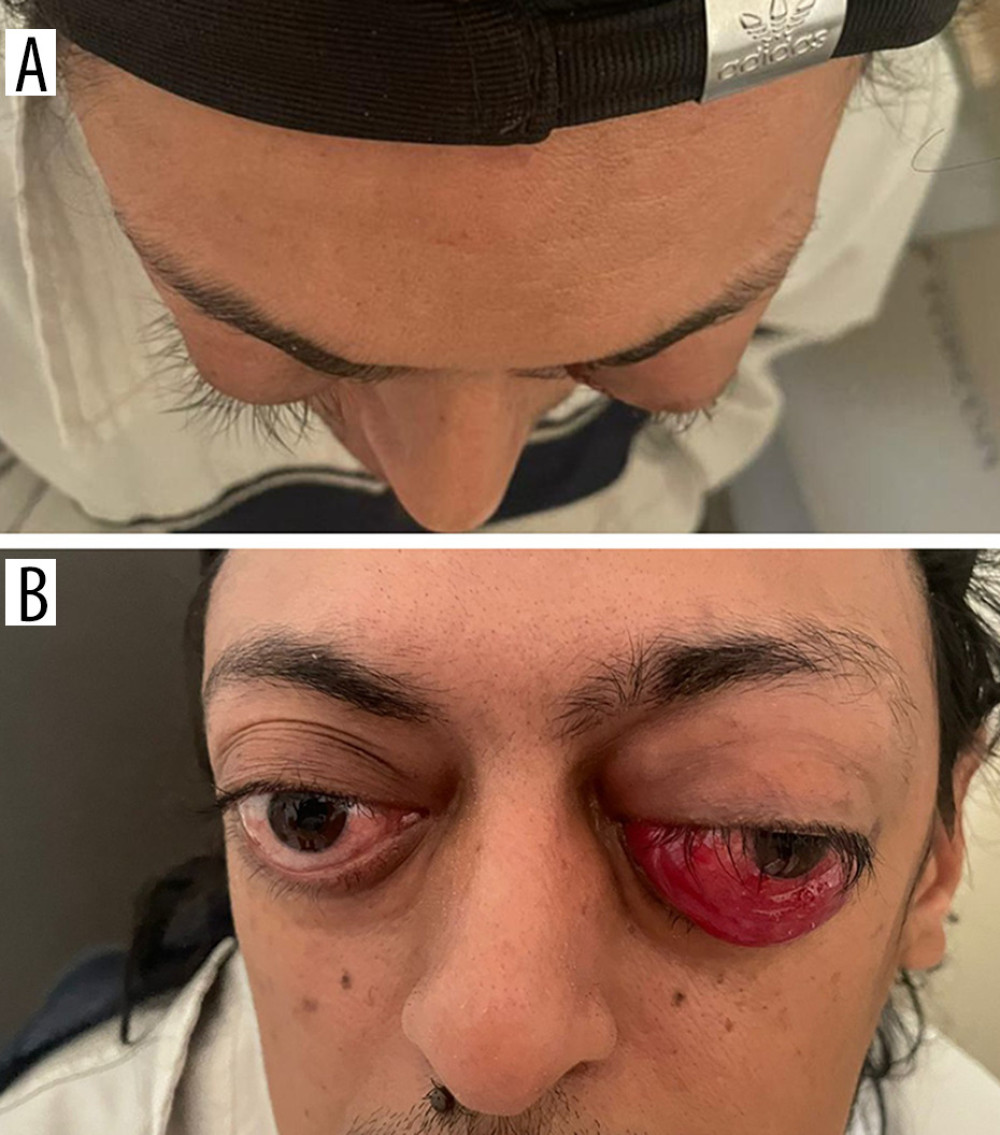 Figure 1. Initial clinical presentation of the patient. (A) Proptosis (OD: 26 mm; OS: 29 mm). (B) Inferior conjunctival chemosis of the left eye.
 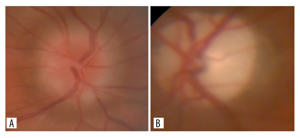 Figure 2. Initial fundus appearance of the patient. (A) Papilledema, Frisén grade II, in the right eye (OD). (B) Total optic disc pallor in the left eye (OS).
 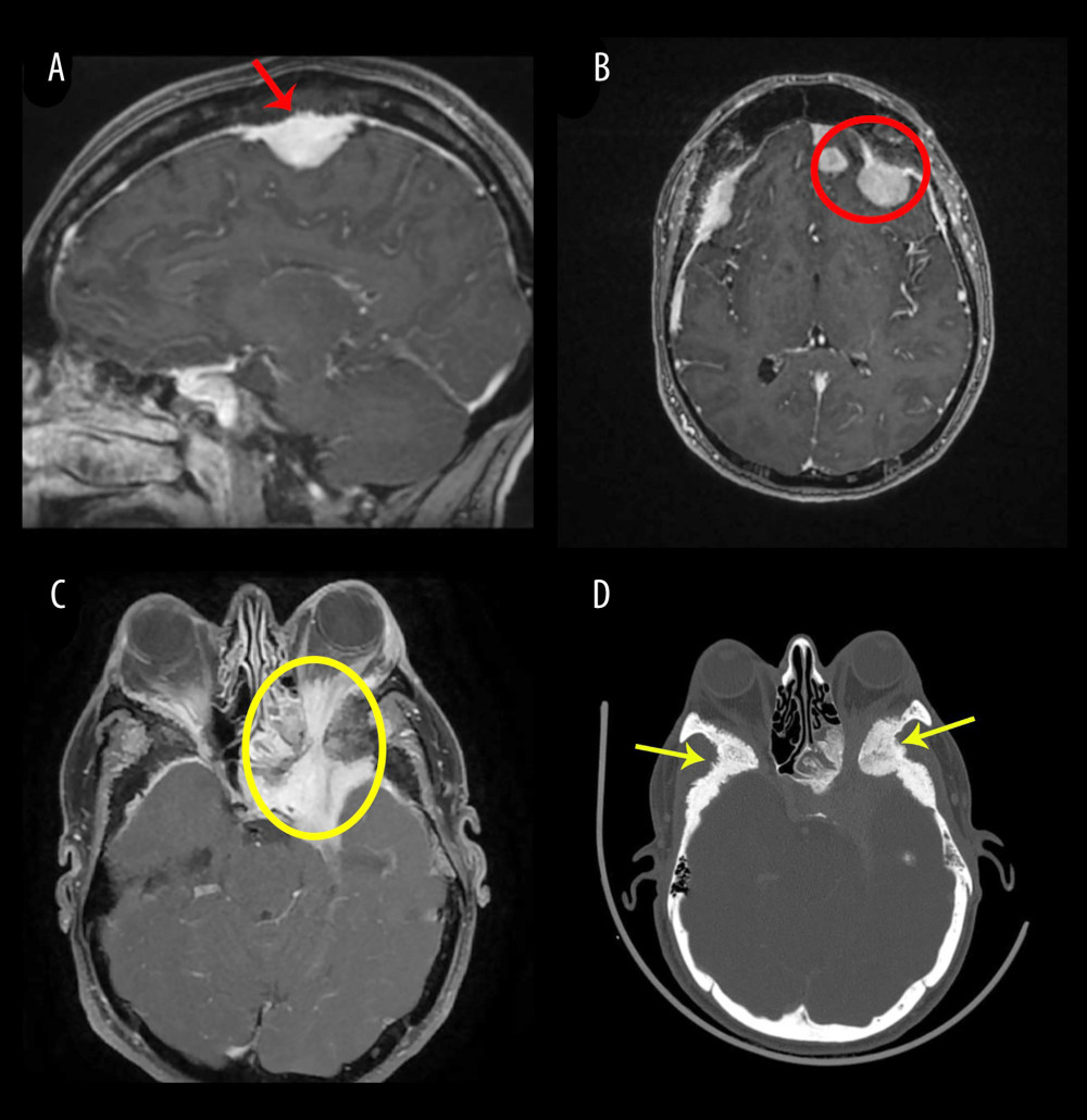 Figure 3. Imaging findings of the patient. (A) Sagittal T1-weighted post-gadolinium magnetic resonance imaging demonstrating a homogeneously enhancing parasagittal extra-axial meningioma with invasion of the superior sagittal sinus (red arrow). (B) Axial T1-weighted post-gadolinium MRI showing multiple left frontal convexity meningiomas adjacent to the falx (red circle). (C) Axial T1-weighted post-gadolinium magnetic resonance imaging demonstrating a meningiomatous lesion involving the left cavernous sinus with extension into the ipsilateral orbit through the optic canal(yellow circle). (D) Axial computed tomography bone window demonstrating marked hyperostotic and sclerotic changes of the sphenoid bone with a spiculated pattern (yellow arrows).
 References
1. Parafita-Fernández A, Sampil M, Cores C, Foster Kennedy syndrome: An atypical presentation: Optom Vis Sci, 2015; 92(12); e425-e30
2. Miller NR, Newman NJ, Biousse V, Kerrison JB: Walsh and Hoyt’s clinical neuro-ophthalmology, 2005, Lippincott Williams & Wilkins
3. Berry S, Lin WV, Sadaka A, Lee AG, Nonarteritic anterior ischemic optic neuropathy: Cause, effect, and management: Eye Brain, 2017; 9; 23-28
4. Watnick RL, Trobe JD, Bilateral optic nerve compression as a mechanism for the Foster Kennedy syndrome: Ophthalmology, 1989; 96(12); 1793-98
5. Giombini S, Solero CL, Lasio G, Morello G, Immediate and late outcome of operations for parasagittal and falx meningiomas: Report of 342 cases: Surg Neurol, 1984; 21(5); 427-35
6. Nakamura M, Struck M, Roser F, Olfactory groove meningiomas: Clinical outcome and recurrence rates after tumor removal through the frontolateral and bifrontal approach: Neurosurgery, 2007; 60(5); 844-52
7. Cowppli-Bony A, Bouvier G, Rué M, Brain tumors and hormonal factors: Review of the epidemiological literature: Cancer Causes Control, 2011; 22(5); 697-714
8. Rutzner S, Fietkau R, Ganslandt T, Electronic support for retrospective analysis in the field of radiation oncology: Proof of principle using an example of fractionated stereotactic radiotherapy of 251 meningioma patients: Front Oncol, 2017; 7; 16
9. Singh SR, Mehta A, Dogra M, Foster Kennedy syndrome: QJM, 2019; 112(8); 623-24
10. Acebes X, Arruga J, Acebes JJ, Intracranial meningiomatosis causing Foster Kennedy syndrome by unilateral optic nerve compression and blockage of the superior sagittal sinus: J Neuroophthalmol, 2009; 29(2); 140-42
11. Assadi M, Juweid ME, Lohmann P, Multidisciplinary management of meningiomas in the era of precision oncology: Nat Rev Clin Oncol, 2026; 23; 539-56
Figures
Figure 1. Initial clinical presentation of the patient. (A) Proptosis (OD: 26 mm; OS: 29 mm). (B) Inferior conjunctival chemosis of the left eye.Figure 2. Initial fundus appearance of the patient. (A) Papilledema, Frisén grade II, in the right eye (OD). (B) Total optic disc pallor in the left eye (OS).Figure 3. Imaging findings of the patient. (A) Sagittal T1-weighted post-gadolinium magnetic resonance imaging demonstrating a homogeneously enhancing parasagittal extra-axial meningioma with invasion of the superior sagittal sinus (red arrow). (B) Axial T1-weighted post-gadolinium MRI showing multiple left frontal convexity meningiomas adjacent to the falx (red circle). (C) Axial T1-weighted post-gadolinium magnetic resonance imaging demonstrating a meningiomatous lesion involving the left cavernous sinus with extension into the ipsilateral orbit through the optic canal(yellow circle). (D) Axial computed tomography bone window demonstrating marked hyperostotic and sclerotic changes of the sphenoid bone with a spiculated pattern (yellow arrows). In Press
Case report 
Am J Case Rep In Press; DOI: 10.12659/AJCR.953543
Case report 
Am J Case Rep In Press; DOI: 10.12659/AJCR.954216
Case report 
Am J Case Rep In Press; DOI: 10.12659/AJCR.954932
Case report 
Am J Case Rep In Press; DOI: 10.12659/AJCR.954146
Most Viewed Current Articles
07 Dec 2021 : Case report
28,712,695
DOI :10.12659/AJCR.934347
Am J Case Rep 2021; 22:e934347
06 Dec 2021 : Case report  180,721
DOI :10.12659/AJCR.934406
Am J Case Rep 2021; 22:e934406
21 Jun 2024 : Case report
124,419
DOI :10.12659/AJCR.944371
Am J Case Rep 2024; 25:e944371
08 May 2026 : Case report
95,462
DOI :10.12659/AJCR.952158
Am J Case Rep 2026; 27:e952158