


02 September 2026: Articles 
Management of a Rare 4-Rooted Maxillary First Molar With 5 Canals and an Endo-Periodontal Lesion: A Case Report
Unusual or unexpected effect of treatment
Yifan Wang ACDEF 1, Hong Jiang ABF 2, Yanhong Hao AB 2, Zhen Fang BC 3, Shige Liu B 2, Jie Pan ACD 4, Huan Jing ABCDG 2*DOI: 10.12659/AJCR.953554
Am J Case Rep 2026; 27:e953554


Abstract
BACKGROUND: Endo-periodontal lesions (EPL), particularly those manifesting as primary periodontal lesions with secondary endodontic involvement, pose unique clinical challenges when accompanied by odontogenic maxillary sinusitis (OMS). Furthermore, limited case reports document the nonsurgical management of such complex conditions in maxillary molars exhibiting rare multi-rooted and multi-canal variations (prevalence <0.103%).
CASE REPORT: A 31-year-old woman presented with pain and grade III mobility in the right maxillary first molar, with an intact crown, a deep palatal periodontal pocket, and a negative cold testing response on clinical examination. Periapical radiography indicated multi-rooted anatomy and extensive bone loss, and cone beam computed tomography (CBCT) confirmed 4 separate roots (mesiobuccal, distobuccal, mesiopalatal, distopalatal), a fifth centrally located pulp chamber canal orifice, a periapical lesion communicating with a periodontal defect, and ipsilateral maxillary sinus mucosal thickening. Nonsurgical root canal treatment for all 5 canals was performed under a dental operating microscope with ultrasonic activation (3% NaOCl) and bioceramic sealer, combined with concurrent supportive periodontal therapy. At the 8-month follow-up, the tooth was asymptomatic with reduced mobility and probing depths; CBCT revealed marked reduction in periapical lesions and significant improvement in maxillary sinus mucosal thickening (ENT consultation advised).
CONCLUSIONS: This case unequivocally demonstrates that meticulous nonsurgical endodontic treatment with ultrasonic activation and bioceramic sealer, in conjunction with targeted periodontal therapy and occlusal management, is a highly effective tooth-preserving strategy for maxillary molars with complex anatomic variations complicated by primary periodontal lesions with secondary endodontic involvement and secondary OMS, achieving excellent clinical and radiological outcomes.
Keywords: Case Reports, Tooth, Female, Molar, Treatment Outcome
Introduction
The maxillary first molar is characterized by 3 roots and 3 or 4 canals, with the second mesiobuccal (MB2) canal representing a common variation. The presence of 4 distinct roots, particularly with 2 separate palatal roots, is an uncommon occurrence. The management of such anatomical complexities becomes significantly more challenging when complicated by endo-periodontal lesion (EPL), where distinct endodontic and periodontal pathologies communicate. A critical consequence of periapical disease affecting maxillary posterior teeth is odontogenic maxillary sinusitis (OMS) due to the proximity of root apices to the sinus floor. EPLs, with their extensive bone defects, pose a high risk for this complication. The accuracy of diagnosis is greatly dependent on the use of three-dimensional imaging, because conventional radiography frequently fails to reveal the full complexity of the root canal system and the spatial relationship between the lesion and the sinus. This case report describes the nonsurgical management of a maxillary first molar exhibiting a rare configuration of 4 roots and 5 canals, associated with a primary periodontal lesion with secondary endodontic involvement and secondary OMS. The core objectives of this report are 2-fold: first, to document and characterize an exceptionally rare anatomic-pathologic entity; and second, to thoroughly discuss the decision-making basis, clinical limits, and rationale for pursuing a nonsurgical, multidisciplinary tooth preservation strategy in a tooth with an initially unfavorable prognosis.
Case Report
A 31-year-old woman was referred to the Department of Endodontics for evaluation of her maxillary right first molar (tooth #16, FDI notation). Her chief complaint was pain on chewing in the right posterior maxilla, with a history of intermittent discomfort over several months. Her medical history was non-contributory.
Clinical examination revealed an intact sound crown with no caries, restorations, or history of trauma. The tooth was tender to percussion and exhibited grade III mobility. A thorough occlusal analysis identified traumatic occlusal interference contributing to this severe mobility. Periodontal probing revealed advanced attachment loss on the palatal aspect, with broad pocket depths of 6 to 8 mm; other sites measured 3 to 4 mm. A vertical root fracture (VRF) was strictly ruled out based on a combination of clinical and radiographic evidence. Clinically, the probing pattern lacked the isolated, narrow ‘drop-off’ defect typical of VRF. Radiographically, a meticulous review of the pre-operative CBCT slices revealed no identifiable fracture lines, signs of root separation, or the characteristic ‘halo’ radiolucency associated with VRF. Finally, no fracture lines or cracks were detected during direct inspection of the pulp chamber floor and canal orifices under a dental operating microscope. The tooth did not respond to cold testing. A periapical radiograph showed unfilled canals, extensive periapical radiolucency, severe horizontal bone loss, and furcation involvement, alongside an unusual 4-root appearance (Figure 1). To reach a definitive diagnosis, a limited field-of-view CBCT scan was obtained. The following information was revealed. We found the roots can be categorized into 4 distinct types: mesiobuccal (MB), distobuccal (DB), mesiopalatal (MP), and distopalatal (DP). A fifth canal orifice, designated “M” for “middle,” was identified in a central position on the pulp chamber floor. There was a large periapical lesion associated with all roots, communicating with a deep periodontal bone defect on the palatal aspect, confirming an EPL. Mucosal thickening of the right maxillary sinus floor was contiguous with the apical lesion, diagnosing secondary OMS (Figure 2).
After a comprehensive evaluation of the patient’s medical history, clinical examinations, and radiographic features, a definitive diagnosis was rendered. Given the intact crown and broad periodontal defects, the pathology was diagnosed as a primary periodontal lesion with secondary endodontic involvement (retrograde pulpitis), alongside secondary odontogenic maxillary sinusitis associated with tooth #16. Written informed consent was obtained from the patient for the publication of this case report and accompanying images. A detailed timeline depicting the episode of care is presented in Figure 3.
The treatment plan comprised nonsurgical root canal treatment of tooth #16 alongside ongoing supportive periodontal therapy. Upon completion of root canal therapy and periodontal treatment, further management of maxillary sinusitis will be determined based on the follow-up assessment of the affected tooth.
Following the initial assessment, under local anesthesia with 4% articaine containing 1: 100 000 adrenaline and rubber dam isolation, an access cavity was prepared under a dental operating microscope (DOM; Zeiss OP-MI 1). Pulp tissue was extirpated. After irrigation with 3% NaOCl, calcium hydroxide paste (Dycal, Dentsply Sirona) was placed as an intracanal medicament, and the access was temporarily sealed with glass ionomer cement (Fuji II; GC Corporation).
Ten days later, the patient returned for the second treatment as scheduled. The tooth was asymptomatic. Under rubber dam isolation and DOM magnification, 5 canal orifices were identified, including the mesiobuccal (MB), distobuccal (DB), mesiopalatal (MP), distopalatal (DP), and an additional centrally located canal. Coronal flaring was performed using ProTaper SX (Dentsply Sirona). Working lengths were determined with an electronic apex locator and confirmed radiographically. Canal preparation was completed using stainless steel hand files (K-files sizes #10–20; Mani) followed by nickel–titanium instruments (WaveOne Gold, medium and large sizes; Dentsply Sirona). Irrigation with 3% NaOCl was performed throughout instrumentation, followed by ultrasonic activation in multiple cycles using an EDDY system (VDW) to enhance irrigant penetration (Figure 4). Calcium hydroxide medication was reapplied as an intracanal medicament, and the access cavity was temporarily restored with glass ionomer cement (Fuji II; GC Corporation).
At the third visit, after removal of the temporary restoration under rubber dam isolation and DOM, working lengths and apical preparation sizes were reconfirmed. A master gutta-percha cone for each root canal was selected and tried in the canal to confirm its fit. The cone was adjusted until it reached the established working length and width with a slight tug-back sensation. A periapical radiograph was then taken to verify the fit of the master cone, ensuring its adaptation to the final apical diameter and the canal morphology (Figure 5). Final irrigation was performed using 3% NaOCl with ultrasonic activation (EDDY; VDW), after which the canals were obturated with gutta-percha cones (B&L Biotech) and a bioceramic sealer (iRoot SP; Innovative Bioceramix, Vancouver, Canada) using the single-cone technique. Post-obturation radiographs confirmed adequate three-dimensional filling (Figure 6A). The tooth was subsequently restored with resin composite (Filtek Z350 XT; 3M ESPE), and the patient was scheduled for combined periodontal and endodontic follow-up.
Throughout the endodontic treatment period, the patient concurrently received comprehensive nonsurgical periodontal therapy. This targeted protocol included deep-site subgingival scaling and root planing (SRP) under local anesthesia to disrupt the subgingival biofilm. Importantly, a precise occlusal adjustment (occlusal reduction) was performed to eliminate the traumatic interference. At the 8-month revisit, the patient exhibited no symptoms. Given the significant but incomplete resolution of the maxillary sinus mucosal thickening at the 8-month follow-up, the patient was advised to consult an otorhinolaryngologist (ENT specialist) to evaluate any potential concomitant rhinogenic factors and determine the need for further rhinologic management. The substantial reduction in tooth mobility from grade III to grade I was primarily attributed to the rapid resolution of periapical inflammation, with the control of periodontal inflammation serving as a crucial secondary factor. Successful endodontic management effectively eradicated the core infection, alleviating abnormal apical hydrostatic pressure and initiating the re-establishment of a firm apical anchorage. Concurrently, the periodontal interventions resolved the lateral periodontal ligament (PDL) edema, facilitating the functional reorganization of the supporting tissues. Together, these mechanisms rapidly restored the mechanical stability of the affected molar. Periapical radiography (Figure 6B) demonstrated a reduction in the previously identified periapical radiolucency of tooth 16 compared to the initial assessment. Notably, CBCT imaging revealed near-complete resolution of the maxillary sinus mucosal thickening and osseous regeneration around the apices (Figure 7). The comprehensive before-and-after comparison of the clinical and imaging indicators is summarized in Table 1. The patient was advised to avoid biting on hard foods with the treated tooth, to undertake regular periodontal maintenance, and to return for a follow-up examination after 6 months.
Discussion
This report documents a successful endodontic and periodontal management strategy for a patient who presented with a rare anatomical variation in their maxillary first molar, exhibiting 4 distinct roots (MB, DB, MP, DP) in conjunction with a fifth centrally located (‘M’) canal. The case was complicated by the presence of an endoperiodontal lesion (EPL) and a secondary odontogenic maxillary sinusitis (OMS). The case highlights several pivotal themes in contemporary endodontics, including the crucial role of advanced imaging in diagnosing complex anatomy, the potential of EPLs to serve as a direct pathway for sinus infection, and the efficacy of careful disinfection in resolving both periapical and sinonasal pathology.
The maxillary first molar is characterized by the presence of 3 roots and 4 canals [1]. Variations involving additional roots are uncommon, and the presence of 2 separate palatal roots has been reported on occasion [2–5]. Large anatomical studies indicate that 4-rooted maxillary first molars account for a very small proportion of cases. In a comprehensive review and meta-analysis of maxillary first molars, Magnucki et al reported that 4 roots were identified in fewer than 0.103% around the world and were mostly described in individual case reports [6]. From an anatomical perspective, the presence of 4 independent roots in maxillary molars is often associated with the ‘Radix’ variation. Although more commonly discussed in mandibular molars, these additional roots in the maxillary arch can be identified as radix mesiolingualis (RML) or radix distolingualis (RDL) based on their position relative to the main root trunk. In this case, the complete separation of the mesiopalatal and distopalatal roots represents a manifestation of these rare morphologic entities. Recognizing such ‘radix’ variants is clinically significant, as their unique trajectories require modified access designs and meticulous instrumentation to ensure the eradication of the intraradicular microbial reservoir. The majority of previously documented variations of maxillary first molars have been found to involve the presence of additional canals within an existing root structure [7]. Typical examples include a second mesiobuccal canal or 2 canals within a single palatal root [8]. A common feature of these configurations is that the canals remain associated with a defined root trunk. In contrast, the present tooth exhibited 4 clearly separated roots and an additional canal located centrally on the pulp chamber floor. This additional canal exhibited no anatomical correlation with any of the 4 roots. Reports from populations exhibiting a high frequency of additional canals suggest that canals without an associated root are uncommon [9,10]. Al-Habib et al noted that while extra canals are frequently detected, their occurrence alongside additional roots is rarely observed [2]. This finding is consistent with the canal arrangement identified in this tooth. Existing root canal classification systems are chiefly descriptive, characterizing canal patterns within individual roots without accounting for canals that are independent of root morphology [11]. Consequently, the combination of 4 separate roots and a centrally positioned fifth canal cannot be readily categorized using commonly applied systems [12,13]. Recent efforts to refine classification models may help to better describe such atypical configurations, but clinical recognition remains essential [14]. Although the presence of additional canals in maxillary molars is a commonly recognized phenomenon, the morphology observed in this study diverges from the established patterns [15,16]. This observation underscores the necessity for careful inspection of the pulp chamber floor, even when all expected roots and canals appear to have been located. However, it should be noted that this specific configuration is extremely rare, and its clinical prevalence cannot be generalized from this single case.
Conventional periapical radiography provided limited information in this case. The number of roots and canals was unidentifiable. Likewise, the extent of bone loss and its connection to the sinus remained obscure. These limitations are well known, especially in maxillary molars, where anatomical structures often overlap. CBCT allowed a more accurate assessment by offering three-dimensional images of the affected area [17]. It clearly demonstrated the spatial arrangement of the 4 roots and the centrally located canal. It also visualized the continuity between the apical and marginal bone defects, which in conjunction with clinical findings supported the diagnosis of an endo-periodontal lesion. In addition, CBCT revealed continuity between the apical lesion and the thickened sinus mucosa, which supported the diagnosis of odontogenic maxillary sinusitis. Previous studies have shown that CBCT is more reliable than conventional radiography for detecting complex root canal anatomy and bone defects. Choi et al reported higher diagnostic accuracy with CBCT when evaluating root canal morphology and periapical pathology [18]. In the present case, CBCT helped confirm the canal configuration and exclude sinus disease unrelated to dental infection. This distinction is important for treatment planning and helps avoid unnecessary referral or intervention. Professional guidelines support the use of CBCT when root canal anatomy cannot be clearly assessed or when endodontic, periodontal, and sinus findings appear to be related [6]. The central canal identified here would likely have been missed on periapical radiographs because of root overlap. This reinforces the value of CBCT in identifying canal orifices that do not follow typical anatomical patterns.
The convergence of independent endodontic and periodontal infections in EPLs establishes a direct ‘pathogenic pathway’ to the maxillary sinus [19]. The maxillary posterior teeth are in proximity to the sinus floor (average distance of 1.2–3.8 mm), making OMS a common complication of periapical/periodontal disease. Wu et al recently reported that EPLs are an overlooked etiology of OMS, accounting for 28% to 32% of unilateral maxillary sinusitis cases [20]. In this case, pulpal necrosis (confirmed via cold testing and clinical signs) and a deep palatal periodontal pocket (6–8 mm) likely formed a consolidated infectious front that breached the Schneiderian membrane, leading to sinus mucosal thickening [21]. This pathogenic mechanism is supported by Peñarrocha-Oltra et al’s meta-analysis, which found a strong association between EPLs and maxillary sinus abnormalities (OR = 4.72, 95% CI: 3.21–6.90) [22]. While the coexistence of EPL-OMS with an extremely rare 4-root, 5-canal anatomy is an uncommon clinical presentation, making it a relevant addition to the literature on odontogenic sinusitis, this interplay remains highly case-dependent. The 5-canal configuration may have further facilitated infection spread by increasing the intraradicular microbial reservoir, underscoring the need to address all canals to eliminate the primary infection.
The management of EPLs prioritizes eliminating the primary endodontic infection, as pulpal toxins and microbes perpetuate periodontal inflammation and hinder healing [23,24]. Schmidt et al conducted a systematic review and confirmed that resolving endodontic infection first (or concurrently with periodontal therapy) results in a 78% higher rate of periodontal attachment gain compared to periodontal therapy alone [25]. In this case, meticulous nonsurgical root canal treatment of all 5 canals—using a dental operating microscope, ultrasonic irrigation (EDDY system), and a bioceramic sealer (iRoot SP)—was critical for eradicating the intracanal microbial reservoir. Concurrently, adequate periodontal therapy was equally essential for managing the deep periodontal pocket and supporting structural healing. The 5-canal anatomy presented unique technical challenges, particularly in locating and disinfecting the centrally positioned “M” canal. The use of magnification (dental operating microscope) and ultrasonic activation of 3% NaOCl ensured adequate irrigant penetration into the narrow “M” canal, which is essential for disinfection in small-diameter canals. Bioceramic sealers were chosen for their favorable biocompatibility, antibacterial properties, and flowability. Varghese et al reported that bioceramic sealers achieve complete obturation in complex canals more effectively than resin-based sealers, with a 92% reduction in microbial load—a critical outcome for the “M” canal, where incomplete obturation could have perpetuated infection [26]. Notably, the successful resolution of OMS without direct sinus intervention or antibiotics aligns with the multidisciplinary consensus that eliminating the dental source is the primary treatment for odontogenic sinusitis [27–34]. Craig et al emphasized that ENT surgical interventions (eg, FESS) are rarely needed if dental treatment is thorough—an observation validated by the 8-month CBCT showing complete sinus mucosal resolution [35]. Finally, the favorable clinical and radiographic outcomes observed reflect a single case, and further studies are needed to evaluate standardized protocols for such complex mixed pathologies.
Conclusions
In conclusion, the successful management of this rare 4-rooted, 5-canal maxillary molar highlights the indispensable role of CBCT and magnification in diagnosing and treating complex endodontic-periodontal conditions. This case unequivocally demonstrates that meticulous nonsurgical endodontic therapy, combined with targeted periodontal and occlusal management, can effectively resolve severe bone loss and secondary odontogenic sinusitis, supporting a conservative, tooth-preserving approach. However, given the inherent limitations of a single case report and the relatively short 8-month follow-up, these findings should be interpreted with caution, and long-term monitoring is required.
Figures
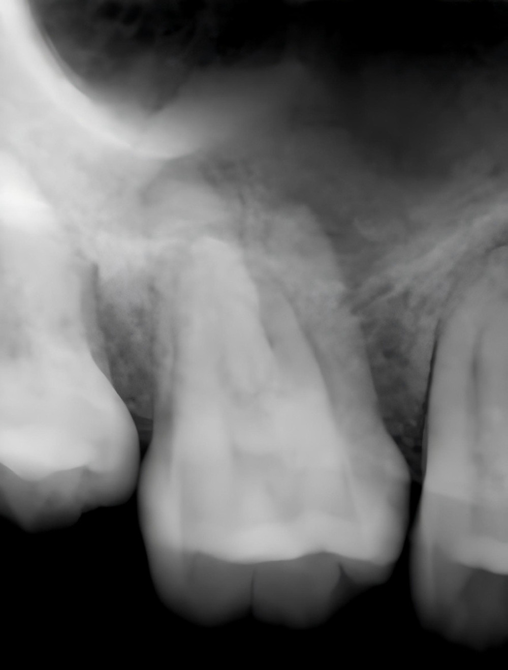 Figure 1. The initial periapical radiograph of the first molar of the right maxilla.
 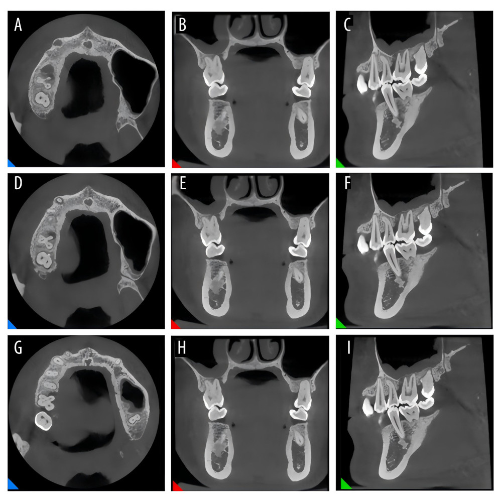 Figure 2. The CBCT imaging of the right maxillary first molar before treatment (A–C: images of the apical third of the root, D–F: images of the middle third of the root, G–I: images of the cervical third of the root).
 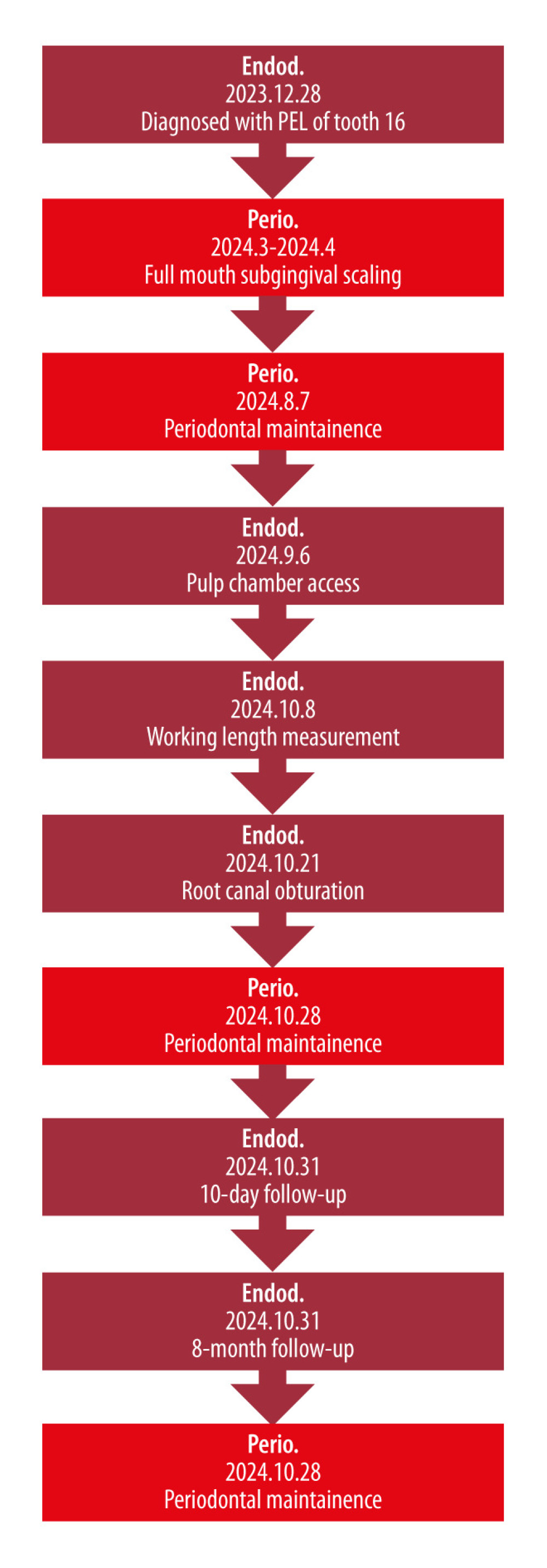 Figure 3. A detailed timeline depicting the episode of care.
 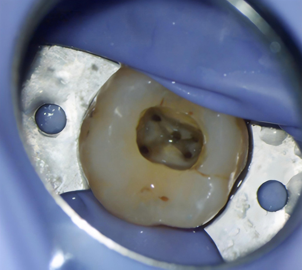 Figure 4. Microscopic evaluation of the spatial distribution of 5 root canal orifices in tooth 16.
 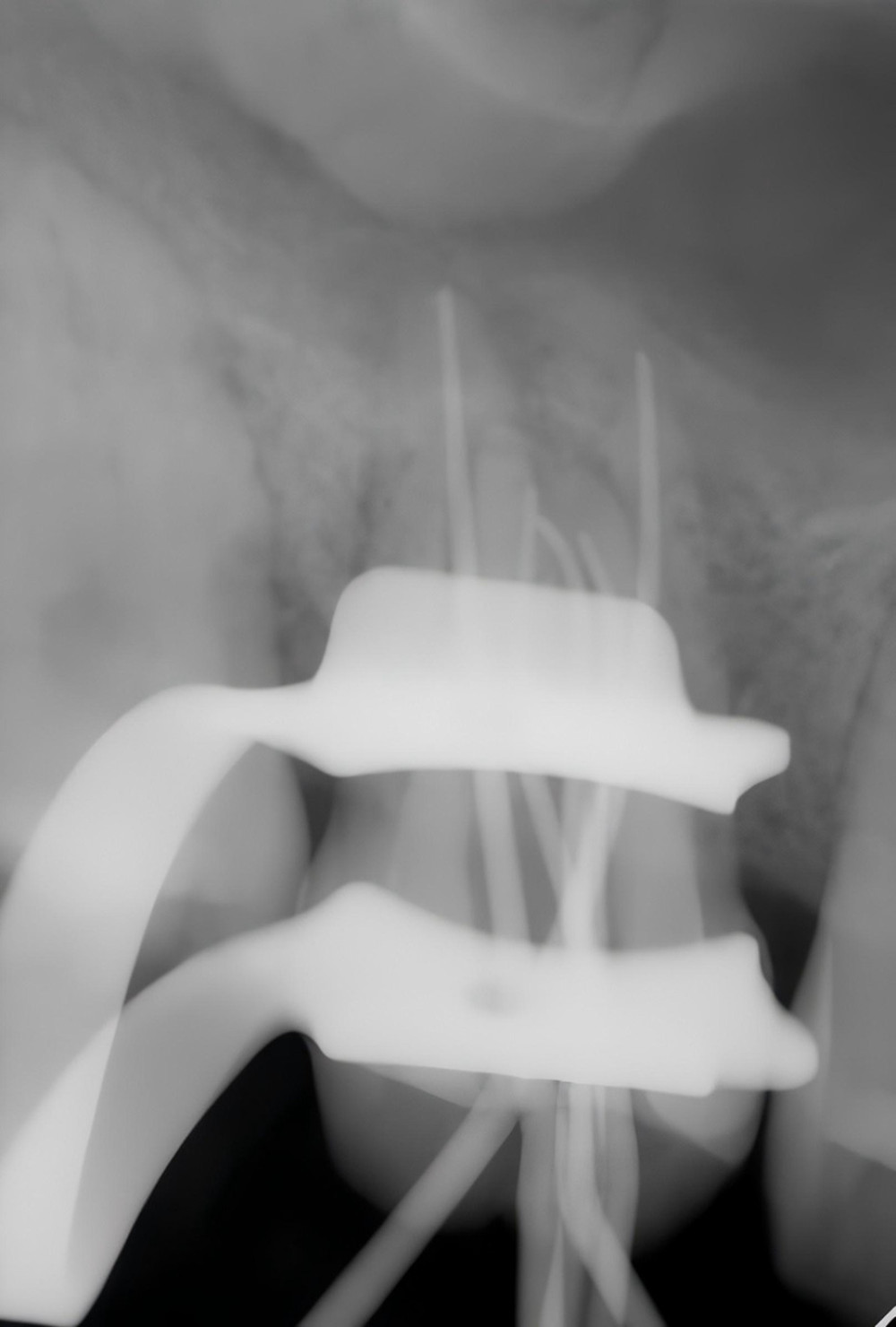 Figure 5. The working length verification radiograph of tooth 16.
 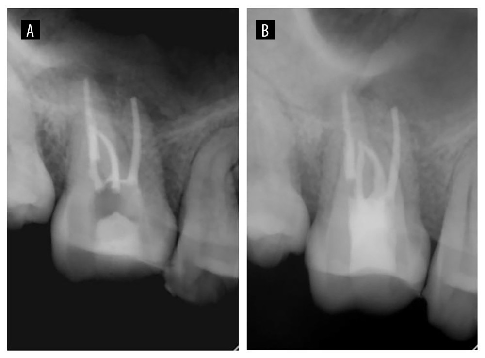 Figure 6. Periapical radiographs taken immediately after root canal filling (A) and after permanent coronal restoration (B).
 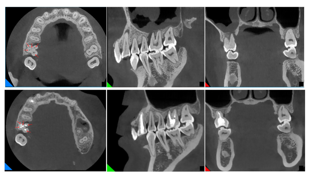 Figure 7. Imaging evaluation of tooth 16 before and after treatment. Cross-sectional view showing the 5 obturated canals (red arrows).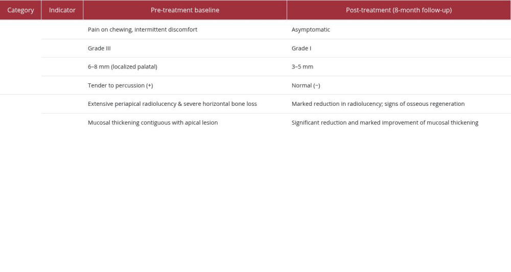
References
1. de Lima CO, de Montan JBM, Barbosa AFA, Micro-computed tomographic evaluation of dentinal microcracks in response to progressive taper enlargement in root canals of maxillary molars: Int Endod J, 2025; 58(2); 357-62
2. Al-Habib M, Howait M, Assessment of mesiobuccal canal configuration, prevalence and inter-orifice distance at different root thirds of maxillary first molars: A CBCT study: Clin Cosmet Investig Dent, 2021; 13; 105-11
3. Gomes Alves CR, Martins Marques M, Stella Moreira M, Second mesiobuccal root canal of maxillary first molars in a Brazilian population in high-resolution cone-beam computed tomography: Iran Endod J, 2018; 13(1); 71-77
4. Peeters HH, Suardita K, Setijanto D, Prevalence of a second canal in the mesiobuccal root of permanent maxillary first molars from an Indonesian population: J Oral Sci, 2011; 53(4); 489-94
5. Fernandes NA, Herbst D, Postma TC, Bunn BK, The prevalence of second canals in the mesiobuccal root of maxillary molars: A cone beam computed tomography study: Aust Endod J, 2019; 45(1); 46-50
6. Magnucki G, Mietling SVK, Four-rooted maxillary first molars: A systematic review and meta-analysis: Int J Dent, 2021; 2021; 8845442
7. Vertucci FJ, Root canal anatomy of the human permanent teeth: Oral Surg Oral Med Oral Pathol, 1984; 58(5); 589-99
8. Holtzman L, Multiple canal morphology in the maxillary first molar: Case reports: Quintessence Int, 1997; 28(7); 453-55
9. Yamada M, Ide Y, Matsunaga S, Three-dimensional analysis of mesiobuccal root canal of Japanese maxillary first molar using Micro-CT: Bull Tokyo Dent Coll, 2011; 52(2); 77-84
10. Khademi A, Zamani Naser A, Bahreinian Z, Root morphology and canal configuration of first and second maxillary molars in a selected Iranian population: A cone-beam computed tomography evaluation: Iran Endod J, 2017; 12(3); 288-92
11. Karobari MI, Parveen A, Mirza MB, Root and root canal morphology classification systems: Int J Dent, 2021; 2021; 6682189
12. Kuzekanani M, Walsh LJ, Amiri M, Prevalence and distribution of the middle mesial canal in mandibular first molar teeth of the Kerman population: a CBCT study: Int J Dent, 2020; 2020; 8851984
13. Talabani RM, Abdalrahman KO, Abdul RJ, Evaluation of radix entomolaris and middle mesial canal in mandibular permanent first molars in an Iraqi subpopulation using cone-beam computed tomography: Biomed Res Int, 2022; 2022; 7825948
14. Ahmed HMA, Rossi-Fedele G, Dummer PMH, Critical analysis of a new system to classify root and canal morphology – A systematic review: Aust Endod J, 2023; 49(3); 750-68
15. Spagnuolo G, Ametrano G, D’Antò V, Microcomputed tomography analysis of mesiobuccal orifices and major apical foramen in first maxillary molars: Open Dent J, 2012; 6; 118-25
16. Saber SEDM, Ahmed MHM, Obeid M, Ahmed HMA, Root and canal morphology of maxillary premolar teeth in an Egyptian subpopulation using two classification systems: A cone beam computed tomography study: Int Endod J, 2019; 52(3); 267-78
17. Venumuddala VR, Moturi S, Satish SV, Endodontic management of a maxillary first molar with seven root canal systems evaluated using cone-beam computed tomography scanning: J Int Soc Prev Community Dent, 2017; 7(5); 297-300
18. Choi E, Pang K, Jeong E, Artificial intelligence in diagnosing dens evaginatus on periapical radiography with limited data availability: Sci Rep, 2023; 13(1); 13232
19. Grygorov S, Poberezhnik G, Grygorova A, Actual issues of odontogenic maxillary sinusitis (review): Georgian Med News, 2018(276); 46-50
20. Wu J, Zheng M, Wang X, Wang S, Endo-periodontal lesions-an overlooked etiology of odontogenic sinusitis: J Clin Med, 2023; 12(21); 6888
21. Nurchis MC, Pascucci D, Lopez MA, Epidemiology of odontogenic sinusitis: An old, underestimated disease, even today. A narrative literature review: J Biol Regul Homeost Agents, 2020; 34(5 Suppl 3); 195-200
22. Peñarrocha-Oltra S, Soto-Peñaloza D, Bagán-Debón L, Association between maxillary sinus pathology and odontogenic lesions in patients evaluated by cone beam computed tomography. A systematic review and meta-analysis: Med Oral Patol Oral Cir Bucal, 2020; 25(1); e34-e48
23. Abbott P, Endodontic management of combined endodontic-periodontal lesions: J N Z Soc Periodontol, 1998(83); 15-28
24. AlRowis RM, Alzahrani AH, Alzuhair SH, Assess the association between periodontitis and maxillary sinusitis: A cross-sectional cone-beam computerized tomography (CBCT) study: Cureus, 2023; 15(11); e48587
25. Schmidt JC, Walter C, Amato M, Weiger R, Treatment of periodontal-endodontic lesions – A systematic review: J Clin Periodontol, 2014; 41(8); 779-90
26. Varghese LL, Bhattacharya A, Basannavar A, Non-surgical endodontic management of odontogenic maxillary sinusitis: BMJ Case Rep, 2021; 14(7); e241821
27. Troeltzsch M, Cagna D, Stähler P, Clinical features of peri-implant medication-related osteonecrosis of the jaw: Is there an association to peri-implantitis?: J Craniomaxillofac Surg, 2016; 44(12); 1945-51
28. Liu S, Chen X, Wang XX, Association between odontogenic conditions and maxillary sinus abnormalities: A retrospective cone-beam computed-tomographic study: Ann Palliat Med, 2023; 12(2); 365-75
29. Vijayakumar S, Balakrishnan SS, Pulimi R, Are we missing something in the CT-PNS report? – An observational study on the rate of reporting the presence of dental disease and the probable etiology of sinusitis on CT scans: J Belg Soc Radiol, 2022; 106(1); 109
30. Zhang ZH, Yao HL, Zhang Y, Wang X, Endodontic management of the maxillary first molar with special root canals: A case report and review of the literature: World J Clin Cases, 2020; 8(12); 2590-96
31. Galli M, De Soccio G, Cialente F, Chronic maxillary sinusitis of dental origin and oroantral fistula: The results of combined surgical approach in an Italian university hospital: Bosn J Basic Med Sci, 2020; 20(4); 524-30
32. Lin J, Wang C, Wang X, Expert consensus on odontogenic maxillary sinusitis multi-disciplinary treatment: Int J Oral Sci, 2024; 16(1); 11
33. Kwiatkowska M, Szczygielski K, Skrzypiec Ł, Jurkiewicz D, Predictive value of tooth and sinuses radiological characteristics in managing odontogenic sinusitis of endodontic origin: Otolaryngol Pol, 2024; 78(5); 33-42
34. Preda MA, Musat G, Sarafoleanu C, Surgical endoscopic treatment of odontogenic sinusitis: Chirurgia (Bucur), 2024; 119(1); 76-86
35. Craig JR, Tataryn RW, Aghaloo TL, Management of odontogenic sinusitis: Multidisciplinary consensus statement: Int Forum Allergy Rhinol, 2020; 10(7); 901-12
Figures
Figure 1. The initial periapical radiograph of the first molar of the right maxilla.Figure 2. The CBCT imaging of the right maxillary first molar before treatment (A–C: images of the apical third of the root, D–F: images of the middle third of the root, G–I: images of the cervical third of the root).Figure 3. A detailed timeline depicting the episode of care.Figure 4. Microscopic evaluation of the spatial distribution of 5 root canal orifices in tooth 16.Figure 5. The working length verification radiograph of tooth 16.Figure 6. Periapical radiographs taken immediately after root canal filling (A) and after permanent coronal restoration (B).Figure 7. Imaging evaluation of tooth 16 before and after treatment. Cross-sectional view showing the 5 obturated canals (red arrows). In Press
Case report 
Am J Case Rep In Press; DOI: 10.12659/AJCR.953543
Case report 
Am J Case Rep In Press; DOI: 10.12659/AJCR.954216
Case report
Am J Case Rep In Press; DOI: 10.12659/AJCR.954932
Case report 
Am J Case Rep In Press; DOI: 10.12659/AJCR.954146
Most Viewed Current Articles
07 Dec 2021 : Case report
28,712,695
DOI :10.12659/AJCR.934347
Am J Case Rep 2021; 22:e934347
06 Dec 2021 : Case report  180,721
DOI :10.12659/AJCR.934406
Am J Case Rep 2021; 22:e934406
21 Jun 2024 : Case report
124,419
DOI :10.12659/AJCR.944371
Am J Case Rep 2024; 25:e944371
08 May 2026 : Case report
95,462
DOI :10.12659/AJCR.952158
Am J Case Rep 2026; 27:e952158