


17 August 2026: Articles 
From Sight to Stridor: Recurrent Angioedema in Systemic Lupus Erythematosus Associated With Complement Activation
Challenging differential diagnosis, Management of emergency care, Rare disease, Educational Purpose (only if useful for a systematic review or synthesis)
Seyed Khalafi ABCDEF 1*, Lanna Felde ABCDEF 1DOI: 10.12659/AJCR.953593
Am J Case Rep 2026; 27:e953593


Abstract
BACKGROUND: Angioedema is a rare manifestation of systemic lupus erythematosus (SLE) that can lead to life-threatening complications, including airway compromise. Acquired angioedema is classically associated with C1 esterase inhibitor (C1-INH) deficiency caused either by increased consumption of C1-INH or the development of autoantibodies against C1-INH. However, emerging reports describe patients with active SLE who develop recurrent angioedema despite preserved C1-INH levels and function, suggesting a possible complement-mediated or bradykinin-associated inflammatory phenotype that remains incompletely characterized.
CASE REPORT: We report the case of a woman in her early 20s with SLE and lupus nephritis who presented with a urinary tract infection and unilateral periorbital edema. Initial laboratory evaluation revealed anemia, thrombocytopenia, and acute-on-chronic kidney injury. She was treated with antibiotics for presumed urinary tract infection and preseptal cellulitis. During hospitalization, she developed laryngeal edema requiring prompt treatment with intravenous dexamethasone, with rapid clinical improvement. Further evaluation demonstrated low C4 and C1q complement levels, elevated C1q complement binding levels, elevated anti-C1q antibodies, and preserved C1-INH antigen and functional levels, raising suspicion for complement-mediated angioedema occurring in the setting of active SLE. Over the ensuing 6 months, she experienced recurrent episodes of angioedema with progressive renal dysfunction and concern for evolving macrophage activation syndrome or severe hyperinflammatory immune activation.
CONCLUSIONS: This case highlights a rare presentation of recurrent angioedema associated with active SLE and complement activation despite preserved C1-INH levels and function. The case underscores the importance of early recognition of airway involvement in patients with lupus and illustrates the diagnostic challenges surrounding complement-mediated angioedema phenotypes in autoimmune disease.
Keywords: angioedema, Complement C1q, Lupus Erythematosus, Systemic, macrophage activation syndrome
Introduction
Angioedema is defined as recurrent, nonpitting, nonpruritic swelling involving the deep dermis, subcutaneous tissues, or mucosal surfaces. The condition may be hereditary or acquired and can result from histaminergic, bradykinin-mediated, or mixed inflammatory pathways [1]. One well-described form of acquired angioedema occurs in the setting of C1 esterase inhibitor (C1-INH) deficiency. C1-INH is an important regulator of both the complement and the kallikrein-kinin systems through inhibition of C1r, C1s, factor XIIa, and kallikrein, thereby limiting excessive bradykinin generation and vascular permeability.
Acquired angioedema associated with C1-INH deficiency is traditionally classified into 2 categories. Type I acquired angioedema results from accelerated consumption of C1-INH and is commonly associated with lymphoproliferative disorders such as non-Hodgkin lymphoma, chronic lymphocytic leukemia, and monoclonal gammopathies. Type II acquired angioedema results from autoantibodies directed against C1-INH and is more commonly associated with autoimmune diseases, including systemic lupus erythematosus (SLE) [2]. Both established forms of acquired angioedema involve acquired C1-INH deficiency or dysfunction. Current international angioedema guidelines therefore recognize acquired angioedema primarily in the setting of C1-INH deficiency [3].
Acquired angioedema most commonly presents with episodic nonpitting edema involving the face, extremities, gastrointestinal tract, or upper airway. Gastrointestinal involvement can manifest with abdominal pain, nausea, vomiting, or diarrhea secondary to bowel wall edema [4]. Laryngeal involvement is uncommon but may rapidly become life-threatening because of airway compromise [1,5,6].
Emerging case reports have described patients with active SLE who develop recurrent angioedema despite preserved C1-INH levels and function, suggesting a possible complement-mediated inflammatory phenotype that remains incompletely characterized [7–11]. These presentations do not fit neatly within currently established classifications of acquired angioedema. We present a case of recurrent facial, laryngeal, and intestinal edema occurring in a patient with active SLE, low complement levels, elevated C1q complement binding levels, elevated anti-C1q antibodies, and preserved C1-INH levels and function.
Case Report
A Black woman in her early 20s with SLE (positive anti-nuclear, double-stranded DNA, and Smith antibodies) complicated by class IV lupus nephritis and treated with hydroxychloroquine, mycophenolate mofetil, and a medium dose of prednisone presented with dysuria, acute renal insufficiency, and chronic nausea, vomiting, and diarrhea. While waiting in the urgent care clinic, she fell asleep and awoke to progressive swelling around her left eye. She denied any family history of angioedema or recurrent swelling. She received intravenous diphenhydramine and prednisone in the emergency department without significant improvement.
On presentation, her vital signs were within normal limits, including a temperature of 37.2°C, blood pressure of 116/73 mm Hg, heart rate of 96 beats/min, respiratory rate of 18 breaths/min, and oxygen saturation of 100% on room air. Physical examination demonstrated an erythematous and edematous left eyelid with conjunctival hemorrhage and mild lip swelling. Computed tomography (CT) of the orbits without contrast showed significant left periorbital swelling and edema that was attributed to preseptal cellulitis (Figure 1). CT of the abdomen and pelvis without contrast was notable for mesenteric edema and a small amount of free fluid, without bowel obstruction (Figure 2).
Admission laboratory studies showed anemia, thrombocytopenia, and acute-on-chronic kidney injury, with a creatinine level of 6.42 mg/dL. Iron studies showed elevated ferritin with normal serum iron, total iron-binding capacity, and percent saturation. She was also noted to have a mildly elevated erythrocyte sedimentation rate and C-reactive protein level (Table 1). Urinalysis was notable for moderate leukocyte esterase, negative nitrites, and many bacteria. The patient was started on a third-generation cephalosporin to cover suspected preseptal cellulitis and urinary tract infection.
On the second hospital day, the patient developed active retching, excessive oral secretions, sore throat, and hoarseness. The otorhinolaryngology service performed bedside laryngoscopy and found an edematous palate, left arytenoid and pharyngeal edema, and a mildly edematous post-cricoid space concerning for angioedema involving the airway. The patient started a 3-day course of intravenous dexamethasone and diphenhydramine, with subsequent clinical improvement over the following days.
Further testing was performed to determine the cause of the angioedema, and results were notable for low C4 complement levels (4 mg/dL; reference range, 10–40 mg/dL), low C1q complement levels (5 mg/dL; range, 12–22 mg/dL), elevated C1q complement binding (7.9 ug Eq/mL; range, 0–3.9 ug Eq/mL), elevated anti-C1q IgG antibody (35 units; range, 0–19 units), and preserved C1-INH function (102%; range, ≥41%) and C1-INH antigen (31 mg/dL; range, 21–38 mg/dL), raising concern for complement-mediated angioedema associated with active SLE. Results showed a low hemoglobin level (7.7 g/dL), and low platelet count (112 × 109/L). The patient was discharged in stable condition with high-dose cetirizine.
Over the ensuing 6 months, the patient was hospitalized twice for recurrent unilateral periorbital and lip edema. During these admissions, renal function progressively worsened, ultimately resulting in end-stage renal disease requiring initiation of peritoneal dialysis. Persistent hypocomplementemia raised concern for ongoing lupus activity. The patient additionally developed worsening cytopenias, elevated ferritin (1824 ng/mL), elevated soluble interleukin-2 receptor levels (3081 pg/mL), and severe neutropenia (absolute neutrophil count 0.29 × 10⁹/L), raising concern for evolving macrophage activation syndrome (MAS) or severe hyperinflammatory immune activation in the setting of active SLE. Belimumab was subsequently initiated in the outpatient setting, after which no additional angioedema episodes have occurred to date.
Discussion
We present a case of recurrent facial, laryngeal, and intestinal edema occurring in a patient with active SLE complicated by lupus nephritis, hypocomplementemia, elevated anti-C1q antibodies, and preserved C1-INH levels and function. This presentation is diagnostically challenging because acquired angioedema has traditionally been classified into Type I and Type II subtypes, both involving acquired C1-INH deficiency. However, multiple case reports have described patients with SLE who develop recurrent angioedema despite preserved C1-INH antigen and functional levels [7–11]. In patients with active lupus, anti-C1q antibodies can contribute to immune complex formation and classical complement pathway activation and are strongly associated with lupus nephritis activity and hypocomplementemia [12,13]. Collectively, these findings suggest that dysregulated activation of the classical complement pathway with possible involvement of the kallikrein-kinin system may contribute to vascular permeability and edema, even in the absence of measurable C1-INH deficiency. A proposed schematic illustrating these interacting pathways is shown in Figure 3. Nevertheless, current allergy and rheumatology guidelines do not recognize this presentation as a formally established subtype of acquired angioedema, and the precise relationship between anti-C1q antibodies, complement consumption, and angioedema development remains incompletely understood [3]. Additional studies are needed to better characterize angioedema presentations occurring in patients with SLE and preserved C1-INH levels.
Although the patient demonstrated laboratory evidence of complement activation, her clinical improvement with corticosteroids and antihistamines suggests that the underlying pathophysiology may not have been exclusively bradykinin-mediated. Distinguishing bradykinin-mediated from histaminergic angioedema can be clinically challenging, particularly in patients with autoimmune disease and overlapping inflammatory processes [14]. A mixed or incompletely characterized inflammatory mechanism involving complement-mediated and histaminergic pathways remains possible. Accordingly, treatment response alone cannot definitively establish the mechanism of angioedema in this case.
Infection has also been proposed as a potential trigger for immune dysregulation in lupus-associated angioedema. SLE alters immune tolerance and may predispose patients to aberrant autoantibody production through mechanisms including molecular mimicry and chronic immune activation [15]. A cross-sectional study by Luo et al noted that patients with SLE-associated angioedema frequently presented with concurrent infections, particularly urinary tract infections [4]. Our patient similarly presented with concern for urinary tract infection during her index hospitalization. However, recurrent episodes occurring alongside persistent hypocomplementemia and progressive renal dysfunction suggested ongoing lupus activity as a major contributor to disease recurrence.
During subsequent admissions, the patient developed worsening cytopenias, markedly elevated ferritin levels, elevated soluble interleukin-2 receptor levels, and progressive renal dysfunction, raising concern for possible evolving MAS in the setting of persistently active lupus. MAS is a severe hyperinflammatory complication of autoimmune disease, particularly active SLE, and is characterized by excessive immune activation and cytokine dysregulation. However, distinguishing MAS from severe lupus flare remains challenging because of substantial overlap in clinical and laboratory findings, including cytopenias, hyperferritinemia, complement consumption, and multiorgan dysfunction [16,17]. Although the temporal association between recurrent angioedema episodes and hyperinflammatory findings is notable, a causal relationship between angioedema and MAS cannot be established from this case alone. Rather, this presentation may reflect severe systemic immune dysregulation associated with persistent lupus activity. Despite this uncertainty, the case highlights the importance of recognizing recurrent angioedema as a potential marker of severe underlying autoimmune disease activity. As demonstrated in prior reports of lupus-associated angioedema with airway involvement, delayed recognition can result in life-threatening respiratory compromise [18,19].
Management of lupus-associated angioedema primarily focuses on the treatment of the underlying autoimmune condition. Acute attacks may be managed with airway protection and targeted therapies such as fresh frozen plasma, C1-INH replacement therapy, icatibant, or ecallantide depending on the suspected mechanism and severity of presentation [3,20]. Corticosteroids and antihistamines are frequently administered when the etiology remains uncertain or when a mixed inflammatory process is suspected [3,15,20]. In our patient, persistent disease activity despite hydroxychloroquine, mycophenolate mofetil, and prednisone prompted escalation of immunosuppressive therapy with belimumab, after which recurrent angioedema episodes resolved.
Conclusions
We present a rare case of recurrent angioedema occurring in association with active SLE, hypocomplementemia, elevated anti-C1q antibodies, and preserved C1-INH levels and function. This case highlights the diagnostic challenges surrounding complement-mediated angioedema phenotypes in autoimmune disease and underscores the importance of early recognition of airway involvement in patients with lupus. Further investigation is needed to better characterize the relationship between complement activation, anti-C1q antibodies, and recurrent angioedema in SLE, particularly in patients with preserved C1-INH levels and function.
Figures
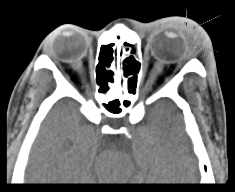 Figure 1. Computed tomography of the orbits and sella without contrast. Significant left periorbital swelling and edema are present without a discrete collection. Convex fluid attenuation and air are noted adjacent to the left lateral globe (arrows).
 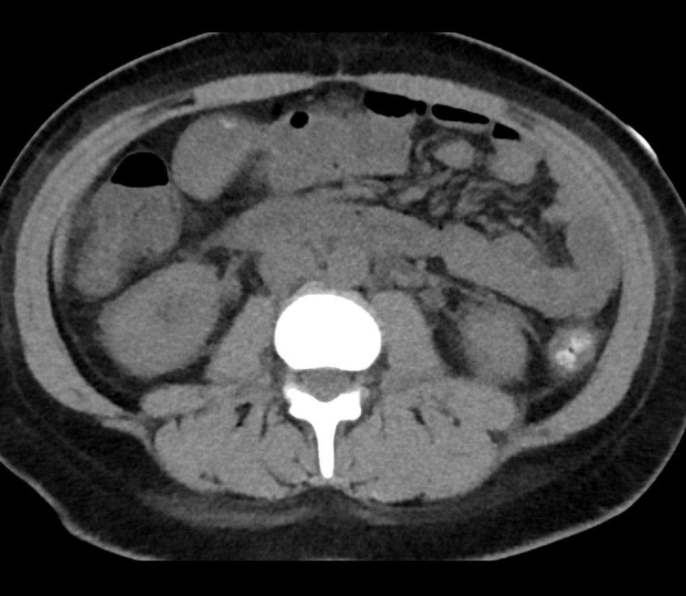 Figure 2. Computed tomography of the abdomen and pelvis without contrast. Mesenteric edema and a small amount of free fluid are present without evidence of bowel obstruction.
 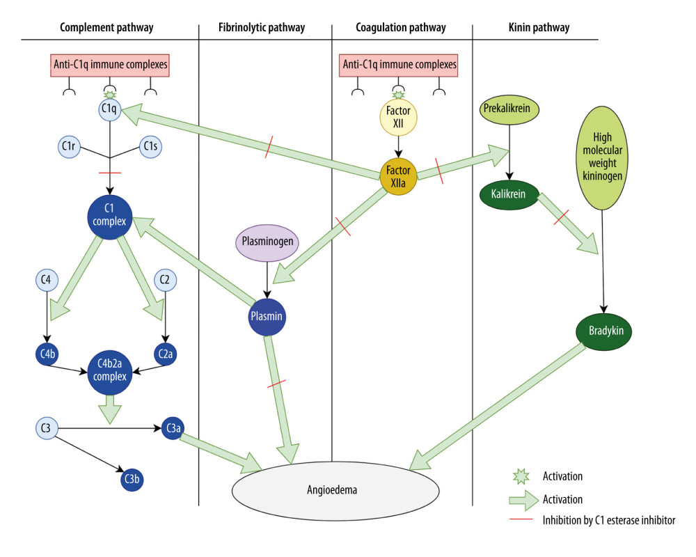 Figure 3. Proposed complement-mediated pathways in lupus-associated angioedema. Autoantibodies directed against C1q form immune complexes that may activate (green starburst/arrow) the classical complement pathway and may also promote contact system (kallikrein-kinin) activation. Binding of anti-C1q immune complexes to C1q may activate the C1 complex (C1r/C1s), leading to cleavage of C4 and C2 and formation of the C3 convertase (C4b2a complex) with generation of anaphylatoxins such as C3a, which may augment vascular permeability. In parallel, contact system activation (Factor XII → XIIa) may promote conversion of prekallikrein to kallikrein, which cleaves high-molecular-weight kininogen to generate bradykinin, a major mediator of edema. Plasmin may further amplify these cascades by enhancing complement activation. Because C1-INH normally inhibits (red dash) C1r/C1s, XIIa, kallikrein, and plasmin, dysregulation of these pathways can result in excess bradykinin and complement mediators that could contribute to angioedema. The precise mechanism underlying this phenotype remains incompletely understood.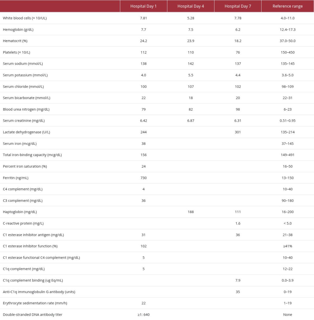
References
1. Cicardi M, Aberer W, Banerji A, Classification, diagnosis, and approach to treatment for angioedema: Consensus report from the Hereditary Angioedema International Working Group: Allergy, 2014; 69(5); 602-16
2. Nettis E, Colanardi MC, Loria MP, Vacca A, Acquired C1-inhibitor deficiency in a patient with systemic lupus erythematosus: A case report and review of the literature: Eur J Clin Invest, 2005; 35(12); 781-84
3. Maurer M, Magerl M, Betschel S, The international WAO/EAACI guideline for the management of hereditary angioedema The 2021 revision and update: Allergy, 2022; 77(7); 1961-90
4. Luo Y, Fan X, Jiang C, Systemic lupus erythematosus and angioedema: A cross-sectional study from the National Inpatient Sample: Arch Rheumatol, 2019; 34(3); 301-7
5. Eskander A, de Almeida JR, Irish JC, Acute upper airway obstruction: N Engl J Med, 2019; 381(20); 1940-49
6. Trainotti S, Johnson F, Hahn J, Acquired angioedema due to C1-inhibitor deficiency (AAE-C1-INH) – A bicenter retrospective study on diagnosis, course, and therapy: J Allergy Clin Immunol Pract, 2023; 11(12); 3772-79
7. Cacoub P, Frémeaux-Bacchi V, De Lacroix I, A new type of acquired C1 inhibitor deficiency associated with systemic lupus erythematosus: Arthritis Rheum, 2001; 44(8); 1836-40
8. Lahiri M, Lim AY, Angioedema and systemic lupus erythematosus – A complementary association?: Ann Acad Med Singap, 2007; 36(2); 142-45
9. Bienstock D, Mandel L, Facial angioedema and systemic lupus erythematosus: Case report: J Oral Maxillofac Surg, 2015; 73(5); 928-32
10. Tekin ZE, Yener GO, Yüksel S, Acquired angioedema in juvenile systemic lupus erythematosus: Case-based review: Rheumatol Int, 2018; 38(8); 1577-84
11. Sami FL, Sami HL, Tahir H, Guan J, Angioedema as a rare presentation of systemic lupus erythematosus: Cureus, 2022; 14(11); e31763
12. Siegert C, Daha M, Westedt M, IgG autoantibodies against C1q are correlated with dsDNA antibodies, hypocomplementemia and nephritis in systemic lupus erythematosus: J Rheumatol, 1991; 18; 230-34
13. Trendelenburg M, Antibodies against C1q in patients with systemic lupus erythematosus: Springer Semin Immunopathol, 2005; 27(3); 276-85
14. Kaplan AP, Angioedema: World Allergy Organ J, 2008; 1(6); 103-13
15. Esposito S, Bosis S, Semino M, Rigante D, Infections and systemic lupus erythematosus: Eur J Clin Microbiol Infect Dis, 2014; 33(9); 1467-75
16. Gavand PE, Serio I, Arnaud L, Clinical spectrum and therapeutic management of systemic lupus erythematosus-associated macrophage activation syndrome: A study of 103 episodes in 89 adult patients: Autoimmun Rev, 2017; 16(7); 743-49
17. Dall’Ara F, Cavazzana I, Frassi M, Macrophage activation syndrome in adult systemic lupus erythematosus: Report of seven adult cases from a single Italian rheumatology center: Reumatismo, 2018; 70(2); 100-5
18. Thong BY, Thumboo J, Howe HS, Feng PH, Life-threatening angioedema in systemic lupus erythematosus: Lupus, 2001; 10(4); 304-8
19. Ko CH, Ng J, Kumar S, Hurst M, Life-threatening angioedema in a patient with systemic lupus: Clin Rheumatol, 2006; 25(6); 917-18
20. Zuraw BL, Bernstein JA, Lang DM, A focused parameter update: Hereditary angioedema, acquired C1 inhibitor deficiency, and angiotensin-converting enzyme inhibitor-associated angioedema: J Allergy Clin Immunol, 2013; 131(6); 1491-93
Figures
Figure 1. Computed tomography of the orbits and sella without contrast. Significant left periorbital swelling and edema are present without a discrete collection. Convex fluid attenuation and air are noted adjacent to the left lateral globe (arrows).Figure 2. Computed tomography of the abdomen and pelvis without contrast. Mesenteric edema and a small amount of free fluid are present without evidence of bowel obstruction.Figure 3. Proposed complement-mediated pathways in lupus-associated angioedema. Autoantibodies directed against C1q form immune complexes that may activate (green starburst/arrow) the classical complement pathway and may also promote contact system (kallikrein-kinin) activation. Binding of anti-C1q immune complexes to C1q may activate the C1 complex (C1r/C1s), leading to cleavage of C4 and C2 and formation of the C3 convertase (C4b2a complex) with generation of anaphylatoxins such as C3a, which may augment vascular permeability. In parallel, contact system activation (Factor XII → XIIa) may promote conversion of prekallikrein to kallikrein, which cleaves high-molecular-weight kininogen to generate bradykinin, a major mediator of edema. Plasmin may further amplify these cascades by enhancing complement activation. Because C1-INH normally inhibits (red dash) C1r/C1s, XIIa, kallikrein, and plasmin, dysregulation of these pathways can result in excess bradykinin and complement mediators that could contribute to angioedema. The precise mechanism underlying this phenotype remains incompletely understood. In Press
Case report 
Am J Case Rep In Press; DOI: 10.12659/AJCR.953543
Case report 
Am J Case Rep In Press; DOI: 10.12659/AJCR.954216
Case report 
Am J Case Rep In Press; DOI: 10.12659/AJCR.954932
Case report
Am J Case Rep In Press; DOI: 10.12659/AJCR.954146
Most Viewed Current Articles
07 Dec 2021 : Case report
28,712,695
DOI :10.12659/AJCR.934347
Am J Case Rep 2021; 22:e934347
06 Dec 2021 : Case report  180,721
DOI :10.12659/AJCR.934406
Am J Case Rep 2021; 22:e934406
21 Jun 2024 : Case report
124,419
DOI :10.12659/AJCR.944371
Am J Case Rep 2024; 25:e944371
08 May 2026 : Case report
95,462
DOI :10.12659/AJCR.952158
Am J Case Rep 2026; 27:e952158