


12 August 2026: Articles 
Syndrome of Inappropriate Antidiuresis and Symptomatic Hyponatremia After Onyx Embolization of a Carotid-Cavernous Fistula: A Case Report
Unusual or unexpected effect of treatment, Rare coexistence of disease or pathology
Amir Nikouei ABCDEF 1, Blake Giarola ACD 1, Shuo Xi CDF 2, Karl Ng AD 2*
AD 2*
DOI: 10.12659/AJCR.953613
Am J Case Rep 2026; 27:e953613


Abstract
BACKGROUND: Carotid-cavernous fistulas (CCFs) are abnormal arteriovenous communications between the carotid artery and cavernous sinus. They may be direct or indirect and commonly present with both ocular and neurovascular symptoms. Endovascular embolization is the main treatment, with high rates of fistula obliteration and clinical improvement. Although generally safe, complications can occur, including cranial nerve dysfunction and vascular injury. Endocrine complications following carotid-cavernous fistula embolization are exceedingly rare. To our knowledge, only 1 case of syndrome of inappropriate antidiuresis (SIAD) after Onyx embolization has been reported in the literature.
CASE REPORT: A 72-year-old woman developed severe symptomatic SIAD 9 days after transarterial Onyx embolization of an indirect CCF. The procedure was successful but associated with worsening of preexisting left abducens nerve paresis. She subsequently re-presented with dizziness, truncal ataxia, and confusion, which resulted in a fall involving wrist fracture but no head strike. Investigations demonstrated profound hypotonic euvolemic hyponatremia with elevated urine osmolality and urine sodium, suggestive of SIAD. The absence of hypovolemia, polyuria, and a negative fluid balance argued against cerebral salt wasting. Adrenal insufficiency, hypothyroidism, and other causes were excluded. Treatment with hypertonic saline and fluid restriction resulted in gradual sodium correction and complete neurological recovery.
CONCLUSIONS: Transient hypothalamic-pituitary dysfunction related to Onyx-associated local mass effect and hemodynamic changes may represent a plausible mechanism. This case highlights delayed-onset SIAD as a rare but clinically significant complication temporally associated with CCF embolization. The findings support consideration of both early and delayed electrolyte monitoring, particularly within the first 1 to 2 weeks after the procedure.
Keywords: Cavernous Sinus, Embolization, Therapeutic, Hyponatremia, Fistula
Introduction
Carotid-cavernous fistulas (CCFs) are pathological arteriovenous shunts between internal or external carotid arteries and the cavernous sinus. Clinical presentation varies widely, ranging from asymptomatic lesions to proptosis, chemosis, diplopia, and pulsatile tinnitus [1]. CCFs are classified according to arterial supply and hemodynamic characteristics (Barrow types A–D); indirect fistulas (Barrow types B–D) typically represent low-flow lesions arising from dural branches. Endocrine complications after carotid-cavernous fistula embolization are exceedingly rare; very few reports have described SIAD following neurointerventional treatment of CCFs.
Endovascular embolization is the standard treatment and may be performed via transarterial, transvenous, or combined approaches using liquid embolic agents such as Onyx. Onyx is a nonadhesive ethylene-vinyl alcohol copolymer that can produce a local mass effect within confined venous structures. Although procedural complications are well described, endocrine disturbances are exceedingly rare [2]. SIAD arises from dysregulated antidiuretic hormone (ADH) secretion, which causes euvolemic hyponatremia through excessive renal water reabsorption. ADH, synthesized in hypothalamic magnocellular neurons and released from the posterior pituitary, maintains osmotic homeostasis through osmoreceptor and baroreceptor inputs [3]. Neurological insults, including vascular disturbances, can precipitate SIAD by disrupting hypothalamic-pituitary perfusion. Recognition of delayed hyponatremia after embolization is clinically important because symptoms can mimic neurological complications of the underlying vascular lesion or procedure.
Case Report
A 72-year-old woman presented with a 2-month history of left frontal and retro-orbital pain and pulsatile tinnitus. Examination revealed mild left proptosis, ptosis, scleral injection, and a partial left abducens palsy, with a normal intraocular pressure of 10 mm Hg. She had no history of trauma, prior cerebrovascular intervention, aneurysm, hypertension, thrombophilia, or connective tissue disease.
Brain magnetic resonance imaging (MRI) demonstrated T2 hyperintensity in the left lateral rectus muscle and asymmetric high-flow signal within the left cavernous sinus on time-of-flight magnetic resonance angiography (Figure 1). Formal catheter angiography confirmed a Barrow type D CCF supplied by bilateral internal and external carotid artery branches, with a discrete fistula point (Figures 2–4).
Endovascular embolization was performed through a transarterial approach using Onyx-18 (Covidien, Mansfield, MA, USA); complete angiographic occlusion was achieved without iatrogenic vessel injury (Figure 5). Pulsatile tinnitus resolved immediately after embolization; however, a complete left abducens palsy developed postoperatively and was attributed to focal mass effect from the Onyx cast within the cavernous sinus.
The patient was euvolemic; her preoperative serum sodium and chloride levels were 136 mmol/L and 102 mmol/L, respectively, with no prior history of hyponatremia. Immediate postprocedural sodium was 133 mmol/L and normalized within 2 days. She was discharged 4 days after the procedure with a sodium level of 134 mmol/L and stable neurological status other than the abducens palsy. Her home medications exclusively consisted of intermittent paracetamol (acetaminophen) for osteoarthritis. She was not taking antidepressants, antiepileptics, diuretics, antipsychotics, chemotherapeutic agents, or other medications known to precipitate SIAD. She did not receive diuretics, hypotonic fluids, or mannitol during the periprocedural period and was administered standard volumes of isotonic crystalloid. After discharge, she maintained her usual oral intake without excessive free water consumption and did not report vomiting, diarrhea, intercurrent illness, polyuria, polydipsia, or substantial pain.
Nine days after embolization, she re-presented with dizziness, severe truncal ataxia, and a fall that resulted in wrist fracture without head strike. On arrival, she was confused but hemodynamically stable and clinically euvolemic, without orthostatic hypotension, tachycardia, edema, signs of dehydration, or a negative fluid balance. Serum sodium was 121 mmol/L and subsequently declined to 117 mmol/L, prompting admission to the intensive care unit.
Biochemical evaluation demonstrated a serum osmolality of 248 mOsm/kg, urine osmolality of 406 mOsm/kg, and urine sodium of 72 mmol/L, suggestive of hypotonic euvolemic hyponatremia with inappropriately concentrated urine, thereby fulfilling the diagnostic criteria for SIAD. Endocrine evaluation showed normal morning cortisol (329 nmol/L), thyroid-stimulating hormone (1.29 mIU/L), follicle-stimulating hormone (40.3 IU/L), and luteinizing hormone (15.7 IU/L) levels, thus excluding adrenal insufficiency, hypothyroidism, and broader hypopituitarism. Renal function and serum glucose were also normal. Additionally, there were no features suggestive of hypovolemia or cerebral salt wasting (CSW), including the absence of high urine output, hemodynamic instability, polyuria, polydipsia, or a negative fluid balance. The patient remained clinically euvolemic throughout admission; she improved with fluid restriction and hypertonic saline but did not require volume replacement. There was no documented hypernatremic or polyuric phase to suggest preceding diabetes insipidus.
She was treated with 3% hypertonic saline and an 800 mL/day fluid restriction, targeting a serum sodium increase of less than 10 mmol/L per 24 hours. Sodium steadily improved over 3 days, paralleling her clinical recovery. Her confusion and truncal ataxia resolved by day 6 after re-presentation; she was discharged with a sodium level of 134 mmol/L and no ongoing therapy.
Follow-up brain MRI demonstrated no infarction, pituitary hemorrhage, or intrinsic pituitary lesion. However, T1-weighted postcontrast imaging showed Onyx filling of the left cavernous sinus adjacent to the pituitary fossa compared with corresponding preoperative sequences (Figures 6, 7). Follow-up catheter angiography confirmed durable occlusion of the fistula. At outpatient review, serum sodium was 140 mmol/L, with near-complete resolution of the abducens palsy and no recurrence of hyponatremia. In the context of the temporal evolution of serum sodium, values were 136 mmol/L preoperatively, 133 mmol/L immediately postoperatively, 136 mmol/L on postoperative day 2, and 134 mmol/L at discharge on day 4. They then fell to 121 mmol/L at re-presentation on day 9, with a nadir of 117 mmol/L in the intensive care unit, before improving to 134 mmol/L at discharge from the second admission and 140 mmol/L at outpatient follow-up (Figure 8).
Discussion
Complication rates following CCF embolization range from 1% to 10% and include thromboembolism, cranial nerve palsy, venous thrombosis, embolic migration, hemorrhage, dissection, and recurrence [4]. Hyponatremia is a recognized complication after neurosurgical and neurointerventional procedures, most commonly due to SIAD or CSW [5]. SIAD following elective neuroendovascular intervention outside the setting of aneurysmal subarachnoid hemorrhage appears exceedingly rare. The literature is largely limited to isolated postembolization case reports, including a case after carotid-cavernous fistula embolization described by Chen et al [2]. In contrast, hyponatremia and SIAD are well recognized following aneurysmal subarachnoid hemorrhage after endovascular treatment, although interpretation is confounded by the high baseline prevalence of subarachnoid-hemorrhage-associated neuroendocrine dysregulation [6]. SIAD and CSW are important differential diagnoses in hyponatremia, given that both can present with elevated urine sodium and concentrated urine. However, SIAD is characterized by inappropriate water retention in a clinically euvolemic state and is managed with fluid restriction, whereas CSW reflects hypovolemic renal sodium loss requiring salt and volume replacement [7].
In the present case, hypotonic euvolemic hyponatremia with elevated urine osmolality and urine sodium, normal adrenal and thyroid function, clinical euvolemia, absence of polyuria or polydipsia, and the absence of diuretics or other precipitating medications supported a diagnosis of SIAD temporally associated with CCF embolization. The temporal pattern is notable. Serum sodium was normal pre-procedurally and during the immediate postoperative period, then followed by abrupt development of severe hyponatremia approximately 9 days later. This delayed onset argues against preexisting or chronic SIAD and supports a procedure-related mechanism.
A previous report by Chen et al [2] described SIAD developing within 2 weeks of Onyx embolization for an indirect CCF, with a similar absence of preexisting endocrine dysfunction. Considered along with the present case, these findings suggest that SIAD represents a rare but reproducible complication of CCF embolization. In our patient, SIAD developed within approximately 2 weeks of Onyx embolization in the absence of preexisting endocrine dysfunction; clinical and biochemical recovery were noted after conservative management. Both cases support a possible association between embolization within the cavernous sinus region and delayed dysregulation of ADH secretion.
In the present case, a mechanical and hemodynamic mechanism may provide a plausible explanation. The proximity of the Onyx cast within the cavernous sinus to the pituitary stalk and posterior pituitary region, as well as the immediate development of a complete abducens palsy after the procedure, supports the presence of local mass effect within this confined anatomical space. The posterior pituitary is supplied by branches of the cavernous internal carotid artery and drains into the cavernous sinus [8]. Therefore, embolic material in this region may transiently alter venous outflow, local pressure, or microvascular perfusion. Such changes may disrupt axonal transport or the regulated release of ADH, resulting in inappropriate secretion without permanent structural damage detectable on imaging. The delayed onset may reflect evolving local inflammatory changes, delayed venous hemodynamic adaptation after fistula occlusion, or progressive local mass effect within the cavernous sinus rather than immediate procedural injury. Complete clinical and biochemical recovery without a persistent endocrine deficit further supports a reversible functional disturbance.
The delayed timing of hyponatremia in the present case resembles delayed hyponatremia observed after transsphenoidal pituitary surgery, which commonly occurs approximately 5 to 8 days postoperatively and is thought to reflect dysregulated ADH release following hypothalamic-posterior pituitary disturbance. Although no direct pituitary manipulation occurred in our patient, a similar delayed neuroendocrine response related to local vascular or mechanical effects may have contributed [9]. Alternative causes of hyponatremia were systematically excluded. There was no clinical or biochemical evidence of hypovolemia, CSW, adrenal insufficiency, hypothyroidism, renal impairment, or cardiac failure. The patient remained clinically euvolemic throughout admission without evidence of persistent polyuria, hypotension, tachycardia, or volume depletion. She was not exposed to SIAD-inducing medications, did not receive hypotonic fluids, and did not report excessive fluid intake. Although postoperative stress can contribute to transient ADH release, the normal sodium levels during the early postoperative period and the delayed deterioration make this explanation less likely.
This report has some limitations. Given its focus on a single case, the present findings cannot definitively establish causality between Onyx embolization and SIAD. Pre-admission outpatient sodium measurements were unavailable, although normal periprocedural sodium values argue against preexisting hyponatremia. Detailed dynamic endocrine testing and serial pituitary imaging during the acute phase were not performed, limiting characterization of transient hypothalamic-pituitary dysfunction. Formal serial fluid balance measurements and body weight trends also were not consistently available retrospectively, hindering detailed assessment of dynamic volume status. Unmeasured factors, including individual susceptibility to ADH dysregulation, cannot be entirely excluded. Dedicated pituitary MRI or magnetic resonance venography may be considered in future similar cases to better characterize transient regional vascular or structural changes involving the hypothalamic-pituitary region. No recurrence of hyponatremia was observed during follow-up; however, the long-term recurrence risk of SIAD after CCF embolization remains uncertain due to the rarity of reported cases.
Conclusions
This case suggests that delayed symptomatic SIAD can occur after Onyx embolization of an indirect carotid-cavernous fistula. The temporal relationship, normal early postprocedural sodium levels, and exclusion of common alternative causes support a possible association with transient hypothalamic-pituitary dysfunction related to local mass effect and hemodynamic changes near the pituitary stalk. Clinicians should consider both early and delayed serum sodium monitoring, particularly within the first 1 to 2 weeks following the procedure, and maintain a low threshold for investigation in patients who present with neurological symptoms after CCF embolization.
Figures
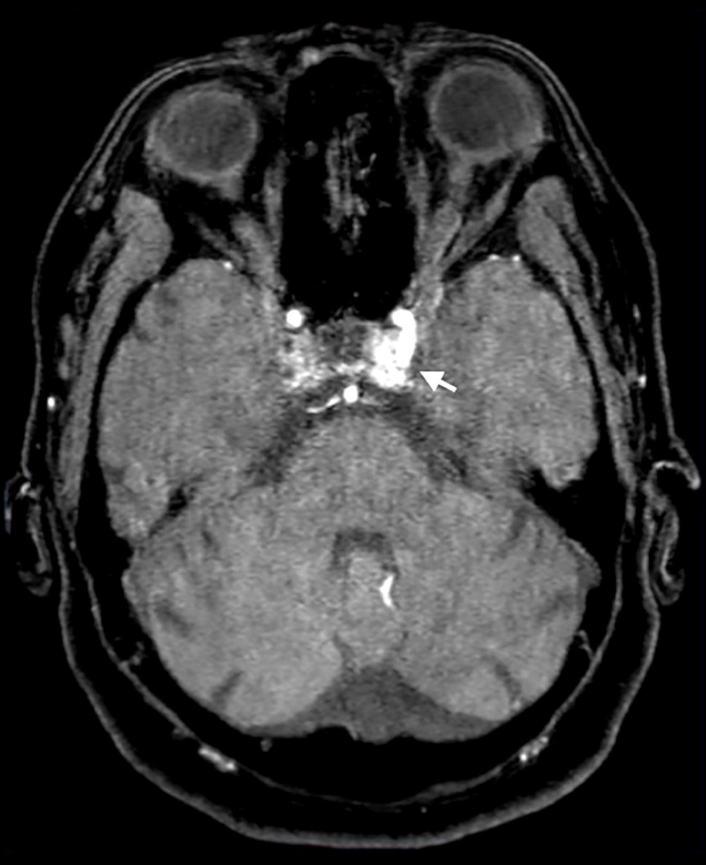 Figure 1. Axial time-of-flight magnetic resonance angiography. Arrow: Arterialized flow within the left cavernous sinus on the time-of-flight sequence.
 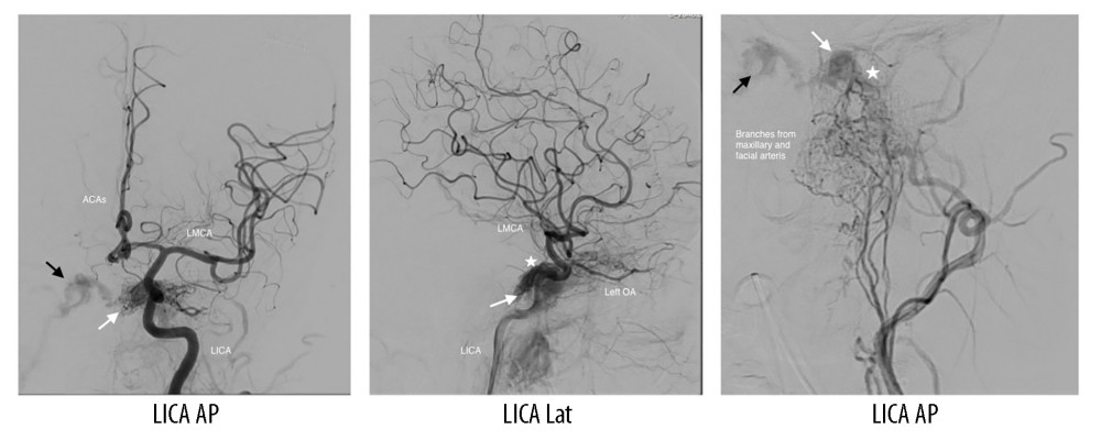 Figure 2. Early opacification of the left cavernous sinus during transarterial injection of the left internal carotid artery (LICA) on anteroposterior (AP) and lateral (Lat) views. Early but less intense opacification is also evident on AP views after left external carotid artery (LECA) injection. White arrow: early filling of the left cavernous sinus on ipsilateral LICA injection; black arrow: early filling of the right cavernous sinus on LICA injection; star: fistula point. Additional abbreviations: LMCA, left middle cerebral artery; ACAs, anterior cerebral arteries; OA, ophthalmic artery.
 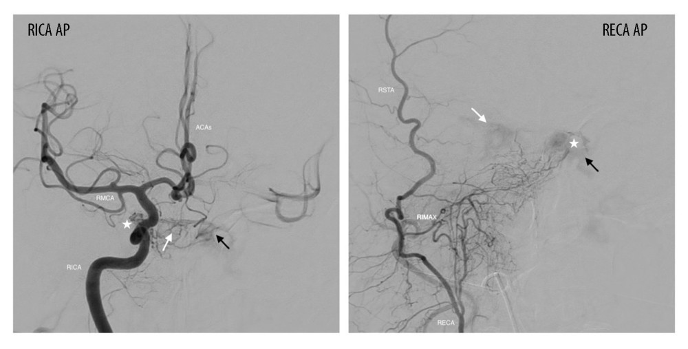 Figure 3. Filling of the left cavernous sinus after right internal carotid artery (RICA) injection on the AP view. Less intense filling is also observed on the AP view after right external carotid artery (RECA) injection. White arrow: early filling of the right cavernous sinus on ipsilateral RICA injection; black arrow: early filling of the left cavernous sinus on RICA injection; star: fistula point. Additional abbreviations: RMCA, right middle cerebral artery; ACAs, anterior cerebral arteries; RSTA, right superficial temporal artery; RIMAX, right internal maxillary artery.
 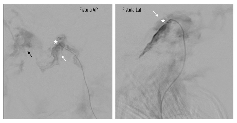 Figure 4. Transvenous injection at the fistula point demonstrates opacification of the left cavernous sinus on anteroposterior (AP) and lateral (Lat) views. White arrow: early filling of the left cavernous sinus on ipsilateral transvenous injection; black arrow: early filling of the right cavernous sinus on left-sided transvenous injection; star: fistula point.
 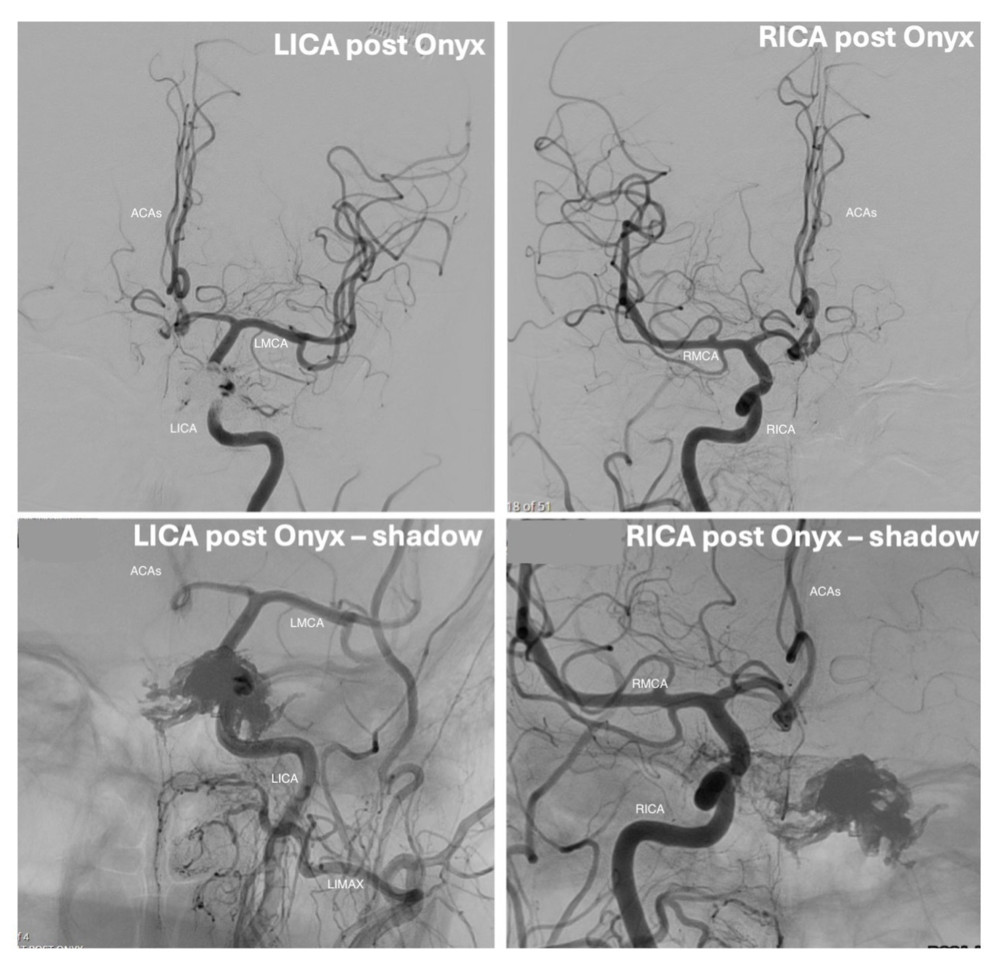 Figure 5. Postembolization digital subtraction angiography demonstrates mixed opacification with the Onyx cast (“shadow”) on transarterial injection. Left internal carotid artery (LICA) injections, including shadow views, show residual flow around the Onyx cast. Right internal carotid artery (RICA) injections, including shadow views, similarly demonstrate partial filling adjacent to the Onyx cast. Additional abbreviations: LMCA, left middle cerebral artery; RMCA, right middle cerebral artery; ACAs, anterior cerebral arteries; LIMAX, left internal maxillary artery.
 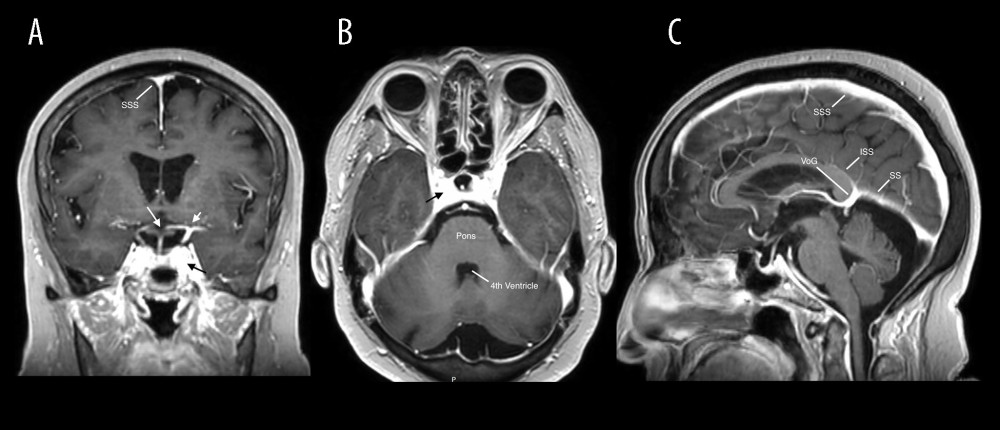 Figure 6. Pre-embolization brain MRI demonstrates the lesion on multiple planes: coronal (A), axial (B), and sagittal (C) T1-weighted post-gadolinium images. Short white arrow: LICA terminus; long white arrow: optic chiasm; black arrow: left cavernous sinus. Additional abbreviations: SSS, superior sagittal sinus; ISS, inferior sagittal sinus; SS, straight sinus; VoG, vein of Galen.
 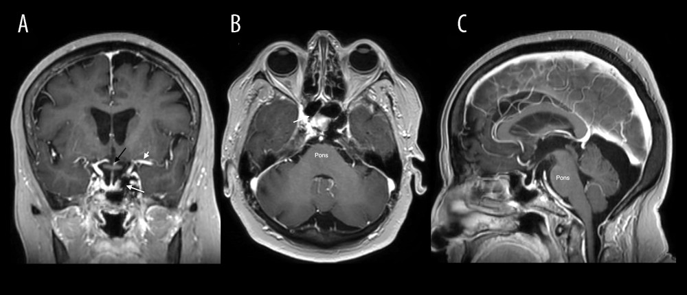 Figure 7. Postembolization brain MRI demonstrates Onyx filling the left cavernous sinus adjacent to the pituitary fossa on coronal (A), axial (B), and sagittal (C) T1-weighted postcontrast images. Short white arrow: LMCA; long white arrow: left cavernous sinus containing Onyx; black arrow: optic chiasm. Additional abbreviation: LMCA, left middle cerebral artery.
 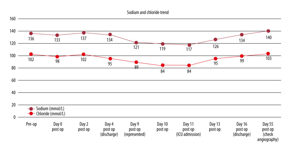 Figure 8. Temporal trends in serum sodium and chloride levels during hospitalization and intervention. Abbreviations: ICU, intensive care unit; Pre-op, preoperative; post op, postoperative.
 References
1. Al-Shalchy A, Al-Wassiti AS, Hashim MA, Neuro-ophthalmic manifestations of carotid cavernous fistulas: A systematic review and meta-analysis: Cureus, 2024; 16(7); e62662
2. Chen T, Kalani MY, Ducruet AF, Development of syndrome of inappropriate antidiuretic hormone secretion after Onyx embolization of a cavernous carotid fistula: BMJ Case Rep, 2016; 2016; bcr2015012104
3. Warren AM, Grossmann M, Christ-Crain M, Syndrome of inappropriate antidiuresis: from pathophysiology to management: Endocr Rev, 2023; 44(5); 819-61
4. Baharvahdat H, Seraj FQ, Al-Raaisi A, Long-term outcome of endovascular treatment for indirect carotid-cavernous fistulas: Neurosurg Focus, 2024; 56(3); E5
5. Bhat R, Baldeweg SE, Wilson SR, Sodium disorders in neuroanaesthesia and neurocritical care: BJA Educ, 2022; 22(12); 466-73
6. Kieninger M, Kerscher C, Bründl E, Acute hyponatremia after aneurysmal subarachnoid hemorrhage: Frequency, treatment, and outcome: J Clin Neurosci, 2021; 88; 237-42
7. Maesaka JK, Imbriano L, Mattana J, Differentiating SIADH from cerebral/renal salt wasting: Failure of the volume approach and need for a new approach to hyponatremia: J Clin Med, 2014; 3; 1373-85
8. Pensak ML, The cavernous sinus: An anatomic study with clinical implication: Laryngoscope Investig Otolaryngol, 2024; 9(2); e1226
9. Lee CC, Wang YC, Liu YT, Incidence and factors associated with postoperative delayed hyponatremia after transsphenoidal pituitary surgery: A meta-analysis and systematic review: Int J Endocrinol, 2021; 2021; 6659152
Figures
Figure 1. Axial time-of-flight magnetic resonance angiography. Arrow: Arterialized flow within the left cavernous sinus on the time-of-flight sequence.Figure 2. Early opacification of the left cavernous sinus during transarterial injection of the left internal carotid artery (LICA) on anteroposterior (AP) and lateral (Lat) views. Early but less intense opacification is also evident on AP views after left external carotid artery (LECA) injection. White arrow: early filling of the left cavernous sinus on ipsilateral LICA injection; black arrow: early filling of the right cavernous sinus on LICA injection; star: fistula point. Additional abbreviations: LMCA, left middle cerebral artery; ACAs, anterior cerebral arteries; OA, ophthalmic artery.Figure 3. Filling of the left cavernous sinus after right internal carotid artery (RICA) injection on the AP view. Less intense filling is also observed on the AP view after right external carotid artery (RECA) injection. White arrow: early filling of the right cavernous sinus on ipsilateral RICA injection; black arrow: early filling of the left cavernous sinus on RICA injection; star: fistula point. Additional abbreviations: RMCA, right middle cerebral artery; ACAs, anterior cerebral arteries; RSTA, right superficial temporal artery; RIMAX, right internal maxillary artery.Figure 4. Transvenous injection at the fistula point demonstrates opacification of the left cavernous sinus on anteroposterior (AP) and lateral (Lat) views. White arrow: early filling of the left cavernous sinus on ipsilateral transvenous injection; black arrow: early filling of the right cavernous sinus on left-sided transvenous injection; star: fistula point.Figure 5. Postembolization digital subtraction angiography demonstrates mixed opacification with the Onyx cast (“shadow”) on transarterial injection. Left internal carotid artery (LICA) injections, including shadow views, show residual flow around the Onyx cast. Right internal carotid artery (RICA) injections, including shadow views, similarly demonstrate partial filling adjacent to the Onyx cast. Additional abbreviations: LMCA, left middle cerebral artery; RMCA, right middle cerebral artery; ACAs, anterior cerebral arteries; LIMAX, left internal maxillary artery.Figure 6. Pre-embolization brain MRI demonstrates the lesion on multiple planes: coronal (A), axial (B), and sagittal (C) T1-weighted post-gadolinium images. Short white arrow: LICA terminus; long white arrow: optic chiasm; black arrow: left cavernous sinus. Additional abbreviations: SSS, superior sagittal sinus; ISS, inferior sagittal sinus; SS, straight sinus; VoG, vein of Galen.Figure 7. Postembolization brain MRI demonstrates Onyx filling the left cavernous sinus adjacent to the pituitary fossa on coronal (A), axial (B), and sagittal (C) T1-weighted postcontrast images. Short white arrow: LMCA; long white arrow: left cavernous sinus containing Onyx; black arrow: optic chiasm. Additional abbreviation: LMCA, left middle cerebral artery.Figure 8. Temporal trends in serum sodium and chloride levels during hospitalization and intervention. Abbreviations: ICU, intensive care unit; Pre-op, preoperative; post op, postoperative. In Press
Case report 
Am J Case Rep In Press; DOI: 10.12659/AJCR.953543
Case report 
Am J Case Rep In Press; DOI: 10.12659/AJCR.954216
Case report 
Am J Case Rep In Press; DOI: 10.12659/AJCR.954932
Case report 
Am J Case Rep In Press; DOI: 10.12659/AJCR.954146
Most Viewed Current Articles
07 Dec 2021 : Case report
28,712,695
DOI :10.12659/AJCR.934347
Am J Case Rep 2021; 22:e934347
06 Dec 2021 : Case report  180,721
DOI :10.12659/AJCR.934406
Am J Case Rep 2021; 22:e934406
21 Jun 2024 : Case report
124,419
DOI :10.12659/AJCR.944371
Am J Case Rep 2024; 25:e944371
08 May 2026 : Case report
95,462
DOI :10.12659/AJCR.952158
Am J Case Rep 2026; 27:e952158