


28 September 2026: Articles 
False Passage During Single-Lung Ventilation in a Tracheostomized Patient: Intraoperative Recognition and Rescue
Unusual clinical course, Challenging differential diagnosis, Diagnostic / therapeutic accidents, Management of emergency care
David Revuelta Quintero E 1, Guillem Urquijo EF 1, Maria Antonia Perelló Llaneras B 1, Álvaro Barranco de Santiago AEF 1*
DOI: 10.12659/AJCR.954119
Am J Case Rep 2026; 27:e954119


Abstract
BACKGROUND: Airway management in tracheostomized patients may become particularly challenging in the presence of recent stomas, local infection, single-lung ventilation, and restricted intraoperative airway access. False passage ventilation is an uncommon but potentially catastrophic complication requiring immediate recognition and rescue.
CASE REPORT: We report the case of a 63-year-old man with a recent surgical tracheostomy performed after emergent cricothyrotomy for upper airway obstruction secondary to cervical cellulitis with mediastinal extension. The patient underwent urgent thoracotomy for mediastinal debridement under general anesthesia with single-lung ventilation achieved using a bronchial blocker through the existing tracheostomy tube. During surgery in the lateral decubitus position, abrupt loss of capnography and inability to ventilate occurred. Bronchoscopic evaluation demonstrated tracheostomy tube malposition with false passage ventilation associated with peristomal tissue dehiscence and severe local infection. In this context, the combination of single-lung ventilation, lateral decubitus positioning, and limited access to the tracheostomy created a particularly challenging intraoperative airway rescue scenario. Given the need for immediate restoration of oxygenation, emergency orotracheal intubation using videolaryngoscopy was successfully performed without prior supination, allowing rapid recovery of ventilation and completion of the procedure.
CONCLUSIONS: This case highlights the diagnostic and rescue challenges posed by false passage ventilation during thoracic surgery in recently tracheostomized patients. It reinforces the importance of early recognition of airway loss and prioritization of oxygenation-restoring rescue strategies in similar high-risk intraoperative settings. This report also underscores the vulnerability of recently created and infected tracheostomies during complex thoracic procedures requiring single-lung ventilation.
Keywords: Tracheostomy, one-lung ventilation, Intraoperative Complications, Anesthesia, General
Introduction
Tracheostomy, a surgically created airway connecting the trachea to the skin, is often considered to be a straightforward and reliable method. However, recently created tracheostomies remain vulnerable to serious complications, particularly during the early postoperative period before maturation of the tracheocutaneous tract. In this setting, tube displacement and false passage formation (creation of an extratracheal tract instead of re-entering the tracheal lumen) may result in rapid loss of effective ventilation, severe hypoxemia, and life-threatening airway loss, requiring immediate and coordinated rescue maneuvers [1,2].
The risks associated with recently created tracheostomies and false passage formation are well recognized. However, evidence addressing their management during thoracic surgery requiring single-lung ventilation remains limited. In particular, the combination of an infected immature stoma, lateral decubitus positioning, restricted stomal access, and the need for uninterrupted lung isolation creates a high-risk scenario in which airway rescue may be particularly demanding.
From an anesthetic perspective, the management of patients with pre-existing tracheostomy undergoing thoracic surgery requires additional perioperative considerations. Intraoperatively, abrupt loss of capnography may represent an early sign of airway failure, but diagnosis can be difficult in complex surgical scenarios [1,3].
We report the case of a recently tracheostomized patient with an infected stoma undergoing thoracic surgery requiring single-lung ventilation in the lateral decubitus position, in whom false passage formation caused sudden ventilatory failure. This case highlights the importance of early suspicion of false passage after abrupt loss of capnography, and supports prompt transition to orotracheal rescue when tracheostomy salvage is unlikely to rapidly restore effective ventilation. This report addresses a clinically relevant scenario that remains underrepresented in the literature.
Case Report
A 63-year-old man with a history of primary hypertension, type 2 diabetes mellitus treated with oral antidiabetic agents, and surgically revascularized ischemic heart disease, was admitted following the diagnosis of pharyngeal cellulitis with abscess formation and mediastinal extension. He developed progressive respiratory distress due to upper airway obstruction and required an emergent cricothyrotomy, which resolved an initial
The clinical course was marked by progressive deterioration and characterized by advancing mediastinal infection and enlargement of a right pleural collection. Therefore, urgent mediastinal debridement via thoracotomy under general anesthesia was indicated. Following anesthetic intravenous induction with propofol, fentanyl, and rocuronium, mechanical ventilation was established through the pre-existing tracheostomy tube (Shiley 7.5) using volume-controlled ventilation, and anesthesia was maintained with total intravenous anesthesia using propofol and remifentanil infusions. Left-sided single-lung ventilation was achieved after fiberoptic-bronchoscopy–guided placement of a 9Fr Ambu® VivaSight™ endobronchial blocker through the tracheostomy tube. Mechanical ventilation was provided in volume-controlled mode with a tidal volume of 350 mL, respiratory rate of 16 breaths/min, positive-end expiratory pressure of 6 cm H2O and inspired oxygen fraction of 0.8. The patient was positioned in the right lateral decubitus position for the procedure, which initially proceeded uneventfully.
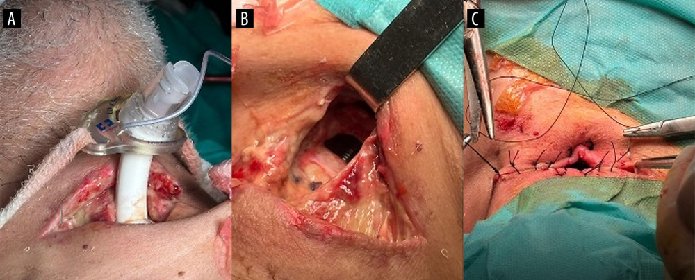
During surgery, a sudden loss of capnography was observed, accompanied by inability to ventilate despite immediate removal of the bronchial blocker. Inspection of the cervical area revealed peristomal dehiscence with disruption of surrounding tissues (Figure 1A), abundant purulent discharge, and granulation tissue. Notably, this occurred without patient repositioning or deliberate manipulation of the tracheostomy tube, and was likely facilitated by tissue friability in the setting of ongoing infection. Inspired oxygen fraction was increased to 1.0, and flexible bronchoscopy was performed through the tracheostomy tube, demonstrating malposition with formation of a false passage into the cervical subcutaneous tissues and inability to re-advance the tube into the tracheal lumen.
Given the acute loss of airway access, and after informing the surgical team of the potential need for emergent supination, the decision was made to proceed with emergency orotracheal intubation while the patient remained in the lateral decubitus position. The tracheostomy tube was left in situ during oral intubation, which was successfully achieved without complications (Figure 1B) using videolaryngoscopy. Correct endotracheal tube placement was immediately confirmed by continuous capnography, bilateral chest expansion, and normalization of ventilatory mechanics. Effective ventilation distal to the false passage was established before the procedure was resumed. Flexible bronchoscopy was then performed through the oral endotracheal tube to confirm intratracheal position and exclude additional airway injury. Once adequate ventilation through the oral endotracheal tube had been secured, the malpositioned tracheostomy tube was removed. In agreement with the surgical team, completion of the procedure was facilitated by alternating periods of double-lung ventilation with controlled apnea, rather than reinserting a bronchial blocker, to minimize further airway manipulation. Following thoracotomy closure, the patient was returned to the supine position, and the otorhinolaryngology team performed stomal repair with the insertion of a new tracheostomy tube (Figure 1C).
Following airway stabilization, effective ventilation and oxygenation were promptly restored, with reappearance of continuous capnography and normalization of ventilatory mechanics. The patient remained hemodynamically stable and was transferred to the intensive care unit for postoperative management under invasive monitoring and close airway surveillance.
Postoperative imaging and clinical assessment were performed to exclude procedure-related complications—such as subcutaneous emphysema, pneumomediastinum, or pneumothorax—and to reassess the extent of mediastinal infection. Subsequent care followed a multidisciplinary approach, including targeted antimicrobial therapy, specialized airway management, and surgical follow-up.
Discussion
This case illustrates that false passage ventilation should be considered immediately after abrupt loss of capnography during single-lung ventilation in a recently tracheostomized patient. When airway rescue is further complicated by lateral decubitus positioning, limited stomal access, and infected peristomal tissues, rapid transition to an alternative airway may be safer than prolonged attempts to salvage the tracheostomy.
Manipulation of immature stomas (typically < 7 days after creation) is consistently associated with an increased risk of false passage formation, inability to ventilate, and thoracic complications such as subcutaneous emphysema or pneumothorax [1]. Accordingly, non-essential tracheostomy tube manipulation or exchange should be avoided, and any unavoidable intervention should ideally be performed under flexible fiberoptic bronchoscopic guidance [1,4]. In our case, local infection with friable peristomal tissues likely predisposed tube displacement, while subsequent positive-pressure ventilation facilitated false passage ventilation and acute loss of tracheal access, resulting in abrupt hypoxemia.
These risks become especially relevant during thoracic surgery, during which single-lung ventilation and lateral decubitus positioning may severely limit access to the stoma and complicate rescue maneuvers. Previous series support bronchial blockers through the existing tracheostomy tube as the preferred technique for lung isolation in recent tracheostomies because they minimize stomal trauma while preserving the established airway [4,5]. This strategy was therefore adopted initially in our patient and provided satisfactory single-lung ventilation under bronchoscopic guidance. However, as illustrated by this case, successful lung isolation does not eliminate the risk of subsequent tracheostomy tube displacement and false passage ventilation.
From a practical perspective, false passage ventilation should be suspected in the presence of sudden capnography loss, inability to ventilate, and rising airway pressures. Immediate verification of tube position using flexible bronchoscopy is recommended whenever feasible. If malposition is confirmed or cannot be rapidly excluded, clinicians should avoid blind reinsertion and proceed promptly to a predefined rescue strategy, prioritizing restoration of oxygenation over attempts to salvage the tracheostomy airway. In situations where stomal access is limited—such as lateral decubitus positioning—early orotracheal intubation using videolaryngoscopy may represent a pragmatic and effective rescue option in selected situations.
The present case highlights the diagnostic and rescue challenges posed by false passage ventilation during thoracic surgery in a recently tracheostomized patient undergoing single-lung ventilation. Under these constrained conditions, abrupt loss of capnography may represent the first sign of airway loss, while limited stomal access can delay conventional rescue maneuvers. In this context, early transition to an alternative airway—prioritizing rapid restoration of oxygenation over attempts to preserve the tracheostomy—allowed effective rescue and safe continuation of the surgery. This management strategy should be interpreted within the specific anatomical, surgical, and logistical circumstances of this case rather than as a universally applicable algorithm.
Conclusions
This case highlights that a pre-existing tracheostomy should not be assumed to provide a definitively secure airway in the perioperative setting, particularly in the presence of an immature or infected stoma and challenging intraoperative positioning. Sudden loss of capnography and inability to ventilate through the tracheostomy tube should immediately raise suspicion of malposition and false passage ventilation.
In thoracic surgery requiring single-lung ventilation, bronchial blockers placed under bronchoscopic guidance represent an effective strategy; however, clinicians must remain prepared for abrupt airway failure. In this context, early transition to an alternative airway—prioritizing restoration of oxygenation over preservation of the tracheostomy airway—is essential.
This case highlights how false passage ventilation in the setting of a recent and infected tracheostomy may rapidly evolve into a high-risk intraoperative airway emergency, particularly when surgical positioning limits conventional rescue options. Although derived from a single clinical scenario, this experience reinforces the importance of early recognition of airway malposition, preparedness for sudden airway loss, and prioritization of oxygenation when airway compromise is suspected in similar high-risk contexts.
References
1. Rosero EB, Hagberg CA, Nussmeier NA, Airway management for anesthesia for the patient with a tracheostomy: UpToDate [Internet] Updated 20 Feb 2025. https://www.uptodate.com/contents/airway-management-for-anesthesia-for-the-patient-with-a-tracheostomy
2. Carr ZJ, Slinger PD, Hagberg CA, Nussmeier NA, Anesthesia for tracheostomy: UpToDate [Internet] Updated 25 July 2025. https://www.uptodate.com/contents/anesthesia-for-tracheostomy
3. Hyzy RC, De Cardenas JL, Feller-Kopman DJ, Finlay G, Tracheostomy in adults: Techniques and intraoperative complications: UpToDate [Internet] Updated May 5, 2026
4. Campos JH, Musselman ED, Hanada S, Ueda K, Lung isolation techniques in patients with early-stage or long-term tracheostomy: A case series report of 70 cases and recommendations: J Cardiothorac Vasc Anesth, 2019; 33(2); 433-39
5. Ng B, Thangavelautham S, Aman M, One-lung ventilation via end tracheostomy for thoracic surgery: A case report: Cureus, 2025; 17(1); e77539
In Press
Case report 
Am J Case Rep In Press; DOI: 10.12659/AJCR.953555
Case report
Am J Case Rep In Press; DOI: 10.12659/AJCR.953169
Case report 
Am J Case Rep In Press; DOI: 10.12659/AJCR.954027
Case report
Am J Case Rep In Press; DOI: 10.12659/AJCR.954380
Most Viewed Current Articles
07 Dec 2021 : Case report  28,713,109
DOI :10.12659/AJCR.934347
Am J Case Rep 2021; 22:e934347
06 Dec 2021 : Case report  180,916
DOI :10.12659/AJCR.934406
Am J Case Rep 2021; 22:e934406
21 Jun 2024 : Case report
124,581
DOI :10.12659/AJCR.944371
Am J Case Rep 2024; 25:e944371
08 May 2026 : Case report
95,842
DOI :10.12659/AJCR.952158
Am J Case Rep 2026; 27:e952158