


26 September 2026: Articles 
When A Fall Isn’t Just a Fall: Delayed Diagnosis of Acquired Hemophilia A in a Nonagenarian Patient
Mistake in diagnosis, Rare disease
Swati Gobhil BCDEF 1*, Arjun Khunger BCDEF 1, Prajeesh Kandoth Karerat BCDEF 1DOI: 10.12659/AJCR.954126
Am J Case Rep 2026; 27:e954126


Abstract
BACKGROUND: Acquired hemophilia A (AHA) is a rare but potentially life-threatening condition caused by autoantibodies against factor VIII. It often presents in older adults and its presentation can range from isolated lab abnormality of prolonged activated partial thromboplastin time (aPTT) without bleeding to mild bleeding or spontaneous vs disproportionate life-threatening bleeding. Delayed diagnosis is frequent due to the rarity of the condition and propensity to attribute bleeding to other common causes, such as falls.
CASE REPORT: A 92-year-old woman with recurrent ground-level falls presented with severe right lower extremity pain of several weeks’ duration. She was not on any anticoagulant or anti-platelet agent. Computed tomography angiography demonstrated bilateral lower extremity intramuscular hematomas. She had 2 prior admissions for falls within 6 months, including a fall leading to intramuscular hematoma, which was conservatively managed without evaluation of coagulopathy. During current admission, prolonged aPTT prompted further workup, revealing low factor VIII activity of 5% and a high factor VIII inhibitor titer of 15.6 Bethesda Units, establishing the diagnosis of acquired hemophilia A. Bleeding control required sequential bypassing agents, corticosteroids, rituximab, cyclophosphamide, and eventually emicizumab.
CONCLUSIONS: This case highlights how recurrent or disproportionate intramuscular hematomas following even minor trauma, such as a ground-level fall, in the absence of risk factors such as anticoagulant use, may prompt evaluation for underlying coagulopathy such as AHA. Basic coagulation testing including prothrombin time and aPTT can help screen patients who warrant additional coagulopathy evaluation. Timely diagnosis is critical to avoid life-threatening bleeding complications.
Keywords: Autoimmune Diseases, Bias, Implicit, Factor VIII, Frail Elderly, Hematoma, Hemophilia A
Introduction
Acquired hemophilia A (AHA) is a rare bleeding disorder caused by autoantibodies against circulating coagulation factor VIII, with an estimated annual incidence of approximately 1.5 cases per million population per year [1,2]. It often affects older adults and can present as asymptomatic isolated prolongation of activated partial thromboplastin time (aPTT) or symptoms can range from minor to life-threatening bleeding which could be spontaneous or disproportionate to the mechanism of injury [1,2]. Isolated prolonged aPTT can also be seen with lupus anticoagulant, congenital factor deficiencies (eg, hemophilia A, B, or factor XI deficiency), anticoagulant exposure (particularly unfractionated heparin and direct thrombin inhibitors), and acquired factor inhibitors [3].
Since bleeding manifestations can be nonspecific and AHA is an uncommon condition, delayed diagnosis is frequent. In patients of advanced age with recurrent falls, bleeding may readily be attributed to trauma, especially in a frail patient. However, recurrent or unexpected bleeding in the absence of anticoagulant or antiplatelet exposure should prompt consideration of other differentials, including coagulopathy, apart from considering purely trauma-based etiology. If such consideration does not occur, the likelihood of this diagnosis being overlooked remains high, especially with atypical presentations. In the absence of appropriate treatment, the mortality rate may reach up to 41% [4].
We present a case of AHA in a 92-year-old patient with recurrent falls and multiple admissions, in whom diagnosis of coagulopathy was delayed due to attribution of bleeding to trauma in a frail patient of advanced age. This case highlights the previously missed opportunities for coagulation evaluation despite repeated bleeding presentations.
Case Report
INDEX ADMISSION:
A 92-year-old female patient with past medical history of coronary artery disease presented to the emergency room with pain in the right lower extremity of several weeks’ duration. She had undergone a stent placement 20 years previously. She was not on any anti-platelet agent, despite having hypertension and long-standing paroxysmal atrial fibrillation. There was no clear reason why she was not on anticoagulation. She had a history of non-traumatic subdural hematoma 8 years previously (when she was on aspirin, 81 mg daily, which was subsequently discontinued). She had no other history of significant bleeding or blood transfusions. She had a surgical history of appendectomy in the remote past. She reported a history of multiple falls, particularly over the preceding 6 months, which were felt to be due to her advanced age and frailty. Her most recent fall had occurred about 6 weeks before the index admission. She described the primary site of pain as the right groin area with radiation to her gluteal musculature as well as bilateral lower extremities, with the right side worse than the left. Upon physical examination, she was found to have stable vital signs and significant asymmetric edema of the right thigh, with severe tenderness to palpation. Computed tomography angiography (CTA) of both lower extremities revealed an intramuscular hematoma within the left gluteus minimus muscle belly measuring 4.1 × 6.3 × 12 cm without active extravasation (Figure 1). In addition, there was a deep intramuscular hematoma within the anterior compartment of the right thigh vastus intermedialis muscle belly and 2 small foci of contrast extravasation noted distally, corresponding to distal intramuscular branches of the descending profunda femoris artery.
Initial laboratory assessment revealed severe anemia with a hemoglobin (Hb) of 6.9 g per dL. Platelet count was normal at 273 000/mcL. Bleeding diathesis (Table 1) revealed a normal prothrombin time (PT) of 10.5 seconds (reference range 9.2–12.2 seconds), internal normalized ratio (INR) of 1.0 (reference range 0.9–1.1), and a prolonged aPTT of 39.9 seconds (reference range 23–29.8 seconds). Following the finding of increased aPTT, further evaluation for acquired coagulopathy was pursued and is outlined in Table 1, which shows the coagulation profile of the patient. It highlights normal PT, prolonged aPTT, reduced factor VIII activity, and increased factor VIII inhibitor levels at 15.6 Bethesda Units (BU) with reference range < 0.5 BU. A coagulation mixing study was done with results reported in Table 2. Mixing of the patient’s plasma with normal pooled plasma in a 1: 1 ratio with immediate testing resulted in coagulation after 31 seconds. Mixing of the patient’s plasma with normal pooled blood plasma with testing after 1 hour of incubation at 37 °C resulted in coagulation after 43 seconds. This indicates the likely presence of a factor VIII inhibitor prolonging aPTT by 12 seconds upon incubation.
Notably, the patient had been hospitalized twice within 6 months prior to this encounter for falls and pain, and she was found to have a large intramuscular hematoma within the anterolateral compartment of the left thigh measuring 6 × 4.3 × 17 cm during her first admission 6 months prior. She had another admission a month prior to index admission following a fall at which time computed tomography (CT) imaging of the leg was not pursued, but magnetic resonance imaging (MRI) of the hip revealed an old hematoma superficial to the left hip. INR (1.0) was normal at both prior admissions, but aPTT was not checked.
An antiphospholipid panel was sent including cardiolipin antibody – Immunoglobulin (Ig) G, IgM, and IgA, lupus anticoagulant, Dilute Russell’s Viper Venom Time screen, beta-2 glycoprotein (IgG, IgA, IgM antibodies) and they were all unremarkable or in normal range. Partial thromboplastin time-lupus anticoagulant (PTT-LA) screen was prolonged, and hexagonal phase confirmed it was positive, which is often a false-positive finding recognized to occur in patients with AHA in the absence of true lupus anticoagulant [5]. A peripheral smear was negative for morphologic abnormalities and revealed normocytic anemia. Platelet function was considered normal based on the platelet function analyzer collagen/epinephrine result. IgG-specific and polyspecific direct antiglobulin tests were negative.
Eventually, based on initial laboratory evaluation, the patient was suspected of having AHA due to unclear etiology. She received fresh frozen plasma while waiting for the coagulation mixing study and then a dose of recombinant factor VIIa (rFVIIa) as well as prednisone 1 mg/kg. She received a total of 4 units of packed red blood cells (PRBCs) during this admission. Factor VIII activity level showed progressive improvement from an initial value of 5% to a plateau at 10%. Eventually, the patient’s Hb stabilized around 8 g/dL, and the patient was discharged home on prednisone 80 mg daily with prophylactic trimethoprim-sulfamethoxazole with plans for close outpatient follow-up with hematology. Extensive workup for the etiology of acquired hemophilia yielded unremarkable results, with no identifiable secondary conditions. Autoimmune workup, including rheumatoid factor, antinuclear antibody screen, and anti-cyclic citrullinated peptide antibody, was negative. Serum protein electrophoresis with immunofixation and quantitative immunoglobulins revealed a normal pattern and no monoclonal proteins. CT of the chest, abdomen, and pelvis to screen for malignancy or lymphadenopathy was also negative.
Figure 2 shows the clinical timeline of the patient’s sequential hospitalizations, key diagnostic findings, treatment interventions, and clinical response. The timeline highlights 2 missed opportunities for coagulation testing prior to diagnosis of AHA, followed by confirmation of factor VIII deficiency with a high inhibitor titer and subsequent clinical improvement after initiation of appropriate therapy.
FIRST READMISSION:
The patient returned to the hospital within 3 days of discharge following an episode of near syncope at home, presenting with right-sided abdominal pain and significant bruising on the knee and abdomen. The patient’s Hb was found to be 5.9 g/dL, representing a decrease from 8.4 g/dL upon recent discharge. CT of the abdomen and pelvis with contrast revealed a new moderate-sized right retroperitoneal hematoma extending from the posterior liver tip to the right lower pelvis anterior to the psoas muscle without evidence of active bleeding at the time of the scan (Figure 3). Additionally, a persistent right anterior thigh quadriceps and left lateral gluteus musculature hematoma, similar to the hematoma seen in her previous admission, was noted. The factor VIII activity and factor VIII inhibitor level came back at 9% and 11.4 BU, respectively. The patient reported that she had not been taking her prednisone at home but took it on the day of readmission. She was treated with a combination of rFVIIa and factor VIII inhibitor bypassing activity (FEIBA) – which is an anti-inhibitor coagulant complex that contains multiple coagulation factors including II, IX, X, and activated VII. During this hospitalization, the patient also developed a spontaneous right forearm hematoma. Due to the ongoing decline in hemoglobin despite steroids and rFVIIa, she was started on rituximab and subsequently cyclophosphamide, but with inadequate response. The patient received 2 doses of each, 1 week apart, during this hospitalization. The hematologist eventually recommended emicizumab, which was not available in the hospital but was scheduled for day 1 following discharge, to be given at the hematology clinic. Factor VIII activity on the day of discharge was 11%. Her Hb eventually stabilized. She required a total of 5 units of PRBCs during this 2-week long hospitalization.
SECOND READMISSION:
Eight days later, she was sent to the emergency room from the oncology clinic when her Hb was found to be 6.7 g/dL. She denied any symptoms but was noted to have new bruising in the left hip area during this admission. She received 1 dose of rFVIIa, 1 dose of FEIBA, 4 units PRBCs, and 1 more dose of rituximab. Her Hb improved, and she was discharged home within 2 days, with plans for another dose of emicizumab and rituximab in 1 week. She received her second dose of emicizumab 2 weeks after the initial dose.
LONG-TERM FOLLOW-UP:
The patient returned to the emergency room a month later, after being found slumped over in a recliner, slow to respond, and with generalized weakness, which was eventually felt to be due to volume depletion and neutropenia secondary to rituximab. There was, however, no evidence of neutropenic fever. She was treated with intravenous fluid replacement and tbo-filgrastim (leukocyte growth factor). Her Hb was stable at 9.3 g/dL, and factor VIII activity was found to be normal at > 182%. No new bleeding was found and she was continued on emicizumab every 4 weeks along with prednisone 50 mg daily and prophylactic trimethoprim-sulfamethoxazole. Rituximab was discontinued following neutropenia. At her hematology follow-up 6 months after the index admission, the patient had experienced no further bleeding episodes and remained independent in activities of daily living. Factor VIII activity had increased to 533% (reference range 50–180%) with a negative factor VIII inhibitor assay. After completing 4 total doses, emicizumab was placed on hold because of concern for thrombotic risk in the setting of inhibitor eradication. She remained on prednisone 10 mg daily with gradual taper over the next 6 weeks, with plans for serial monitoring of factor VIII inhibitor levels with consideration of resuming emicizumab if the inhibitor levels again rose to abnormal level.
Discussion
AHA is a potentially life-threatening autoimmune coagulopathy precipitated by the development of inhibitory autoantibodies directed against coagulation factor VIII. The clinical presentation typically involves spontaneous bleeding, a finding that encompasses a broad differential diagnosis. Our case illustrates the diagnostic complexities inherent in the acute clinical setting, with potential for anchoring bias. The incidence of AHA increases with age, with more than 80% of cases occurring in individuals aged 65 years or older [4]. Similar to other reported cases of AHA in older adults, our patient presented with predominantly soft tissue bleeding, including extensive hematomas rather than the hemarthroses classically associated with congenital hemophilia [2]. The patient was a 92-year-old woman who presented with multiple expanding hematomas in the soft tissues involving different anatomical locations. She did not have any documented hemorrhagic diathesis. However, she was at significant risk for trauma-induced soft tissue hematomas, as evidenced by prior hospital visits for recurrent falls. Further, she was not on any antiplatelet or anticoagulation medications. She did have a history of non-traumatic subdural hematoma, more than 5 years earlier, when she was on low-dose aspirin for secondary cardiovascular prevention, but this was also subsequently discontinued. In about 50% of cases, no clear etiology is found [1]. This was consistent in our case as no definitive etiology or underlying causative factor was identified.
The diagnosis of AHA was initially suspected upon detection of an isolated prolonged aPTT [2,6–7]. However, if PT and INR are within normal limits, and there is no prior history of unexpected bleeding, aPTT laboratory testing is not routinely pursued [2,6]. The same diagnostic oversight occurred in our case, in which aPTT was not assessed in initial presentations. After isolated aPTT prolongation was recognized and appropriately worked up, low factor VIII activity and failure to correct in coagulation mixing studies strongly suggested AHA. In our patient, initial factor VIII activity was 3% (reference range 50–180%) and aPTT remained elevated in coagulation mixing studies at 31 seconds and 43 seconds. Finally, the confirmatory test involves the Bethesda assay to quantify factor VIII inhibitor titers and to exclude lupus anticoagulant. Our patient had abnormally high factor VIII inhibitor levels of 15.6 BU (reference range < 0.5 BU). In our patient, the autoimmune panel was negative while the PTT-LA screen was prolonged and the hexagonal phase confirmed that it was positive. However, lupus anticoagulant testing can be falsely positive in AHA, and this is a well-recognized diagnostic pitfall [5]. This is because factor VIII inhibitor itself can cause a false-positive result [5].
Treatment of AHA includes 2 different therapeutic approaches that are used concurrently [6,8]. The first involves hemostatic therapy with the use of bypassing agents such as rFVIIa or activated prothrombin complex concentrate to control bleeding [6,8]. The second component is the eradication of the inhibitor to achieve remission and bleeding risk reduction. This is accomplished through immunosuppressive therapy using agents such as corticosteroids, rituximab, or cyclophosphamide [6,8]. Our patient, during multiple hospitalizations, received first- and second-line therapy with inadequate response. She received hemostatic therapy in the form of rVIIa and was started on immunosuppressive therapy with corticosteroids during the index admission. This is first-line therapy in patients with factor VIII ≥ 1% and inhibitor ≥ 20 BU [8].
She had an inadequate response as evidenced by recurrence of spontaneous hemorrhage during her first readmission. During this admission, apart from hemostatic therapy with rVIIa and FEIBA, the patient also received immunosuppressive therapy in the form of cyclophosphamide (first-line therapy) and rituximab (second-line therapy) but still had low factor VIII levels and ongoing spontaneous hematoma. Emicizumab was subsequently considered as an adjunctive strategy to provide effective factor VIII-mimetic activity while allowing reduction of prolonged immunosuppressive exposure [9]. Emicizumab is a bispecific monoclonal antibody that bridges activated factor IX and factor X, functionally mimicking the action of factor VIII, but bypasses factor VIII inhibitors [8]. In our patient, initiation of emicizumab was associated with stabilization of bleeding manifestations and eventual normalization of factor VIII activity levels over several weeks (> 182%). Its subcutaneous administration, reduction in immunosuppressive therapy-related adverse effects, and favorable bleeding control profile make it an increasingly attractive adjunctive therapy in difficult-to-manage AHA cases [9]. While emicizumab has traditionally been reserved for refractory AHA, recent prospective and real-world data support earlier integration into treatment algorithms [2,10,11]. However, factors opposing its off-label use include cost and therefore limited availability, laboratory monitoring challenges, and the continued need for immunosuppression to achieve inhibitor eradication [2,10,11].
Although spontaneous hematomas are a recognized manifestation of AHA, this case highlights a clinically important diagnostic context in which the patient’s advanced age, frailty, and predisposition to recurrent falls obscured the recognition of disproportionate bleeding. If bleeding is recurrent and out of proportion to presenting circumstances and pre-existing risk factors, acquired coagulopathy such as AHA should be considered in differential diagnosis, and aPTT checked along with PT and INR. Additionally, an isolated unexplained aPTT prolongation, even in the absence of bleeding, should always prompt further evaluation for underlying coagulopathy as it could lead to diagnosis before hemorrhagic manifestations become evident [1]. The educational value of this report lies less in rarity of diagnosis and more in recognizing how repeated attribution of bleeding to trauma delayed evaluation for coagulopathy despite multiple prior presentations.
Conclusions
AHA is a rare but potentially fatal bleeding disorder that may mimic trauma-related hemorrhage in elderly patients with recurrent falls. This case highlights how repeated attribution of bleeding to trauma delayed recognition of an underlying coagulopathy despite multiple prior opportunities for evaluation. In patients with recurrent or disproportionate soft tissue hematomas, particularly in the absence of anticoagulant or antiplatelet therapy, basic coagulation studies including PT and aPTT should be obtained to identify those requiring further hematologic evaluation. Early diagnosis and timely treatment including involvement of hematology to guide immunosuppressive and hemostatic management can significantly reduce morbidity and mortality [6]. Incorporating coagulation screening into the evaluation of fall patients of advanced age with unexplained or disproportionate soft tissue hematomas may help reduce diagnostic delays and improve outcomes.
Figures
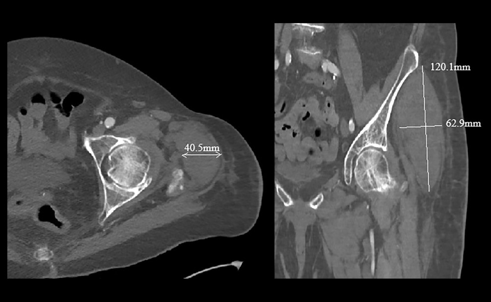 Figure 1. CTA of left lower extremity demonstrating intramuscular hematoma within the left gluteus minimus muscle belly measuring 4.1 × 6.3 × 12 cm in axial (left image) and sagittal (right image) views. CTA, computed tomography angiography.
 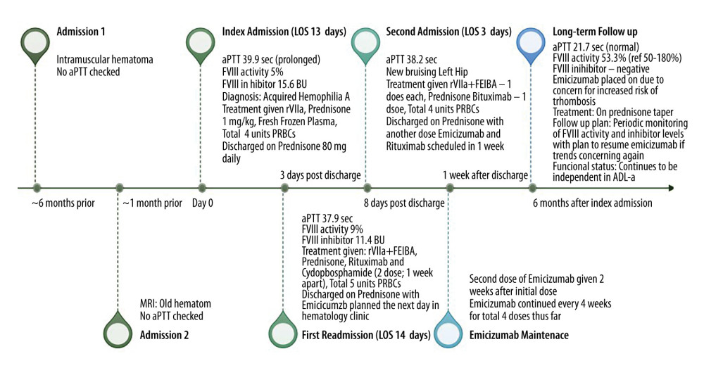 Figure 2. Clinical timeline showing the patient’s sequential hospitalizations, key diagnostic findings, treatment interventions, and clinical response. The timeline highlights 2 missed opportunities for coagulation testing prior to diagnosis of acquired hemophilia A. Factor VIII deficiency was confirmed via a high inhibitor titer and subsequent clinical improvement after initiation of appropriate therapy. aPTT, activated partial thromboplastin time; FVIII, factor VIII; BU, Bethesda units; rFVIIa, recombinant activated factor VII; FEIBA, factor 8 inhibitor bypassing activity (activated prothrombin complex concentrate); MRI, magnetic resonance imaging; PRBCs, packed red blood cells; ADLs, activities of daily living.
 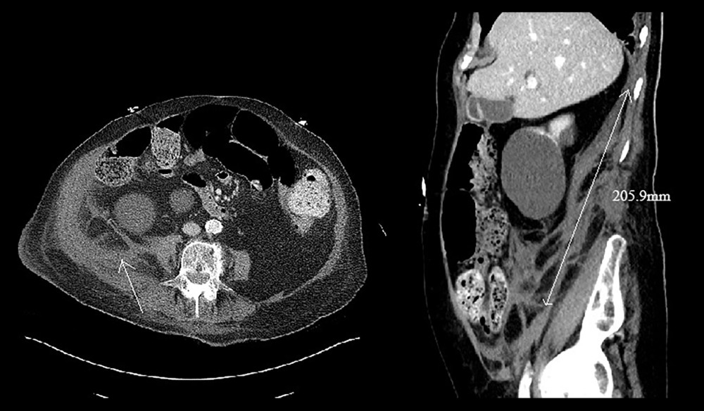 Figure 3. Abdominal and pelvic CT with contrast demonstrating right retroperitoneal hematoma extending from the posterior tip of the liver to the right lower pelvis. The hematoma measured 20.5 cm in the craniocaudal dimension, as is demonstrated on the sagittal plane in the right-side image. The pointer line on the left-side image demonstrates the same, in axial view.
 Tables
Table 1. Coagulation profile of the patient during index admission. It highlights normal PT, prolonged aPTT, reduced factor VIII activity, and increased factor VIII inhibitor levels. D-dimer elevation was felt to be secondary to large soft tissue hemorrhage.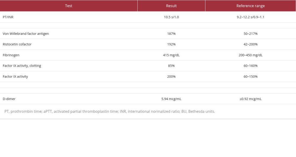 Table 2. Coagulation mixing study results. Immediate testing of a 1: 1 mix of patient and normal pooled plasma resulted in an activated partial thromboplastin time (aPTT) of 31 seconds, whereas testing of the same mix after a 1-hour incubation at 37 °C resulted in an aPTT of 43 seconds, which indicates likely presence of a factor VIII inhibitor prolonging aPTT by 12 seconds upon incubation.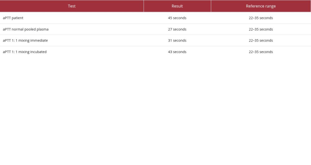
References
1. Huth-Kühne A, Baudo F, Collins PJ, International recommendations on the diagnosis and treatment of patients with acquired hemophilia: A Haematologica, 2009; 94(4); 566-75
2. Zanon E, Acquired hemophilia A: An update on the etiopathogenesis, diagnosis, and treatment: Diagnostics (Basel), 2023; 13(3); 420
3. Rasmussen KL, Philips M, Tripodi A, Goetze JP, Unexpected, isolated activated partial thromboplastin time prolongation: A practical mini-review: Eur J Haematol, 2020; 104; 519-25
4. Mazzucconi MG, Baldacci E, Ferretti A, Santoro C, Acquired haemophilia A: An intriguing disease: Mediterr J Hematol Infect Dis, 2020; 12(1); e2020045
5. Adcock DM, Favaloro EJ, Pearls and pitfalls in factor inhibitor assays: Int J Lab Hematol, 2015; 37(Suppl 1); 52-60
6. Kruse-Jarres R, Kempton CL, Baudo F, Acquired hemophilia A: Updated review of evidence and treatment guidance: Am J Hematol, 2017; 92(7); 695-705
7. Rinaldi I, Prasetyawaty F, Fazlines S, Diagnosis and management of acquired hemophilia A: case reports and a literature review: Case Rep Med, 2021; 2021; 5554664
8. Tiede A, Collins P, Knoebl P, International recommendations on the diagnosis and treatment of acquired hemophilia A: Haematologica, 2020; 105(7); 1791-801
9. Knoebl P, Thaler J, Jilma P, Emicizumab for the treatment of acquired hemophilia A: Blood, 2021; 137(3); 410-19
10. Tiede A, Hart C, Knöbl P, Emicizumab prophylaxis in patients with acquired haemophilia A (GTH-AHA-EMI): An open-label, single-arm, multicentre, phase 2 study: Lancet Haematol, 2023; 10(11); e913-e21
11. Pfrepper C, Klamroth R, Oldenburg J, Emicizumab for the treatment of acquired hemophilia A: Consensus recommendations from the GTH-AHA Working Group: Hamostaseologie, 2024; 44(6); 466-71
Figures
Figure 1. CTA of left lower extremity demonstrating intramuscular hematoma within the left gluteus minimus muscle belly measuring 4.1 × 6.3 × 12 cm in axial (left image) and sagittal (right image) views. CTA, computed tomography angiography.Figure 2. Clinical timeline showing the patient’s sequential hospitalizations, key diagnostic findings, treatment interventions, and clinical response. The timeline highlights 2 missed opportunities for coagulation testing prior to diagnosis of acquired hemophilia A. Factor VIII deficiency was confirmed via a high inhibitor titer and subsequent clinical improvement after initiation of appropriate therapy. aPTT, activated partial thromboplastin time; FVIII, factor VIII; BU, Bethesda units; rFVIIa, recombinant activated factor VII; FEIBA, factor 8 inhibitor bypassing activity (activated prothrombin complex concentrate); MRI, magnetic resonance imaging; PRBCs, packed red blood cells; ADLs, activities of daily living.Figure 3. Abdominal and pelvic CT with contrast demonstrating right retroperitoneal hematoma extending from the posterior tip of the liver to the right lower pelvis. The hematoma measured 20.5 cm in the craniocaudal dimension, as is demonstrated on the sagittal plane in the right-side image. The pointer line on the left-side image demonstrates the same, in axial view. Tables
Table 1. Coagulation profile of the patient during index admission. It highlights normal PT, prolonged aPTT, reduced factor VIII activity, and increased factor VIII inhibitor levels. D-dimer elevation was felt to be secondary to large soft tissue hemorrhage.Table 2. Coagulation mixing study results. Immediate testing of a 1: 1 mix of patient and normal pooled plasma resulted in an activated partial thromboplastin time (aPTT) of 31 seconds, whereas testing of the same mix after a 1-hour incubation at 37 °C resulted in an aPTT of 43 seconds, which indicates likely presence of a factor VIII inhibitor prolonging aPTT by 12 seconds upon incubation.Table 1. Coagulation profile of the patient during index admission. It highlights normal PT, prolonged aPTT, reduced factor VIII activity, and increased factor VIII inhibitor levels. D-dimer elevation was felt to be secondary to large soft tissue hemorrhage.Table 2. Coagulation mixing study results. Immediate testing of a 1: 1 mix of patient and normal pooled plasma resulted in an activated partial thromboplastin time (aPTT) of 31 seconds, whereas testing of the same mix after a 1-hour incubation at 37 °C resulted in an aPTT of 43 seconds, which indicates likely presence of a factor VIII inhibitor prolonging aPTT by 12 seconds upon incubation. In Press
Case report 
Am J Case Rep In Press; DOI: 10.12659/AJCR.953543
Case report 
Am J Case Rep In Press; DOI: 10.12659/AJCR.954216
Case report 
Am J Case Rep In Press; DOI: 10.12659/AJCR.954932
Case report
Am J Case Rep In Press; DOI: 10.12659/AJCR.954146
Most Viewed Current Articles
07 Dec 2021 : Case report
28,712,695
DOI :10.12659/AJCR.934347
Am J Case Rep 2021; 22:e934347
06 Dec 2021 : Case report  180,721
DOI :10.12659/AJCR.934406
Am J Case Rep 2021; 22:e934406
21 Jun 2024 : Case report
124,419
DOI :10.12659/AJCR.944371
Am J Case Rep 2024; 25:e944371
08 May 2026 : Case report
95,462
DOI :10.12659/AJCR.952158
Am J Case Rep 2026; 27:e952158