


19 September 2026: Articles 
Pulsed Radiofrequency of the Sacral Dorsal Root Ganglia as a Potential Rescue Therapy for Refractory Chronic Pelvic Pain: A Case Series
Unknown etiology, Challenging differential diagnosis, Unusual or unexpected effect of treatment, Clinical situation which can not be reproduced for ethical reasons
Paweł Gogol ABCDEF 1,2,3*, Małgorzata Gierczak B 4, Robert Szczepaniak E 1,2, Rafał Wiśniewski E 1,2, Michał Sobstyl F 5, Rafał Pasztaleniec E 6, Beniamin Oskar Grabarek CE 7
DOI: 10.12659/AJCR.954251
Am J Case Rep 2026; 27:e954251


Abstract
BACKGROUND: Chronic pelvic pain (CPP) is a prevalent, multifactorial condition with neuropathic, visceral, myofascial, and psychogenic components; a subset of patients remain refractory to multimodal pharmacotherapy and conventional interventional procedures. Pulsed radiofrequency (PRF) of the dorsal root ganglia (DRG) is a minimally invasive neuromodulatory technique that can attenuate pathological nociceptive transmission while preserving neural integrity. We describe 3 patients with severe, treatment-refractory CPP of predominantly neuropathic origin who underwent sacral DRG PRF as rescue therapy.
CASE REPORT: Case 1 was a 46-year-old man with a 7-year history of neuropathic penile pain following circumcision. Case 2 was a 54-year-old woman with an 11-year history of urethral burning pain and urgency after anal sphincterotomy. Case 3 was a 39-year-old woman with an 18-year history of CPP related to interstitial cystitis, vulvodynia, and pelvic floor myofascial dysfunction. All 3 had failed extensive pharmacological, nerve block, and neuromodulation therapies. Bilateral PRF of the S2-S4 DRG produced reductions in pain intensity (Numeric Rating Scale, NRS) of 55.6-62.5% at 3 months, partially sustained at 6 months (42.9-50.0%), with parallel improvements in neuropathic pain scores, quality of life, and reduced analgesic use, without procedure-related complications.
CONCLUSIONS: In these 3 refractory CPP cases, sacral DRG PRF was associated with clinically meaningful, although partially attenuating, symptomatic improvement. Given the small, uncontrolled sample, these findings represent preliminary clinical observations suggesting possible benefit in carefully selected patients and should be regarded as hypothesis-generating rather than evidence of established clinical effectiveness.
Keywords: chronic pain, Neuralgia, Pulsed Radiofrequency Treatment
Introduction
CPP is defined as pain perceived in structures related to the pelvis, lasting at least 3 to 6 months. It affects roughly 5% to 26% of women worldwide, and a smaller but still clinically important share of men [1]. The etiology is rarely simple: neuropathic, visceral, myofascial, and psychogenic components typically overlap, and the condition is frequently associated with irritable bowel syndrome, interstitial cystitis/bladder pain syndrome, endometriosis, pelvic floor dysfunction, prior pelvic surgery or trauma, and comorbid mood disorders [1,2]. Diagnosis rests mainly on history and physical examination, including assessment for cutaneous allodynia and pelvic floor muscle tenderness; imaging and laboratory testing help identify or rule out structural, infectious, or neoplastic causes but are often inconclusive [1]. Management is usually stepwise and multidisciplinary: patient education, pelvic floor physiotherapy, and pharmacological therapy (eg, nonsteroidal anti-inflammatory drugs [NSAIDs], gabapentinoids, tricyclic antidepressants, and serotonin–norepinephrine reuptake inhibitors [SNRIs]) come first, with nerve blocks, botulinum toxin injection, and neuromodulation reserved for patients who remain refractory [1]. In some patients, pain becomes consolidated at both peripheral and central levels through peripheral sensitization, central sensitization, and maladaptive neuroplasticity [2].
This consolidation is what tends to blunt the response to pharmacological therapy and to standard interventional procedures, including peripheral nerve blocks, autonomic plexus blocks, and various neuromodulation techniques [3]. The S2–S4 dorsal root ganglia (DRG) relay and modulate sensory input from the perineum, pelvic organs, and external genitalia, which makes them a logical neuromodulatory target. Pulsed radiofrequency (PRF) applied to the DRG is one such approach: it attenuates pathological nociceptive signaling while sparing the surrounding neural tissue [4,5]. Because PRF delivers short bursts of high-frequency current without producing neurodestructive thermal lesions, it carries a favorable safety profile and a low risk of permanent neural injury [6]. In patients with severe, refractory CPP, sacral DRG PRF has therefore been proposed as an option after conventional pharmacological and interventional strategies have failed [7]. Clinical experience with it remains limited, but modulation of sensory transmission at the S2–S4 level appears particularly relevant for patients with a substantial neuropathic pain component [8]. Small case series of PRF applied to the pudendal nerve or sacral nerve roots for refractory pelvic pain have reported favorable short- and long-term symptomatic improvement, which supports further evaluation of DRG-targeted approaches in this setting [9]. What is still missing is a clearer picture of the durability of response and the patient characteristics that predict it.
The objective of the present case series was to describe clinical outcomes following bilateral pulsed radiofrequency treatment of the S2–S4 dorsal root ganglia in a highly selected group of patients with refractory CPP characterized by a predominant neuropathic component. Specifically, we sought to evaluate whether treatment was associated with short-term changes in pain intensity, neuropathic symptom burden, patient-reported functional status, and quality of life. Given the descriptive nature of the study and limited sample size, the findings are intended to be exploratory and hypothesis-generating rather than confirmatory regarding treatment efficacy.
Case Reports
CASE 1:
Case 1 was a 46-year-old White man with a 7-year history of severe CPP that developed following circumcision. He had post-circumcision neuropathic penile pain with a prominent burning, allodynic component, without an identifiable structural or infectious cause. He reported tearing pain localized to the glans penis with a prominent neuropathic component characterized by burning sensations and hyperesthesia. During exacerbations, pain occasionally radiated to the testes. Symptoms were consistently aggravated by exposure to cold, sexual activity, and ejaculation, frequently precluding satisfactory sexual function and markedly impairing quality of life. The patient denied tobacco smoking, alcohol abuse, and illicit substance use, and no major comorbidities were identified.
Comprehensive urological assessment and imaging studies demonstrated no structural abnormalities except for small varicoceles considered clinically insignificant as a pain source. Electrophysiological evaluation of the pudendal nerves revealed no pathological abnormalities within the motor component. Previous interventions included peripheral nerve blocks, plexus blocks, and pulsed radiofrequency procedures targeting pelvic and perineal pain pathways, none of which resulted in meaningful or sustained clinical improvement. Uroproctological rehabilitation aggravated symptoms without therapeutic benefit.
Prior pharmacological treatment included nonsteroidal anti-inflammatory drugs (NSAIDs), including ibuprofen 400 mg orally twice daily, diclofenac 50 mg orally twice daily, and etoricoxib 90 mg orally once daily, all ineffective in reducing pain intensity. Opioid treatment with tramadol/acetaminophen 37.5 mg/325 mg orally and buprenorphine 0.2 mg orally 3 times daily produced minimal analgesic benefit. Gabapentinoids, including pregabalin 225 mg orally twice daily and gabapentin 600 mg orally 3 times daily, worsened glans hypersensitivity and neuropathic symptoms. Serotonin–norepinephrine reuptake inhibitors (SNRIs), including venlafaxine 150 mg orally once daily and duloxetine 90 mg orally once daily, as well as amitriptyline 25 mg orally twice daily, were ineffective and associated with excessive somnolence and reduced libido. Given the refractory nature of neuropathic penile pain, the patient qualified for bilateral PRF treatment targeting the S2–S4 dorsal root ganglia (Figure 1).
At baseline, the patient’s NRS score was 8/10, with elevated DN4 (7/10) and painDETECT (26/38) scores and an SF-36 score of 38, consistent with severe pain and marked neuropathic and quality-of-life impairment. Following bilateral PRF, NRS fell to 3 at 3 months and 4 at 6 months, with parallel improvement in DN4 (to 3, then 4) and painDETECT (to 11, then 14) scores and a rise in the SF-36 score to 65 and 58, respectively (Table 1). The patient rated the outcome “much improved” on the PGIC at 3 months and “moderately improved” at 6 months. Analgesic and adjuvant medication use fell by approximately 60% at 3 months and 40% at 6 months (Table 2), and no procedure-related complications occurred.
CASE 2:
Case 2 was a 54-year-old White woman who had experienced CPP characterized by persistent urinary urgency and burning sensations localized to the urethral region since 2011. She had neuropathic urethral burning pain and urgency arising after anal sphincterotomy, with clinical and neurophysiological features consistent with pudendal nerve irritation. Symptoms developed following proctological treatment for anal fissure consisting of lateral sphincterotomy. Postoperative proctological symptoms resolved completely. The patient had a history of chronic constipation and denied tobacco smoking, alcohol abuse, and substance use. Persistent pelvic symptoms significantly impaired daily functioning, sexual health, and quality of life, leading to depression and long-term psychiatric treatment.
Comprehensive imaging and neurophysiological investigations revealed no focal lesions or evidence of pudendal nerve injury. Findings were limited to signs of irritation involving both sensory and motor neural components. Previous unsuccessful interventions included superior and inferior hypogastric plexus blocks, test sacral neuromodulation at the S3 level complicated by technical difficulties during DRG root stimulation, intravesical botulinum toxin administration, multiple peripheral nerve blocks, and pulsed radiofrequency procedures targeting the pudendal nerves, ganglion impar, piriformis muscles, and the ischial tuberosity region. Uroproctogynecological physiotherapy provided no clinical improvement.
Pharmacological treatment included pregabalin 150 mg orally 3 times daily, gabapentin 800 mg orally 3 times daily, duloxetine 90 mg orally once daily, and amitriptyline 25 mg orally in the morning and 50 mg orally in the evening, all without clinically meaningful symptom reduction. Partial improvement of nocturnal urinary urgency was achieved with tolperisone. Due to refractory CPP with neuropathic characteristics, the patient qualified for bilateral PRF treatment of the S2–S4 dorsal root ganglia (Figure 2).
At baseline, the patient’s NRS score was 9/10, with a DN4 score of 8/10, a painDETECT score of 30/38, and an SF-36 score of 42. After bilateral PRF, NRS decreased to 4 at 3 months and 5 at 6 months, DN4 to 4 and 5, and painDETECT to 15 and 18, while the SF-36 score rose to 64 and 56, respectively (Table 1). PGIC was “moderately improved” at 3 months and “slightly improved” at 6 months. Pharmacological burden decreased by roughly 60% at 3 months and 50% at 6 months (Table 2), without procedure-related adverse events.
CASE 3:
Case 3 was a 39-year-old White woman with an 18-year history of CPP associated with chronic interstitial cystitis and vulvodynia. She had mixed neuropathic–myofascial chronic pelvic pain associated with interstitial cystitis, vulvodynia, and increased pelvic floor muscle tone, exacerbated by ganglion impar neurolysis.
Initial symptoms were associated with recurrent infections treated with multiple courses of antibiotics. During the preceding 3 years, substantial clinical deterioration occurred following prolonged antibiotic therapy involving multiple antibiotic classes, including vancomycin prescribed by treating urologists. The patient denied tobacco smoking, alcohol abuse, and illicit substance use.
Following neurolysis of the ganglion impar, the patient experienced marked exacerbation of pain accompanied by urinary incontinence and bladder spasms. Pudendal nerve blocks combined with pulsed radiofrequency procedures did not provide clinical improvement. Botulinum toxin injections administered into pelvic floor muscles also failed to reduce symptoms. A diagnosis of CPP of mixed etiology, involving neuropathic and myofascial components with increased pelvic floor muscle tone, was established. Uroproctogynecological physiotherapy worsened symptoms rather than alleviating them. Due to severe persistent symptoms and lack of response to previous interventions, the patient required long-term psychiatric care.
Pharmacological management included pregabalin 300 mg orally twice daily, which improved sleep quality and urinary comfort without adequate analgesic benefit. Additional medications included amitriptyline 10 mg orally in the morning and 25 mg orally in the evening, venlafaxine 225 mg orally once daily, duloxetine 90 mg orally once daily, and diclofenac 50 mg orally twice daily, none of which significantly reduced pain intensity. Given incapacitating refractory CPP, the patient qualified for bilateral PRF treatment of the S2–S4 dorsal root ganglia (Figure 3).
At baseline, the patient’s NRS score was 7/10, with a DN4 score of 6/10, a painDETECT score of 24/38, and an SF-36 score of 35. Bilateral PRF reduced NRS to 3 at 3 months and 4 at 6 months, DN4 to 2 and 3, and painDETECT to 9 and 12, with the SF-36 score increasing to 63 and 55, respectively (Table 1). The patient reported being “much improved” on the PGIC at 3 months and “moderately improved” at 6 months. Medication use fell by about 60% at 3 months and 50% at 6 months (Table 2), with no procedure-related complications.
Discussion
This case series suggests that sacral DRG PRF are a further rescue option worth considering in patients with severe, treatment-refractory CPP once standard pharmacological and interventional therapies have failed. In these 3 patients, treatment was followed by a clinically meaningful, although partially attenuating, reduction in pain intensity and improvement in functional status. Given the design of this series, these observations should be regarded not as evidence of treatment efficacy but as a preliminary finding warranting further controlled investigation. CPP is a complex and multifactorial clinical condition characterized by persistent pain lasting longer than 6 months and involving both peripheral and central sensitization mechanisms. These syndromes frequently exhibit a mixed nociceptive–neuropathic phenotype and are associated with substantial impairment in functional status, psychological well-being, and quality of life. Importantly, CPP often demonstrates limited responsiveness to conventional pharmacological therapies, including gabapentinoids, serotonin–norepinephrine reuptake inhibitors (SNRIs), tricyclic antidepressants, and opioids, as well as to interventional approaches such as nerve blocks and neuromodulation procedures [10]. The present case series describes clinical outcomes following PRF treatment targeting the DRG at the S2–S4 levels in a small group of patients with refractory CPP. Improvements were observed across several patient-reported outcome measures, including pain intensity, neuropathic symptom burden, and quality of life. However, given the uncontrolled design and limited sample size, these findings should be considered exploratory and hypothesis-generating rather than confirmatory evidence of treatment efficacy. Nevertheless, the observed clinical responses support further investigation of sacral DRG-targeted PRF in carefully selected patients with refractory CPP [10].
The contribution of the present case series should be interpreted within the context of its exploratory design. Rather than providing confirmatory evidence of treatment efficacy, this report offers an incremental extension of the existing literature on DRG-targeted neuromodulation by describing clinical outcomes following bilateral S2–S4 PRF treatment in a highly selected subgroup of patients with refractory CPP. All included patients had previously failed multiple conservative and interventional strategies, including pharmacotherapy, peripheral nerve blocks, autonomic plexus interventions, neuromodulation attempts, and specialized physiotherapy. Consequently, the principal value of this study lies in documenting consistent clinical improvement in a difficult-to-treat population for whom therapeutic options were limited. These observations support the feasibility and potential clinical relevance of sacral DRG-targeted PRF in this setting and provide a rationale for future prospective studies designed to more rigorously evaluate its efficacy, durability, and optimal clinical application.
All patients included in this series presented with severe, long-standing CPP refractory to multimodal pharmacotherapy and prior interventional strategies. Notably, these patients had undergone advanced treatments, including autonomic plexus blocks, pudendal nerve blocks, and sacral neuromodulation, without achieving sustained clinical benefit. In this context, PRF treatment was associated with reductions in pain intensity, attenuation of neuropathic pain features, improvement in patient-reported functional status, and a reduction in pharmacological treatment burden during follow-up. These observations are of interest because PRF provides neuromodulation without requiring permanent implantation of hardware [10].
The observed clinical improvements are consistent with the proposed neuromodulatory mechanism of PRF, which differs fundamentally from destructive ablative techniques. PRF delivers short bursts of high-frequency current while maintaining tissue temperatures below neurodestructive thresholds (≤ 42 °C), thereby preserving neural integrity while inducing functional modulation of nociceptive transmission [6]. Experimental and translational studies have demonstrated that PRF influences multiple molecular and cellular targets involved in pain signaling. These include modulation of voltage-gated ion channels such as Na+/K+-ATPase and hyperpolarization-activated cyclic nucleotide-gated (HCN) channels, regulation of purinergic receptors such as P2X3, and alterations in synaptic receptor function involving AMPA and GABA-B receptors [11–13]. These molecular effects have been associated with reduced neuronal hyperexcitability and suppression of ectopic discharges originating within the DRG, which are recognized contributors to neuropathic pain.
Furthermore, PRF appears to exert anti-inflammatory and neuroimmune regulatory effects. Preclinical studies have demonstrated that PRF reduces microglial activation and modulates inflammatory cytokine signaling within the peripheral and central nervous system, thereby attenuating neuroinflammation-associated sensitization [11–13]. At the structural level, PRF has been shown to promote normalization of neuronal ultrastructure and mitochondrial integrity in DRG neurons, suggesting a restorative rather than destructive effect. Additionally, emerging molecular evidence indicates that PRF can influence gene expression patterns within sensory neurons, potentially contributing to longer-term modulation of nociceptive signaling pathways [11–13].
Beyond peripheral neuromodulation, PRF may also influence central pain-processing networks. Functional neuroimaging studies suggest that modulation of afferent input at the level of the DRG can alter central nervous system activity within key brain regions involved in pain perception, including the thalamus, insular cortex, anterior cingulate cortex, and somatosensory cortex [2]. By reducing aberrant afferent signaling and central sensitization, PRF may facilitate normalization of maladaptive neural network activity associated with chronic pain states. Although such mechanisms provide biologically plausible explanations for the clinical observations reported here, the present case series was not designed to evaluate these mechanisms directly.
Implantable neuromodulation and DRG-targeted PRF represent distinct interventional approaches with different procedural characteristics and clinical objectives. Unlike implantable neuromodulation systems, PRF does not require permanent hardware placement. However, the present study was not designed to compare these modalities, and no conclusions regarding relative efficacy, safety, durability, cost-effectiveness, or clinical superiority can be drawn from the current observations. Future comparative studies will be required to determine the relative role of sacral DRG PRF within the broader spectrum of neuromodulatory interventions for CPP [14,15].
The findings observed in this series are broadly consistent with reports evaluating DRG-targeted PRF in other chronic neuropathic pain conditions. Clinical studies evaluating PRF for lumbosacral radicular pain have reported reductions in pain intensity and disability, with clinical success rates of approximately 50% to 57% at 6 months following treatment [16]. Similarly, retrospective analyses have reported improved outcomes when PRF is combined with adjunctive interventional strategies [17]. A similar pattern turns up in a case series of PRF applied to the pudendal nerve for pudendal neuralgia: 79% of patients rated their condition “(very) much better” at 3 months, rising to 89% at long-term follow-up (median 4 years), with no serious adverse events [9]. That series targeted the pudendal nerve rather than the sacral DRG and used a different outcome metric (Patient Global Impression of Improvement), so the comparison is only approximate, but the overall result of sustained benefit that fades somewhat over time without procedural complications matches what we observed here. Although studies specifically addressing CPP remain limited, observations from broader neuropathic pain populations provide a rationale for further evaluation of DRG-targeted PRF in this clinical setting [10].
The present findings also highlight the potential importance of the DRG in the pathophysiology of CPP. The DRG serves as a key anatomical and functional hub integrating peripheral sensory input and transmitting nociceptive signals to the central nervous system. Pathological changes within the DRG, including neuronal hyperexcitability, ion channel dysregulation, and neuroinflammatory processes, have been implicated in the development and maintenance of neuropathic pain states. Consequently, modulation of DRG activity is a biologically plausible therapeutic strategy in selected patients with neuropathic features of CPP.
Despite the encouraging observations reported in this case series, several limitations must be acknowledged. The small sample size and uncontrolled design limit the ability to draw definitive conclusions regarding treatment efficacy and generalizability. Additionally, the absence of a control group precludes exclusion of placebo effects or spontaneous symptom fluctuations. Nevertheless, the consistency of improvement across multiple outcome measures, including pain intensity, neuropathic pain scores, quality of life, and pharmacological treatment burden, supports further investigation of sacral DRG-targeted PRF in this patient population.
Future research should focus on prospective, controlled clinical trials with larger patient cohorts to evaluate the safety and clinical utility of PRF targeting the S2–S4 DRG in CPP. Further studies are also needed to identify optimal patient selection criteria, procedural parameters, and predictors of treatment response. Integration of neurophysiological, molecular, and neuroimaging biomarkers may help to better characterize the mechanisms underlying PRF-mediated neuromodulation and facilitate personalized treatment approaches.
Conclusions
Sacral DRG PRF was followed by a clinically meaningful, if partially fading, improvement in pain intensity, neuropathic symptom burden, functional status, and analgesic use in these 3 patients with severe, treatment-refractory chronic pelvic pain. We present this as a preliminary observation suggesting possible benefit in carefully selected patients, not as evidence that the treatment is established or broadly effective—the sample is small, the design uncontrolled, and the follow-up short.
Larger, prospective, controlled studies are needed to confirm these observations, define which patients are most likely to benefit, refine the procedural parameters, and clarify how durable the effect is and how sacral DRG PRF compares with other neuromodulatory options for refractory chronic pelvic pain.
Figures
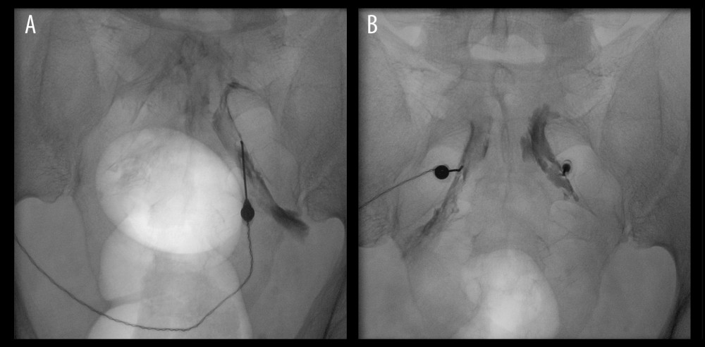 Figure 1. Fluoroscopic guidance during bilateral pulsed radiofrequency (PRF) treatment of the sacral (S2–S4) dorsal root ganglia (DRG) in Case 1. (A) Anteroposterior fluoroscopic view showing unilateral placement of a curved-tip radiofrequency needle within the S2 posterior sacral foramen; a Foley catheter balloon is visible within the bladder and was used as an anatomical landmark. (B) Anteroposterior fluoroscopic view after bilateral needle placement within the S2–S4 posterior sacral foramina, prior to sensory and motor stimulation testing and delivery of PRF current. PRF, pulsed radiofrequency; DRG, dorsal root ganglion; S2–S4, second to fourth sacral spinal levels.
 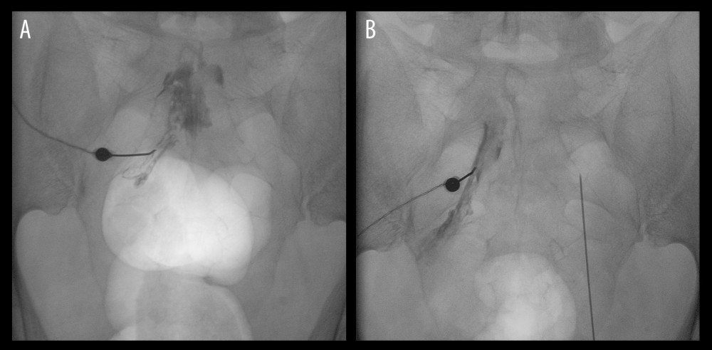 Figure 2. Fluoroscopic guidance during bilateral pulsed radiofrequency (PRF) treatment of the sacral (S2–S4) dorsal root ganglia (DRG) in Case 2. (A) Anteroposterior fluoroscopic view showing radiofrequency needle placement with contrast medium outlining the posterior sacral foramen and confirming epineural spread around the target nerve root; a Foley catheter balloon is visible within the bladder and was used as an anatomical landmark. (B) Anteroposterior fluoroscopic view after bilateral needle placement within the sacral posterior foramina, prior to sensory and motor stimulation testing and delivery of PRF current. PRF, pulsed radiofrequency; DRG, dorsal root ganglion; S2–S4, second to fourth sacral spinal levels.
 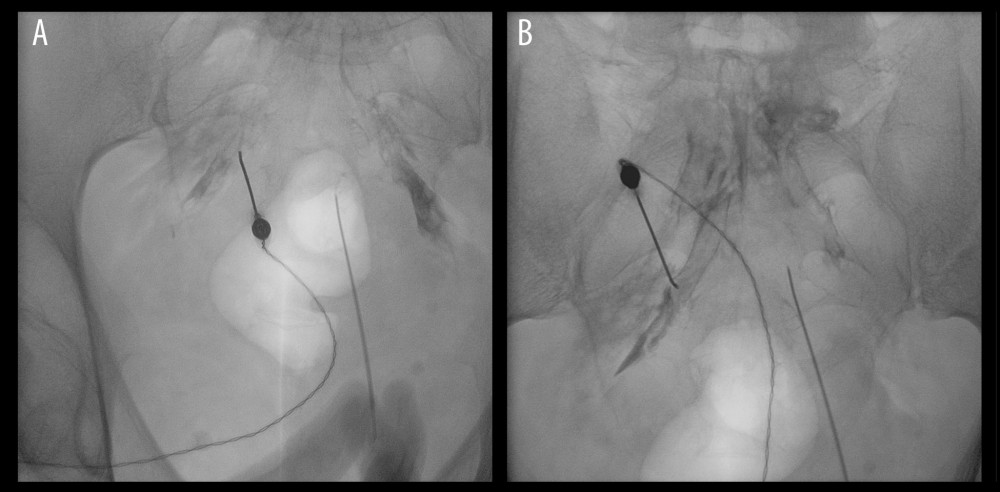 Figure 3. Fluoroscopic guidance during bilateral pulsed radiofrequency (PRF) treatment of the sacral (S2–S4) dorsal root ganglia (DRG) in Case 3. (A) Anteroposterior fluoroscopic view showing unilateral placement of a curved-tip radiofrequency needle within the posterior sacral foramen; a Foley catheter balloon is visible within the bladder and was used as an anatomical landmark. (B) Anteroposterior fluoroscopic view after bilateral needle placement within the sacral posterior foramina, prior to sensory and motor stimulation testing and delivery of PRF current. PRF, pulsed radiofrequency; DRG, dorsal root ganglion; S2–S4, second to fourth sacral spinal levels.
 References
1. Dydyk AM, Singh C, Gupta N, Chronic pelvic pain. [Updated 2025 May 2]: StatPearls [Internet], 2026, Treasure Island (FL), StatPearls Publishing Available from: https://www.ncbi.nlm.nih.gov/books/NBK554585/
2. Ploteau S, Labat JJ, Riant T, New concepts on functional chronic pelvic and perineal pain: Pathophysiology and multidisciplinary management: Discov Med, 2015; 19(104); 185-92
3. Gish B, Langford B, Sobey C, Neuromodulation for the management of chronic pelvic pain syndromes: A systematic review: Pain Pract, 2024; 24(2); 321-40
4. Ghorayeb JH, Chitneni A, Rupp A, Dorsal root ganglion stimulation for the treatment of chronic pelvic pain: A systematic review: Pain Pract, 2023; 23(7); 838-46
5. Hao D, Yurter A, Chu R, Neuromodulation for management of chronic pelvic pain: A comprehensive review: Pain Ther, 2022; 11(4); 1137-77
6. De la Cruz J, Benzecry Almeida D, Silva Marques M, Elucidating the mechanisms of pulsed radiofrequency for pain treatment: Cureus, 2023; 15(9); e44922
7. Hong LW, Chen KT, A real-world evidence of a consecutive treatment of 42 spine-related pain using dorsal root ganglion-pulsed radiofrequency (DRG-PRF): Clin Neurol Neurosurg, 2020; 197; 106186
8. Sun J, Gu J, Ding Y, Dorsal root ganglion-targeted analgesic delivery for effective relief of neuropathic pain: Mater Today Bio, 2025; 33; 102025
9. Krijnen EA, Schweitzer KJ, van Wijck AJM, Withagen MIJ, Pulsed radiofrequency of pudendal nerve for treatment in patients with pudendal neuralgia. A case series with long-term follow-up: Pain Pract, 2021; 21(6); 703-7
10. Vuka I, Marciuš T, Došenović S, Efficacy and safety of pulsed radiofrequency as a method of dorsal root ganglia stimulation in patients with neuropathic pain: A systematic review: Pain Med, 2020; 21(12); 3320-43
11. Sam J, Catapano M, Sahni S, Ma F, Pulsed radiofrequency in interventional pain management: Cellular and molecular mechanisms of action – An update and review: Pain Physician, 2021; 24(8); 525-32
12. Liu Y, Feng Y, Zhang T, Pulsed radiofrequency treatment enhances dorsal root ganglion expression of hyperpolarization-activated cyclic nucleotide-gated channels in a rat model of neuropathic pain: J Mol Neurosci, 2015; 57(1); 97-105
13. Fu M, Meng L, Ren H, Luo F, Pulsed radiofrequency inhibits expression of P2X3 receptors and alleviates neuropathic pain induced by chronic constriction injury in rats: Chin Med J (Engl), 2019; 132(14); 1706-12
14. Lee C, Pizarro-Berdichevsky J, Clifton MM, Vasavada SP, Sacral neuromodulation implant infection: Risk factors and prevention: Curr Urol Rep, 2017; 18(2); 16
15. Shih C, Miller JL, Fialkow M, Reoperation after sacral neuromodulation therapy: A single-institution experience: Female Pelvic Med Reconstr Surg, 2013; 19(3); 175-78
16. Van Boxem K, de Meij N, Kessels A, Pulsed radiofrequency for chronic intractable lumbosacral radicular pain: A six-month cohort study: Pain Med, 2015; 16(6); 1155-62
17. Gu R, Huang Y, Li Y, Dorsal root ganglion pulsed radiofrequency with scar radiofrequency ablation combined for chronic postoperative abdominal pain: A retrospective study: J Pain Res, 2025; 18; 3933-42
Figures
Figure 1. Fluoroscopic guidance during bilateral pulsed radiofrequency (PRF) treatment of the sacral (S2–S4) dorsal root ganglia (DRG) in Case 1. (A) Anteroposterior fluoroscopic view showing unilateral placement of a curved-tip radiofrequency needle within the S2 posterior sacral foramen; a Foley catheter balloon is visible within the bladder and was used as an anatomical landmark. (B) Anteroposterior fluoroscopic view after bilateral needle placement within the S2–S4 posterior sacral foramina, prior to sensory and motor stimulation testing and delivery of PRF current. PRF, pulsed radiofrequency; DRG, dorsal root ganglion; S2–S4, second to fourth sacral spinal levels.Figure 2. Fluoroscopic guidance during bilateral pulsed radiofrequency (PRF) treatment of the sacral (S2–S4) dorsal root ganglia (DRG) in Case 2. (A) Anteroposterior fluoroscopic view showing radiofrequency needle placement with contrast medium outlining the posterior sacral foramen and confirming epineural spread around the target nerve root; a Foley catheter balloon is visible within the bladder and was used as an anatomical landmark. (B) Anteroposterior fluoroscopic view after bilateral needle placement within the sacral posterior foramina, prior to sensory and motor stimulation testing and delivery of PRF current. PRF, pulsed radiofrequency; DRG, dorsal root ganglion; S2–S4, second to fourth sacral spinal levels.Figure 3. Fluoroscopic guidance during bilateral pulsed radiofrequency (PRF) treatment of the sacral (S2–S4) dorsal root ganglia (DRG) in Case 3. (A) Anteroposterior fluoroscopic view showing unilateral placement of a curved-tip radiofrequency needle within the posterior sacral foramen; a Foley catheter balloon is visible within the bladder and was used as an anatomical landmark. (B) Anteroposterior fluoroscopic view after bilateral needle placement within the sacral posterior foramina, prior to sensory and motor stimulation testing and delivery of PRF current. PRF, pulsed radiofrequency; DRG, dorsal root ganglion; S2–S4, second to fourth sacral spinal levels. Tables
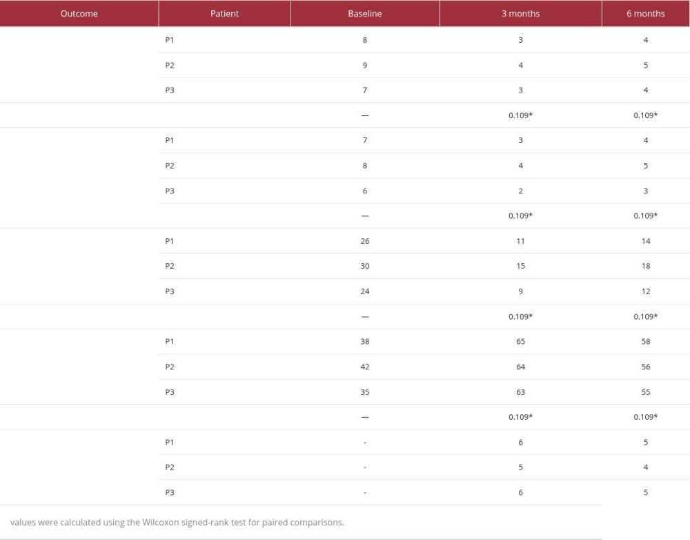 Table 1. Clinical outcomes following pulsed radiofrequency of the S2–S4 dorsal root ganglia.
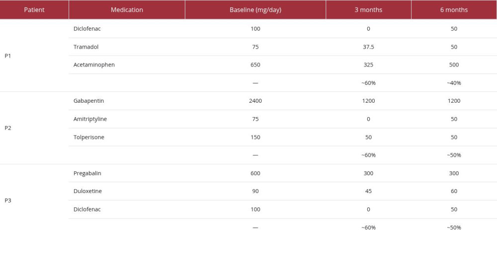 Table 2. Pharmacological treatment before and after PRF of the S2–S4 dorsal root ganglia.Table 1. Clinical outcomes following pulsed radiofrequency of the S2–S4 dorsal root ganglia.Table 2. Pharmacological treatment before and after PRF of the S2–S4 dorsal root ganglia.
 In Press
Case report 
Am J Case Rep In Press; DOI: 10.12659/AJCR.953317
Case report 
Am J Case Rep In Press; DOI: 10.12659/AJCR.954643
Case report
Am J Case Rep In Press; DOI: 10.12659/AJCR.954074
Case report 
Am J Case Rep In Press; DOI: 10.12659/AJCR.953592
Most Viewed Current Articles
07 Dec 2021 : Case report
28,712,429
DOI :10.12659/AJCR.934347
Am J Case Rep 2021; 22:e934347
06 Dec 2021 : Case report  180,530
DOI :10.12659/AJCR.934406
Am J Case Rep 2021; 22:e934406
21 Jun 2024 : Case report
124,276
DOI :10.12659/AJCR.944371
Am J Case Rep 2024; 25:e944371
08 May 2026 : Case report
95,045
DOI :10.12659/AJCR.952158
Am J Case Rep 2026; 27:e952158