


10 August 2026: Articles 
Massive Inferior Vena Cava Thrombosis and Pulmonary Embolism Secondary to Hepatic Hydatid Cyst: A Fatal Case
Unusual clinical course, Challenging differential diagnosis, Management of emergency care
Abdulrahman Alozaymi ABEF 1*, Roaa Amer BEF 1, Abdulaziz Fahad Alsayari BEF 1
DOI: 10.12659/AJCR.952041
Am J Case Rep 2026; 27:e952041


Abstract
BACKGROUND: Hydatid disease, caused by Echinococcus granulosus, is an endemic parasitic infection that predominantly affects the liver. Although slow-growing and asymptomatic, large hepatic cysts lead to catastrophic vascular complications, including inferior vena cava (IVC) thrombosis and pulmonary embolism (PE). Although rare, life-threatening events require prompt recognition and multidisciplinary management to optimize outcomes.
CASE REPORT: A 68-year-old woman with paroxysmal atrial fibrillation presented with right upper-quadrant pain, fever, and poor oral intake for several weeks. Examination revealed abdominal distension, hepatomegaly, and mottled, cold lower limbs. Laboratory findings showed severe metabolic acidosis and leukocytosis, indicating significant systemic involvement and possible sepsis. Computed tomography (CT) angiography demonstrated a pulmonary embolism, a giant (29×18 cm) right hepatic cyst exerting a mass effect, and extensive thrombosis extending from the infrarenal IVC to both external iliac veins. Echocardiography showed extrinsic cardiac compression without signs of tamponade but with functional compromise related to mass effect. Management included anticoagulation, albendazole, broad-spectrum antibiotics, vasopressors, and continuous renal replacement therapy. Ultrasound-guided percutaneous drainage of the cyst yielded 4 L of purulent fluid, with transient improvement in limb perfusion suggesting partial decompression of venous outflow obstruction. An IVC filter was inserted, but despite intensive therapy, the patient developed refractory shock and died due to progressive hemodynamic collapse and multiorgan failure.
CONCLUSIONS: Massive hepatic hydatid cysts can precipitate IVC thrombosis and PE through mechanical compression or infection-related inflammation. Early diagnosis, multidisciplinary coordination, and timely surgical or percutaneous decompression are vital to prevent death.
Keywords: Echinococcosis, Pulmonary Embolism, Vena Cava, Inferior
Introduction
Hydatid disease, caused by
The life cycle of
The natural clinical history of hepatic cystic echinococcosis is characterized by an indolent course. The parasite develops into a fluid-filled cyst that grows at a rate of approximately 1 cm per year [6]. This slow growth often allows the host to remain asymptomatic for a long time, until the cyst reaches a massive size or a complication occurs [7]. Cysts are considered “giant” when they exceed 10 cm in diameter; these lesions are significantly more likely to cause mass effect-related complications in adjacent organs, including the gallbladder, stomach, and major vascular structures like the portal vein and the IVC [8].
The most common genotypes among canids are
The intermediate host in Saudi Arabia varies among hosts according to site, season, animal age, and sex; the prevalence of infection is 32.9% in camels, 8.3% in cattle, 12.6% in sheep, and 6.56% in goats. It was reported that sheep and goats are the most important intermediate hosts of
Hepatic hydatid cysts result from infection with
Case Report
A 68-year-old woman with a history of paroxysmal atrial fibrillation presented to the emergency department with 3 months of right-upper-quadrant abdominal pain and recent worsening fever. She denied respiratory, urinary, or neurological symptoms. While in the waiting area, she suddenly collapsed, gasping for air. Her oxygen saturation dropped to 75% despite high-flow oxygen, and her Glasgow Coma Scale (GCS) score was 8/15. She was transferred to the resuscitation unit and intubated.
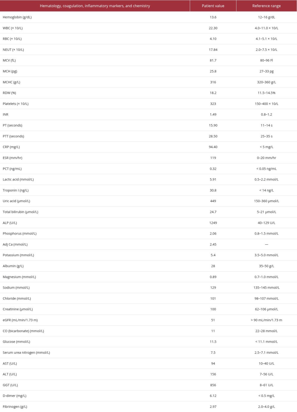
On examination, the abdomen was distended with palpable hepatomegaly. Air entry was decreased bilaterally. Both lower limbs were cold; the right leg was swollen with purple mottling. Venous blood gas analysis revealed severe metabolic acidosis. Laboratory tests are shown in Tables 1 and 2.
Contrast-enhanced computed tomography (CT) pulmonary angiography demonstrated segmental and subsegmental PEs, a giant (29 × 18 cm) right-lobe hepatic cyst, and mass effect on the right hemithorax with leftward displacement (Figure 1). Abdominal CT revealed extensive thrombosis extending from the infrarenal IVC to both external iliac veins, more pronounced on the right- prothrombotic state (Figure 2). Echocardiography showed a small pericardial effusion and extrinsic right-atrial compression without tamponade or pulmonary hypertension. The diagnosis of
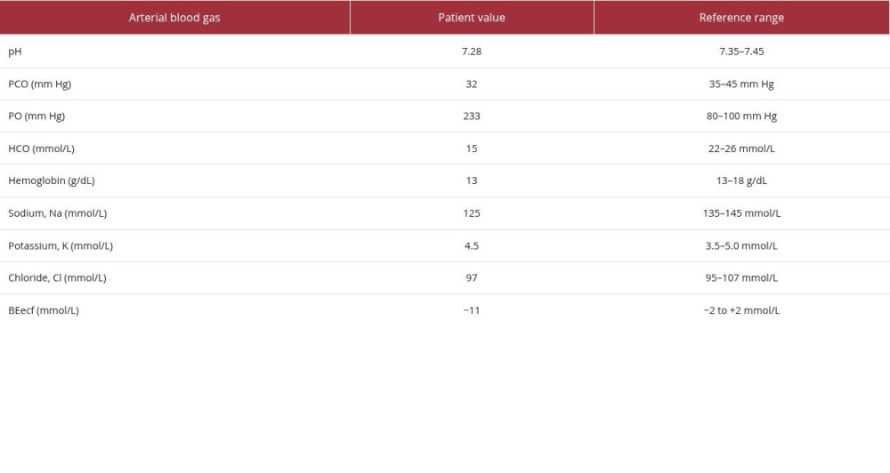
The pulmonary embolism response team initiated intravenous heparin, albendazole, and broad-spectrum antibiotics. Vascular surgery advised conservative management as Doppler studies demonstrated triphasic arterial flow without ischemia. The patient was admitted to the intensive care unit with worsening acidosis (arterial blood gas results are shown in Table 3). She received vasopressors, sedation, piperacillin–tazobactam, vancomycin, meropenem, albendazole, and continuous renal replacement therapy for acute kidney injury.
Given persistent shock and concern for the infected cyst, bedside decompression was performed. Under ultrasound guidance, a 10-Fr catheter was inserted, draining 4 L of purulent material that cultured
Discussion
This case highlights a rare but devastating complication of hepatic hydatid disease—IVC thrombosis with PE. The formation of a thrombus in the IVC is governed by the principles of Virchow’s triad: stasis, endothelial injury, and hypercoagulability [15]. In this case, all 3 elements of Virchow’s triad contributed to the development of IVC thrombosis [16]. First, stasis was present due to mechanical compression from a large cyst located on the retrohepatic segment of the IVC. Second, endothelial injury resulted from localized inflammation caused by an infected CE3b cyst. Third, hypercoagulability was driven by systemic sepsis, paroxysmal atrial fibrillation, and metabolic acidosis. These factors collectively created a profound prothrombotic state, culminating in the patient’s presentation.
Current recommendations suggest that in the presence of extensive IVC thrombosis, the filter should be placed before any invasive manipulation of the cyst [15,17]. Decompressing a giant cyst relieves the tamponade effect on the IVC. While this is necessary for restoring venous return, sudden restoration of flow through a previously compressed vessel can mobilize unstable thrombotic material or fragments of the cyst wall, leading to catastrophic pulmonary embolism [18].
The pathophysiology likely involves extrinsic compression by the enlarging cyst, compounded by inflammatory changes and infection. Similar cases have been reported, but the combination of massive cyst size (29 × 18 cm), septic thrombosis, and embolic phenomena underscores the clinical severity [14]. Mahajan et al described acute IVC thrombosis as an unusual manifestation of hydatid cysts [19], while Ismail et al reported direct cyst–IVC fistulization [12]. Our patient’s presentation was consistent with extrinsic compression rather than fistulization. Kantorová et al emphasized early recognition and surgical management in preventing fatal outcomes; however, our patient presented in extremis, precluding definitive surgery [13]. Management requires individualized multidisciplinary care. Surgical cyst excision is the standard treatment, but it may be infeasible in unstable patients. Percutaneous drainage can offer temporary decompression, as seen here, but carries risks of infection and recurrence [5,13]. Albendazole therapy and vigilant anticoagulation are essential adjuncts, but anticoagulation can be challenging in septic or coagulopathic patients.
Conclusions
Massive hepatic hydatid cysts can cause severe vascular complications, including IVC thrombosis and PE. Cyst complications arise because of the pressure effect of a large cyst on adjacent vascular structures, leading to mechanical obstruction and potentially triggering embolization. The potential life-threatening outcomes of a large hepatic hydatid cyst underscore the importance of vigilant clinical monitoring in affected patients.
Timely recognition of the vascular complications is essential. Early diagnosis enables prompt intervention, which may involve decompression procedures or definitive surgical management. Therefore, prompt action is critical to improving patient outcomes and survival. Multidisciplinary care is often required, and the approach should be tailored to the clinical status of the patient.
Figures
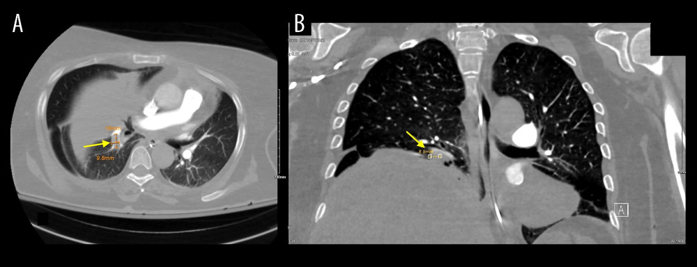 Figure 1. CT pulmonary angiography showing segmental and subsegmental pulmonary emboli (yellow arrows) and a giant right hepatic cyst measuring 29 × 18 cm, exerting right-hemithoracic mass effect with leftward mediastinal displacement. (A) Axial image. (B) Coronal image
 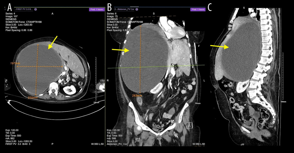 Figure 2. CT abdomen, showing a hydatid cyst with mass effect with IVC thrombosis in A: axial, B: coronal, and C: sagittal planes. The arrows point to the hydatid cyst in the liver.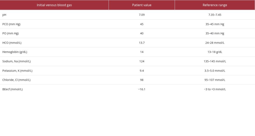
References
1. Greco S, Cannella R, Giambelluca D, Complications of hepatic echinococcosis: Multimodality imaging approach: Insights Imaging, 2019; 10; 113
2. Alabousi A, Patlas MN, Khalili K, Haider EA, Parasitic liver infections: Imaging findings and strategies for timely diagnosis: Curr Probl Diagn Radiol, 2020; 49; 447-51
3. Brunetti E, Tamarozzi F, Macpherson C, Ultrasound and cystic echinococcosis: Ultrasound Int Open, 2018; 4; E70-78
4. Frey H, Barth TF: Visceral Medicine, 2025; 42(2); 85-91
5. Naser A, Alqader RS, Ghanem L, Inferior vena cava thrombosis secondary to liver hydatid cyst: Case report: Clin Surg J, 2024; 5; 1-5
6. Staudacher M, Hotz JF, Ramharter M, Lagler H, Cystic echinococcosis (hydatid disease): Current insights into epidemiology, diagnosis, therapy, and prophylaxis: Br Med Bull, 2026; 157; ldag008
7. Govindasamy A, Bhattarai PR, John J, Liver cystic echinococcosis: A parasitic review: Ther Adv Infect Dis, 2023; 10; 20499361231171478
8. Shaprinskiy V, Verba A, Formanchuk T, Surgical treatment of echinococcosis of the liver and its complications: Wiad Lek, 2022; 75; 244-50
9. Shams M, Khazaei S, Naserifar R: Parasitology, 2022; 149; 1147-59
10. Karshima SN, Ahmed MI, Adamu NB: Parasit Vectors, 2022; 15; 357
11. Ibrahim MM, Study of cystic echinococcosis in slaughtered animals in Al Baha region, Saudi Arabia: Interaction between some biotic and abiotic factors: Acta Tropica, 2010; 113; 26-33
12. Ismail IB, Sghaier M, Boujmil K, Hydatid cyst of the liver fistulized into the inferior vena cava: Int J Surg Case Rep, 2022; 94; 107060
13. Kantorová E, Vajdová V, Hnilicová P, Cystic echinococcosis causing thrombosis of the inferior vena cava in a patient with multiple sclerosis treated with alemtuzumab: Ther Adv Neurol Disord, 2020; 13; 1756286420954081
14. Koçak S, Bumin C, Erdem E, Unusual complication of hydatid cysts: Acute inferior vena caval thrombosis: Dig Surg, 1993; 10; 114-15
15. Musielak MC, Singh R, Hartman E, Bernstein J, Simple hepatic cyst causing inferior vena cava thrombus: Int J Surg Case Rep, 2014; 5; 339-41
16. Kushner A, West WP, Suheb MZK, Pillarisetty LS, Virchow triad: StatPearls [Internet], 2024, StatPearls Publishing
17. Aloun A, Conventional and minimally invasive treatment of liver hydatid disease: Hydatid disease: Diagnosis, treatment and follow up strategies, 2025; 105-14, Springer
18. Aili A, Peng L, Zhang J, Hydatid pulmonary embolism: A case report and literature review: Am J Case Rep, 2021; 22; e934157
19. Mahajan A, Verma M, Kapila S, Spontaneous rupture of hepatic hydatid cyst into the inferior vena cava and hepatic vein: Indian J Thorac Cardiovasc Surg, 2025; 41; 219-22
Figures
Figure 1. CT pulmonary angiography showing segmental and subsegmental pulmonary emboli (yellow arrows) and a giant right hepatic cyst measuring 29 × 18 cm, exerting right-hemithoracic mass effect with leftward mediastinal displacement. (A) Axial image. (B) Coronal imageFigure 2. CT abdomen, showing a hydatid cyst with mass effect with IVC thrombosis in A: axial, B: coronal, and C: sagittal planes. The arrows point to the hydatid cyst in the liver. In Press
Case report 
Am J Case Rep In Press; DOI: 10.12659/AJCR.953543
Case report 
Am J Case Rep In Press; DOI: 10.12659/AJCR.954216
Case report 
Am J Case Rep In Press; DOI: 10.12659/AJCR.954932
Case report 
Am J Case Rep In Press; DOI: 10.12659/AJCR.954146
Most Viewed Current Articles
07 Dec 2021 : Case report
28,712,695
DOI :10.12659/AJCR.934347
Am J Case Rep 2021; 22:e934347
06 Dec 2021 : Case report  180,721
DOI :10.12659/AJCR.934406
Am J Case Rep 2021; 22:e934406
21 Jun 2024 : Case report
124,419
DOI :10.12659/AJCR.944371
Am J Case Rep 2024; 25:e944371
08 May 2026 : Case report
95,462
DOI :10.12659/AJCR.952158
Am J Case Rep 2026; 27:e952158