


21 September 2026: Articles 
Simultaneous Thyroid Eye Disease and IgG4-Related Orbital Disease in a Patient With Diabetes Mellitus: A Case Report
Challenging differential diagnosis, Diagnostic / therapeutic accidents, Management of emergency care, Educational Purpose (only if useful for a systematic review or synthesis), Rare coexistence of disease or pathology
Brenda Hayatulhaya ABCDEF 1*, Tomoyuki Kashima ABCDEFG 2DOI: 10.12659/AJCR.952068
Am J Case Rep 2026; 27:e952068


Abstract
BACKGROUND: Thyroid eye disease (TED) and IgG4-related orbital disease are distinct inflammatory conditions that rarely occur simultaneously. Their coexistence can pose diagnostic and therapeutic challenges, particularly in patients with systemic comorbidities. This report describes a rare case of concurrent TED and IgG4-related orbital disease in a patient with diabetes mellitus and details the surgical approach and postoperative results.
CASE REPORT: A 72-year-old man with a history of TED and diabetes had been experiencing progressive proptosis and orbital swelling for approximately 17 years. Despite consulting several doctors, he was refused surgery due to the associated high risks. He had facial changes, exophthalmos, and dry eyes, which led him to visit our clinic. Clinical evaluation and orbital imaging revealed changes consistent with TED, along with a localized orbital tumor extending into the deep orbital region. To alleviate the proptosis, we first performed an orbital tumor resection, along with orbital fat and bone decompression surgery. Five months later, we conducted an additional surgical resection of the residual right superior orbital tumor. Histopathological examination and immunohistochemical staining of the resected tissue, together with serological findings, confirmed the diagnosis of IgG4-related orbital disease.
CONCLUSIONS: This case highlights the importance of considering multiple etiologies in patients with atypical or long-standing TED. While orbital decompression is commonly performed for TED-associated proptosis, the presence of a localized IgG4-related orbital lesion can require surgical excision. This report suggests that combined surgical management can be considered in selected cases.
Keywords: Case Reports, IgG4-Related Disease, Ophthalmology, Orbital Neoplasms, proptosis, Thyroid Eye Disease
Introduction
Thyroid eye disease (TED) is the most common cause of orbital inflammation and proptosis in adults. This condition is an important type of orbital tissue inflammation associated with autoimmune disorders linked to thyroid dysfunction, predominantly Graves’ disease. It is characterized by inflammatory infiltration, and fibrosis of orbital tissues, resulting in extraocular muscle enlargement, orbital fat expansion, and varying degrees of visual and cosmetic impairment [1–6]. In contrast, IgG4-related disease (IgG4-RD) is a systemic fibroinflammatory condition defined by dense lymphoplasmacytic infiltration rich in IgG4-positive plasma cells, and variable degrees of fibrosis. Orbital involvement, referred to as IgG4-related orbital disease (IgG4-ROD), most commonly affects the lacrimal glands, extraocular muscles, infraorbital and supraorbital nerves, and orbital soft tissues. Clinically and radiologically, IgG4-ROD can mimic inflammatory, neoplastic, or thyroid-associated orbital disorders, often complicating the diagnostic process [7–9].
The coexistence of TED and IgG4-related orbital disease (IgG4-ROD) within the same patient is exceedingly rare and poses significant diagnostic challenges due to overlapping clinical and radiologic features. Few cases have been reported in the literature [2,3]. Both conditions can present with extraocular muscle enlargement, orbital soft tissue infiltration, and proptosis, making distinction based solely on imaging or clinical findings difficult. Failure to recognize dual pathology can lead to incomplete diagnosis or suboptimal management. From a clinical perspective, this overlap has important implications. IgG4-ROD can mimic or coexist with TED, yet its diagnosis relies on histopathological confirmation and can necessitate different therapeutic considerations, including systemic immunomodulatory therapy or targeted surgical excision. Awareness of this possibility is therefore essential, particularly in atypical, refractory, or mass-forming orbital disease.
The surgical management of TED presents considerable challenges due to the variety of available operative techniques. Orbital decompression remains a cornerstone in the surgical management of TED, particularly for patients with disfiguring proptosis or dysthyroid optic neuropathy. Multiple surgical techniques have been described, including fat decompression, bony decompression, or a combination of both, with the choice guided by disease severity, orbital anatomy, and the presence of diplopia [10–13]. For patients with disfiguring exophthalmos accompanied by diplopia in the inactive stage of the disease, fatty decompression is the preferred surgical approach for those with mild proptosis. In cases of disfiguring exophthalmos without prior diplopia, both fatty decompression and balanced decompression can be effective treatment options. Fatty decompression is suitable for patients with mild to moderate proptosis, while balanced decompression is more appropriate for those exhibiting severe proptosis. However, the presence of coexisting orbital pathology can significantly influence both surgical planning and outcomes [11–13].
In this report, we present a rare case of long-standing TED complicated by coexisting IgG4-related orbital disease. This case highlights the diagnostic complexity, the role of multimodal evaluation, including imaging and detailed histopathology, and the challenges of surgical decision-making in a patient with significant systemic comorbidities. Furthermore, it raises important considerations for future research into the immunopathological relationship between TED and IgG4-RD and their implications for clinical practice.
Case Report
SURGERY TECHNIQUE:
To address the patient’s significant proptosis and orbital mass, a staged surgical approach was planned. Orbital decompression was performed to reduce orbital volume and relieve pressure symptoms. The orbit was approached via a continuous conjunctival approach from the inferior fornix to the superior medial fornix. The first surgery was performed on the right eye. The orbital fat was removed, and any tumor that appeared in the surgical field was also removed, paying close attention to whether the tumor had adhered to muscles or nerves. At the same time, the medial orbital wall was resected to enhance the decompression effect. This surgery resulted in the removal of 9.8 cc of orbital fat and tumor. After confirming that there were no abnormalities in the right eye’s visual function, a second surgery was performed 2 weeks later, in which 14.0 cc of fat and tumor were removed from the left side, along with resection of the medial orbital wall. The resected specimen was sent for pathological and immunohistochemical examination. Five months later, a third surgery was performed to remove the remaining tumor in the right upper eyelid. The resected specimen was then sent for histopathological and immunohistochemical analysis. Access to the orbital tumor was secured through an upper eyelid incision, allowing for careful dissection from surrounding tissue. The histopathological specimens confirming IgG4-RD are shown in Figures 4–6.
PATHOLOGICAL FINDINGS:
Histopathological examination of the resected orbital specimens revealed characteristic features consistent with IgG4-related disease. Hematoxylin and eosin (H&E) staining showed a dense lymphoplasmacytic infiltrate in the orbital soft tissue, with a predominance of plasma cells distributed in both a diffuse and focally nodular pattern. The staining demonstrated significant infiltration of small lymphocytes and numerous mature plasma cells within a fibrotic stroma. Areas of storiform-type fibrosis were identified, characterized by a whorled arrangement of spindle-shaped fibroblasts. No significant cytological atypia or features suggestive of malignancy were observed.
Immunohistochemical analysis showed strong IgG positivity in plasma cells, with a significantly increased number of IgG4-positive plasma cells. Quantitative assessment demonstrated > 50 IgG4-positive plasma cells per high-power field, with an IgG4/IgG ratio exceeding 40%, supporting the diagnosis of IgG4-related disease. Additionally, the inflammatory infiltrate included scattered CD3-positive T lymphocytes and CD20-positive B lymphocytes, consistent with a mixed inflammatory background. No evidence of granulomatous inflammation or necrosis was found. Areas of fibrosis were identified, displaying an irregular to partially storiform pattern. Although classic obliterative phlebitis was not observed, small to medium-sized vessels were surrounded by inflammatory cells without evidence of vasculitic destruction, a finding consistent with reported patterns in IgG4-related orbital disease. According to the laboratory test results, there was a high level of IgG4 in the serum (123 mg/dL).
In summary, the combination of characteristic histomorphological features and immunohistochemical findings fulfilled the pathological criteria for IgG4-related orbital disease. The mildly elevated serum IgG4 concentration (123 mg/dL) was considered a supportive, but not diagnostic, finding. Histopathological criteria for IgG4-related orbital disease are shown in Table 1.
RESULT:
After the surgery, the patient showed significant improvements. There was a notable reduction in proptosis, and symptoms related to orbital compression improved. Additionally, eyelid closure was significantly better, which helped alleviate exposure keratopathy and dry eye symptoms. Histopathological evaluation confirmed the diagnosis of IgG4-RD, with high level of IgG4 serum (123 mg/dL). This condition is characterized by a dense infiltrate of lymphocytes and plasma cells, with positive IgG4 plasma cells and a negative malignant lymphoma titer. Despite the patient having diabetes mellitus, there were no major complications during the perioperative period, such as vision loss, severe hematoma, or infection. On follow-up, his condition remained stable, with only mild residual diplopia present preoperatively, and he had satisfactory functional and cosmetic outcomes.
Discussion
In patients with presumed TED who exhibit atypical imaging features, mass-forming lesions, or an incomplete response to conventional therapy, clinicians should suspect additional inflammatory or infiltrative orbital disorders. Early consideration of dual pathology can prompt timely tissue diagnosis, reduce diagnostic delay, and prevent inappropriate or incomplete treatment [2].
From a diagnostic standpoint, this case highlights the limitations of clinical presentation, imaging findings, and serum IgG4 levels. Advances in orbital imaging techniques, including quantitative MRI analysis and radiomics, may improve the ability to distinguish TED from IgG4-related orbital disease and other mimickers in the future. However, histopathological evaluation revealed findings characteristic of IgG4-related disease, including dense plasma cell infiltration, storiform fibrosis, and an elevated IgG4/IgG ratio (> 40%)
Therapeutically, the coexistence of TED and IgG4-related disease raises important questions regarding optimal management strategies. While orbital decompression remains an established intervention for TED-related proptosis, IgG4-related orbital disease can respond to systemic corticosteroids, immunosuppressive agents, or emerging biologic therapies targeting B cells and plasma cells.
In the present case, although imaging findings were initially suggestive of TED, the presence of a localized orbital mass prompted further investigation. Definitive diagnosis of IgG4-related orbital disease was established through histopathological and immunohistochemical analysis, underscoring the importance of tissue biopsy in atypical or refractory cases.
While serum IgG4 levels may be normal in a substantial proportion of patients with IgG4-related disease, tissue-based diagnosis remains the gold standard. In this patient, elevated serum IgG4 levels and characteristic histopathological findings supported the diagnosis. Importantly, immunohistochemistry excluded lymphoma, a known association with IgG4-related orbital disease [12–14].
Orbital decompression is an established treatment for TED, primarily addressing functional and cosmetic concerns. In contrast, IgG4-related disease is typically managed with systemic immunosuppressive therapy; however, surgical intervention plays a crucial role when mass lesions cause compression, diagnostic uncertainty exists, or tissue confirmation is required. In this case, combined fat and medial wall decompression with staged tumor excision resulted in symptomatic improvement and provided diagnostic clarity.
Given the limited number of reported cases, strong conclusions regarding optimal surgical management cannot be drawn. Nevertheless, this case suggests that carefully planned surgical intervention can be feasible and beneficial in selected patients with coexisting TED and IgG4-related orbital disease, even in the presence of systemic comorbidities such as diabetes mellitus. Studies are needed to clarify the role and timing of surgical intervention relative to medical therapy in patients with overlapping disease, particularly in those with significant systemic comorbidities [15–18].
Conclusions
This case highlights the diagnostic and therapeutic challenges posed by the rare coexistence of TED and IgG4-ROD. A combined surgical approach of orbital decompression and tumor removal can be safe and effective, even in patients with systemic comorbidities such as diabetes mellitus. Clinicians should maintain a high index of suspicion for multiple etiologies in patients with atypical orbital findings, and surgical intervention remains an effective and safe option even in medically complex patients. The constellation of dense lymphoplasmacytic infiltrate, storiform fibrosis, and elevated IgG4-positive plasma cells with a high IgG4/IgG ratio is consistent with IgG4-related orbital disease, correlating with the clinical presentation. Definitive confirmation of IgG4-ROD in this patient relied on detailed histopathological and quantitative immunohistochemical analysis, emphasizing the importance of contemporary diagnostic standards. Careful evaluation and individualized surgical planning remain essential in achieving good functional and aesthetic outcomes.
Figures
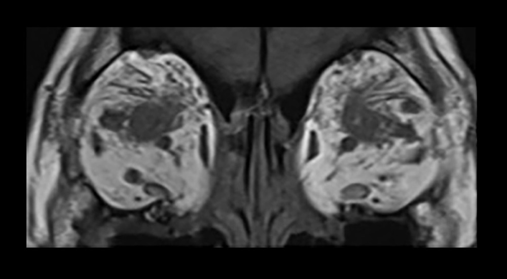 Figure 1. Coronal orbital magnetic resonance imaging findings (T1-weighted and fat-suppressed images using short-tau inversion recovery sequences); bilateral extraocular muscle enlargement with tendon sparing (TED) and diffuse orbital soft tissue /lacrimal gland infiltration with high STIR signal (IgG4-RD).
 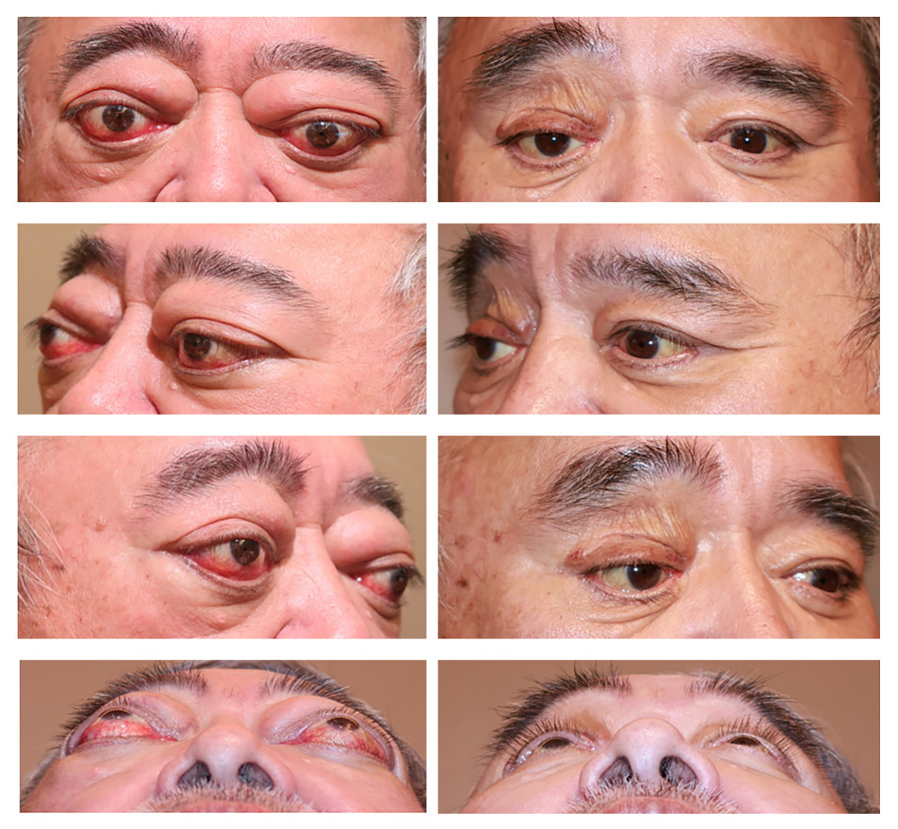 Figure 2. Clinical photographs of the patient. Left: Pre-surgery view. Right: Six months after surgery, showing a reduction in proptosis, orbital pain, and overall improvement in appearance.
 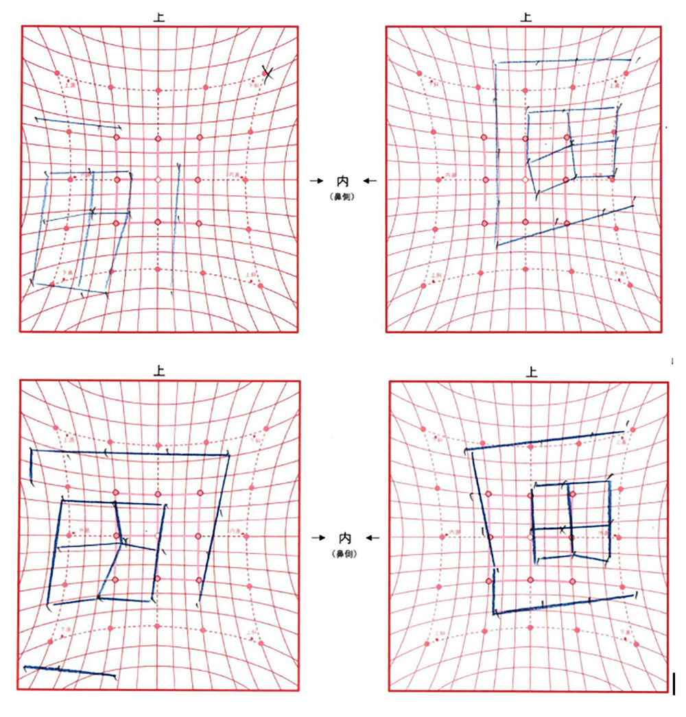 Figure 3. Hess chart analysis of ocular motility: At first visit (above) the left panel represents the right eye (RE), and the right panel represents the left eye (LE). There are marked right inferior rectus restriction with significant field contraction in the RE and expansion in the left superior field. At 2-week follow-up (below), there is partial improvement in ocular motility with a less contracted right eye field and reduced overaction of the left superior rectus, indicating recovery of restrictive strabismus.
 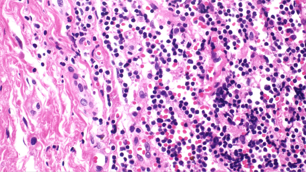 Figure 4. Low-power view of orbital tissue showing diffuse lymphoplasmacytic infiltration with associated fibrosis (H&E stain, original magnification ×40). The inflammatory infiltrate is distributed in a patchy to diffuse pattern within the stromal background.
 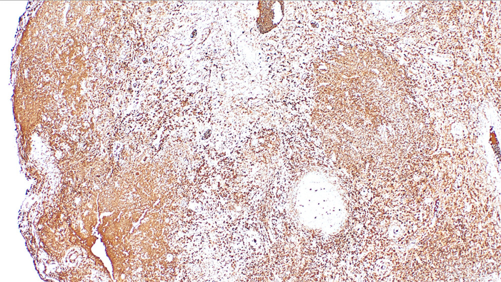 Figure 5. Immunohistochemical staining for IgG demonstrating numerous IgG-positive plasma cells diffusely distributed throughout the lesion (original magnification ×100), consistent with a plasma cell-rich inflammatory process.
 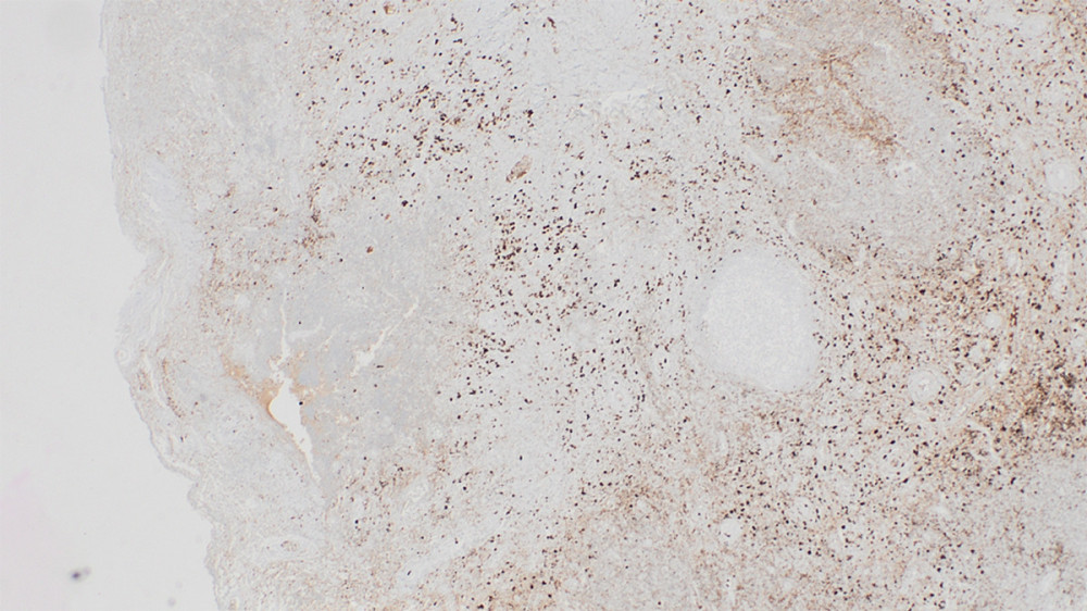 Figure 6. Immunohistochemical staining for IgG4 revealing a markedly increased number of IgG4-positive plasma cells (original magnification ×100). Quantitative assessment showed > 50 IgG4-positive plasma cells per high-power field with an IgG4/IgG ratio > 40%, supporting the diagnosis of IgG4-related disease.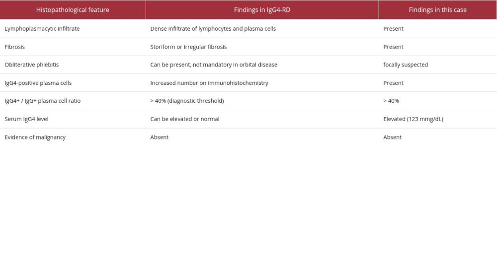
References
1. Yamana Y, Kashima T, Dysthyroid optic neuropathy dramatically improved by sheath-guided triamcinolone orbital injection: A case report: Cureus, 2024; 16(9); e70437
2. Ye H, Xiao W, Chen R, Elevated immunoglobulin G4 levels in patients with thyroid eye disease and their clinical implications: Invest Ophthalmol Vis Sci, 2020; 61(5); 57
3. Douglas RS, Bahn RS, Smith TJ, Advances in the pathogenesis and treatment of thyroid eye disease: Emerging role of biologics: Lancet Diabetes Endocrinol, 2022; 10(3); 207-17
4. Khandji J, Campbell AA, Callahan AB, Sirinek P, Kazim M, IgG4-related orbital disease masquerading as thyroid eye disease, vice versa, or both?: Orbit, 2018; 37(4); 239-42
5. Smith TJ, Hegedüs L, Graves’ disease: N Engl J Med, 2016; 375; 1552-65
6. Kahaly GJ, Douglas RS, Holt RJ, Management of thyroid eye disease: Update on surgical and medical therapy: Curr Opin Endocrinol Diabetes Obes, 2021; 28(5); 463-70
7. Andrew NH, Sladden N, Kearney DJ, Selva D, IgG4-related orbital disease: A meta-analysis and review: Acta Ophthalmol, 2020; 98(2); 121-30
8. Umehara H, Okazaki K, Masaki Y, IgG4-related disease: Updated 2020 comprehensive diagnostic criteria: Mod Rheumatol, 2021; 31(3); 529-33
9. Cheuk W, Yuen HK, Chu SY, Ocular adnexal IgG4-related disease: A clinicopathologic study of 116 cases: Am J Surg Pathol, 2017; 41(5); 663-75
10. Wiersinga WM, Bartalena L, Kahaly GJ, Clinical management of Graves’ orbitopathy: Update on diagnosis and treatment: Endocr Rev, 2021; 42(6); 706-40
11. Cheng AMS, Wei YH, Liao SL, Strategies in surgical decompression for thyroid eye disease: Oxid Med Cell Longev, 2020; 2020; 3537675
12. Kuriyan AE, Woog JJ, Kazim M, Surgical management of thyroid eye disease: Surv Ophthalmol, 2020; 65(6); 789-803
13. Jefferis JM, Jones RK, Currie Z, Clinical outcomes following orbital decompression surgery for thyroid eye disease: Eye (Lond), 2019; 33(6); 1021-28
14. Sah RP, Chari ST, Serologic issues in IgG4-related systemic disease and autoimmune pancreatitis: Curr Opin Rheumatol, 2011; 23(2); 108-13
15. Yamamoto M, Takahashi H, Tabeya T, Risk of malignancies in IgG4-related disease: Mod Rheumatol, 2012; 22(3); 414-18
16. Umehara H, Okazaki K, Masaki Y, Comprehensive diagnostic criteria for IgG4-related disease: Mod Rheumatol, 2012; 22(1); 21-30
17. Sato Y, Ohshima K, Takata K, Yoshino T, IgG4-related disease: A cross-sectional study of 114 cases: Am J Surg Pathol, 2013; 37(10); 1548-56
18. Stone JH, Zen Y, Deshpande V, IgG4-related disease: N Engl J Med, 2012; 366(6); 539-51
Figures
Figure 1. Coronal orbital magnetic resonance imaging findings (T1-weighted and fat-suppressed images using short-tau inversion recovery sequences); bilateral extraocular muscle enlargement with tendon sparing (TED) and diffuse orbital soft tissue /lacrimal gland infiltration with high STIR signal (IgG4-RD).Figure 2. Clinical photographs of the patient. Left: Pre-surgery view. Right: Six months after surgery, showing a reduction in proptosis, orbital pain, and overall improvement in appearance.Figure 3. Hess chart analysis of ocular motility: At first visit (above) the left panel represents the right eye (RE), and the right panel represents the left eye (LE). There are marked right inferior rectus restriction with significant field contraction in the RE and expansion in the left superior field. At 2-week follow-up (below), there is partial improvement in ocular motility with a less contracted right eye field and reduced overaction of the left superior rectus, indicating recovery of restrictive strabismus.Figure 4. Low-power view of orbital tissue showing diffuse lymphoplasmacytic infiltration with associated fibrosis (H&E stain, original magnification ×40). The inflammatory infiltrate is distributed in a patchy to diffuse pattern within the stromal background.Figure 5. Immunohistochemical staining for IgG demonstrating numerous IgG-positive plasma cells diffusely distributed throughout the lesion (original magnification ×100), consistent with a plasma cell-rich inflammatory process.Figure 6. Immunohistochemical staining for IgG4 revealing a markedly increased number of IgG4-positive plasma cells (original magnification ×100). Quantitative assessment showed > 50 IgG4-positive plasma cells per high-power field with an IgG4/IgG ratio > 40%, supporting the diagnosis of IgG4-related disease. In Press
Case report 
Am J Case Rep In Press; DOI: 10.12659/AJCR.954074
Case report 
Am J Case Rep In Press; DOI: 10.12659/AJCR.953592
Case report 
Am J Case Rep In Press; DOI: 10.12659/AJCR.954225
Case report 
Am J Case Rep In Press; DOI: 10.12659/AJCR.952870
Most Viewed Current Articles
07 Dec 2021 : Case report
28,712,481
DOI :10.12659/AJCR.934347
Am J Case Rep 2021; 22:e934347
06 Dec 2021 : Case report  180,580
DOI :10.12659/AJCR.934406
Am J Case Rep 2021; 22:e934406
21 Jun 2024 : Case report
124,319
DOI :10.12659/AJCR.944371
Am J Case Rep 2024; 25:e944371
08 May 2026 : Case report
95,142
DOI :10.12659/AJCR.952158
Am J Case Rep 2026; 27:e952158