


21 August 2026: Articles 
Histiocytic Sarcoma With Striking Clinical Improvement
Challenging differential diagnosis, Unusual or unexpected effect of treatment, Rare disease
Nur Farhanah EF 1*, Jessica Novia Hadiyanto F 2, Erlieza Roosdhania F 3, Dhesi Ariembi B 4, Enny Puji Astuti B 5, Subiyakto Bustomi F 6, Eny Dyah Kurniawati F 7, Hermawan Istiadi F 8, Budi Setiawan F 9
DOI: 10.12659/AJCR.952214
Am J Case Rep 2026; 27:e952214


Abstract
BACKGROUND: Histiocytic sarcoma (HS) is a rare and aggressive hematopoietic malignancy characterized by histiocytic cell proliferation. Its rarity and nonspecific presentation create significant diagnostic and therapeutic challenges, particularly in HIV-positive patients.
CASE REPORT: An HIV-positive man in his late 20s from a rural area and low-income background, referred from a district hospital, presented with asymptomatic nodular and crateriform lesions on the face, thorax, and back, characterized by central keratosis, crust, and erythematous-violaceous infiltrated edges. Histopathological and immunohistochemical analysis confirmed the diagnosis of histiocytic sarcoma, with tumor cells positive for CD68. Considering the patient’s multisystem involvement, 6 to 8 cycles of CHOP chemotherapy were recommended. However, the patient declined systemic therapy and opted for a watch-and-wait approach. No invasive procedures such as bone marrow biopsy were performed, as the patient exhibited no constitutional symptoms or cytopenias. Clinical follow-up over 6 months revealed significant improvement in tumor size and overall condition. This favorable outcome may have been partially influenced by immune reconstitution associated with ongoing antiretroviral therapy (ART), although a direct causal relationship cannot be established. Management of histiocytic sarcoma remains controversial due to the lack of established guidelines and variability in natural history. For this patient, a non-aggressive, observational approach yielded favorable outcomes, challenging the conventional use of systemic chemotherapy in all cases of multisystem involvement. Regular clinical follow-up has been integral to monitoring disease progression.
CONCLUSIONS: This report underscores the diagnostic and therapeutic challenges associated with histiocytic sarcoma and highlights the importance of a multidisciplinary team approach involving infectious disease specialists and hematology-oncology experts in achieving optimal clinical outcomes. Despite declining systemic treatment, the patient had a favorable clinical outcome.
Keywords: Histiocytic Sarcoma, HIV Infections, Immunohistochemistry, Hematology, Sarcoma, Histiocytic, Skin Neoplasms, Watchful Waiting, Case Reports, CHOP Protocol
Introduction
Histiocytic sarcoma (HS) is a rare and aggressive hematopoietic malignancy characterized by the malignant proliferation of histiocytic cells. It accounts for less than 1% of all hematopoietic malignancies with a poor prognosis, particularly in cases involving multisystem disease [1]. The diagnosis of HS is challenging due to its nonspecific presentation and overlapping features with other malignancies [2]. Immunohistochemical evaluation is critical, with positive expression of histiocytic markers such as CD68, CD163, and lysozyme, along with the exclusion of Langerhans cell markers (CD1a and langerin) and other lineage-specific markers, supporting the diagnosis of histiocytic sarcoma [3].
HS occurring in patients with human immunodeficiency virus (HIV) is extremely rare, with few cases reported in the literature. The coexistence of HS and HIV presents additional diagnostic and therapeutic challenges because of immunosuppression, increased susceptibility to opportunistic infections, and the need to balance oncologic treatment with antiretroviral therapy (ART). In HIV-associated hematologic malignancies, immune recovery following ART has been associated with improved immune function and can contribute to more favorable clinical outcomes in selected patients. Furthermore, the prognosis of HS can vary according to disease extent, with localized or cutaneous HS generally demonstrating a more indolent course compared with disseminated or multisystem disease, which is associated with poorer outcomes [3].
In HIV-positive patients, the management of HS is further complicated by immunosuppression and the need to balance treatment efficacy with patient preferences [4]. This case report discusses the clinical presentation, diagnosis, and management of an HIV-positive man in his late 20s with histiocytic sarcoma who opted for a non-aggressive management approach.
Case Report
An HIV-positive man in his late 20s from a rural, low-income background was referred from a district hospital with asymptomatic nodular and crateriform lesions involving the face, thorax, and back. The lesions were characterized by central keratosis, crusts, and erythematous-violaceous infiltrated edges. The patient reported a gradual increase in the size and number of lesions over several months but denied any constitutional symptoms such as fever, weight loss, or fatigue (Figure 1).
Laboratory investigations revealed anemia, with a hemoglobin level of 8.3 g/dL, and leukopenia, with a leukocyte count of 2.1 × 10³/μL. Initial HIV evaluation demonstrated poorly controlled disease, with an HIV viral load of 356 000 copies/mL and a CD4+ T-cell count of 48 cells/μL, indicating severe immunosuppression at the time of diagnosis. MSCT of the paranasal sinuses with contrast showed multiple enhancing polypoid lesions in the nasal cavity, choana, nasopharyngeal mucosal space, maxillary sinuses, and ethmoid sinuses, raising suspicion of malignancy, with Kaposi sarcoma as a differential diagnosis. Areas of hypoattenuation suggested possible bleeding. Serological and microbiological tests for VDRL and TPHA were negative. A fungal culture from the nasal cavity showed no fungal growth, while acid-fast bacilli (BTA) staining was also negative. Microscopic examination revealed minimal epithelial cells and leukocytes, and fungal staining confirmed the absence of yeast cells.
Histopathological examination of the nasal cavity biopsy revealed a diffuse proliferation of large pleomorphic cells with abundant eosinophilic cytoplasm, irregular nuclei, and marked cytologic atypia (Figure 2). In accordance with the WHO classification, the diagnosis of histiocytic sarcoma was established based on these characteristic morphologic features combined with the positive expression of histiocytic markers (CD68, CD163, and lysozyme), alongside the exclusion of lymphoid and Langerhans cell lineages (CD3, CD20, CD1a, langerin, and S100) (Figures 3, 4). Due to significant anemia, the patient received a transfusion of 2 units of packed red cells (PRC), increasing the hemoglobin level to 12 g/dL. Treatment options, including systemic chemotherapy with 6 to 8 cycles of the CHOP regimen, were discussed, but the patient opted for a watch-and-wait approach.
Despite forgoing chemotherapy, the patient remained adherent to his antiretroviral therapy (ARV), consisting of a combination of Dolutegravir 50 mg, Lamivudine 300 mg, and Tenofovir Disoproxil Fumarate 300 mg. In addition, he was given Cotrimoxazole 960 mg daily as prophylaxis for opportunistic infections. Over the next 6 months of follow-up, he showed significant clinical improvement, with a noticeable reduction in the size of the lesions and no signs of systemic progression. After 6 months of follow-up, repeat HIV evaluation showed marked improvement, with the viral load decreasing to < 40 copies/mL and the CD4+ T-cell count increasing to 312 cells/μL. Hematologic parameters also improved progressively, accompanied by a noticeable reduction in lesion size and no evidence of systemic progression. Currently, the patient continues to attend monthly follow-ups and remains in excellent clinical condition, with no recurrence of the lesions to date.
Discussion
HS is an exceptionally rare malignancy, with limited data guiding its optimal management [1]. In cases with multisystem involvement, systemic chemotherapy is generally recommended due to the aggressive nature of the disease [5]. However, the patient’s decision to decline systemic therapy posed a significant management challenge. Remarkably, he had spontaneous clinical improvement under a watch-and-wait strategy, challenging the conventional use of aggressive treatment in all cases of HS [6]. Several reports in the literature have also described indolent or partially regressive courses of cutaneous HS, particularly in localized disease, suggesting that the biological behavior of HS can be heterogeneous and not uniformly aggressive [7].
The absence of constitutional symptoms or cytopenias justified the decision to postpone invasive staging procedures such as bone marrow biopsy and avoid high-risk interventions [8]. It is also possible that this case was a more localized or biologically less aggressive subtype of HS, and the possibility of partial spontaneous regression or pathological underestimation should be considered when interpreting the favorable clinical course. This case highlights the potential for non-aggressive approaches to achieve favorable outcomes in select patients. Further research is needed to better understand the natural history of HS and identify predictors of treatment response [9,10].
Patients with HIV face unique challenges in the management of malignancies like HS due to their compromised immune system, despite improved immune function with antiretroviral therapy [11]. In the present case, the marked virologic suppression and substantial CD4+ T-cell recovery observed during follow-up coincided with significant regression of the lesions. Although a causal relationship cannot be established, immune reconstitution associated with effective antiretroviral therapy may have contributed to enhanced immune surveillance and potentially influenced the biological behavior of the tumor. A multidisciplinary approach involving oncologists, infectious disease specialists, and other relevant experts is crucial to ensure comprehensive care [12]. This collaborative strategy enables the development of personalized treatment plans that integrate effective HIV management with appropriate cancer therapies, tailoring decisions on invasive procedures, systemic treatment, and palliative care to the patient’s overall condition and specific needs [13,14]. In recent years, emerging therapies for refractory or advanced HS, including targeted therapy and immune checkpoint inhibitors such as programmed cell death protein 1 (PD-1) inhibitors, have shown promising results in selected cases, particularly in tumors harboring actionable molecular alterations. Although evidence remains limited, these approaches may provide future therapeutic options for patients who are unsuitable for conventional chemotherapy [3].
Conclusions
An HIV-positive man in his late 20s presented with multiple asymptomatic nodular and crateriform lesions on the face, thorax, and back, with central keratosis, crust, and erythematous-violaceous infiltrated edges. Histopathology and immunohistochemistry confirmed histiocytic sarcoma. Despite multisystem involvement, he declined systemic chemotherapy and opted for a watch-and-wait approach while continuing antiretroviral therapy and prophylaxis for opportunistic infections. Due to the absence of constitutional symptoms or cytopenias, no invasive staging procedures were performed. Over 6 months of observation, there was marked reduction in lesion size and significant improvement in overall condition without signs of systemic progression. This case shows that non-aggressive management can yield favorable outcomes in select patients with histiocytic sarcoma, especially when guided by patient preference and close multidisciplinary follow-up. In particular, for patients with localized or predominantly cutaneous HS, well-controlled HIV infection, and no significant systemic symptoms, careful observation and regular follow-up can be considered as an individualized management strategy. This report highlights the importance of individualized care and the need for further research to define optimal management strategies for this rare malignancy.
Figures
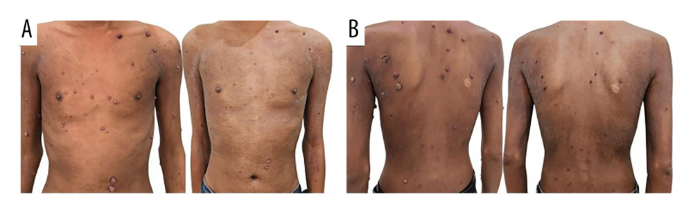 Figure 1. The lesions are observed on the thorax (A) and back (B). Nodular and crateriform lesions characterized by central keratosis, crust, and erythematous-violaceous infiltrated edges. Clinical presentation of the lesions before and after 6 months of observation under a watch-and-wait approach. There is a significant reduction in lesion size and improvement in the skin’s overall condition.
 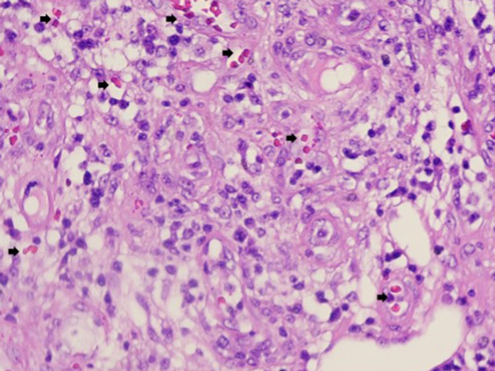 Figure 2. The morphological features of the nasal cavity biopsy highlighting the pleomorphic cells (black arrow) and lack of necrosis, consistent with a diagnosis of histiocytic sarcoma.
 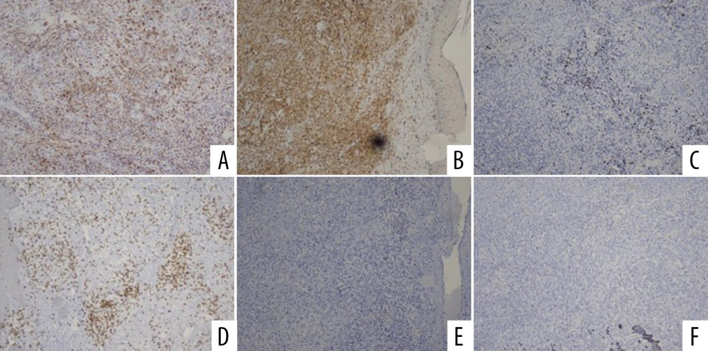 Figure 3. The immunohistochemical findings support the diagnosis of histiocytic sarcoma, with positive staining for (A) CD68 (histiocytic marker), (B) CD45 (hematopoietic origin), and a high proliferation index with (C) Ki67 (60–70%). Negative results for (D) CD3, (E) CD20, (F) CD30, CD34, and S100 exclude lymphoma, dendritic neoplasms, and Langerhans cell tumors.
 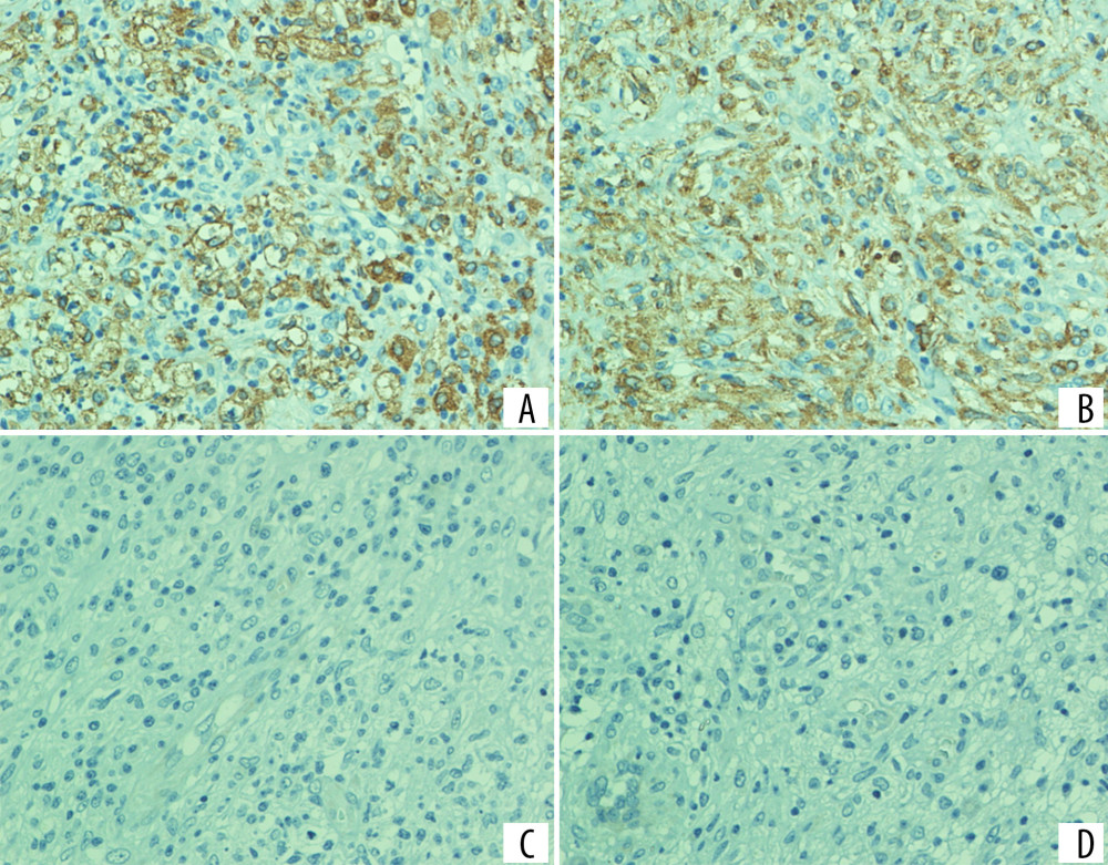 Figure 4. Immunohistochemical profile of the lesion; (A) CD163 showing positive cytoplasmic staining in tumor cells; (B) lysozyme showing positive staining, supporting histiocytic differentiation; (C) CD1a showing negative staining; (D) langerin (CD207) showing negative staining. The positivity of CD163 and lysozyme, together with the absence of CD1a and langerin expression, supports the diagnosis of histiocytic sarcoma and excludes Langerhans cell histiocytosis.
 References
1. May JM, Waddle MR, Miller DH, Primary histiocytic sarcoma of the central nervous system: A case report with platelet derived growth factor receptor mutation and PD-L1/PD-L2 expression and literature review: Radiat Oncol, 2018; 13(1); 167
2. Imoto Y, Yamadera M, Ueno H, Massive enteric necrosis caused by histiocytic sarcoma embolism: A case report: Surg Case Rep, 2024; 10(1); 228
3. Huff D, Fortin Ensign S, Ryan MS, Histiocytic sarcoma treated with pembrolizumab: A case report and literature review: J Immunother Precis Oncol, 2023; 6(4); 198-202
4. An K, He Y, Tang Y, Histiocytic sarcoma following CAR T-cell therapy: A case report: Int J Hematol, 2024; 119(3); 338-41
5. Zhang W, Li H, Li B, Long-term response to radiotherapy in localized histiocytic sarcoma: A case report and review of the literature: Front Oncol, 2021; 11; 755893
6. Goyal G, Young JR, Koster MJ, The Mayo Clinic Histiocytosis Working Group consensus statement for the diagnosis and evaluation of adult patients with histiocytic neoplasms: Erdheim-Chester disease, Langerhans cell histiocytosis, and Rosai-Dorfman disease: Mayo Clin Proc, 2019; 94(10); 2054-71
7. Trevisan F, Xavier CA, Pinto CA, Case report of cutaneous histiocytic sarcoma: Diagnostic and therapeutic dilemmas: An Bras Dermatol, 2013; 88(5); 807-10
8. The ASCO Post Staff, FDA approves oral MEK inhibitor cobimetinib: The ASCO Post, 2022
9. Vos JA, Abbondanzo SL, Barekman CL, Histiocytic sarcoma: A study of five cases including the histiocyte marker CD163: Mod Pathol, 2005; 18(5); 693-704
10. Feldman AL, Dogan A, Smith DI, Novel insights into the biology and treatment of histiocytic sarcoma: Nat Rev Clin Oncol, 2020; 17(8); 494-507
11. Laurent C, Pissaloux D, Xerri L, Comprehensive genetic characterization of nodal histiocytic/dendritic cell sarcomas: Am J Surg Pathol, 2021; 45(3); 305-16
12. Wei S, Wang Y, Li X, Multisystem histiocytic sarcoma presenting with cutaneous lesions: J Cutan Pathol, 2022; 49(8); 734-38
13. Andersen KF, Hager HH, Hansen JA, Histiocytic sarcoma: Challenging course, dismal outcome: Diagnostics (Basel), 2021; 11(2); 310
14. Wall SA, Ferris M, Malone ER, Multidisciplinary approach to older adults with hematologic malignancies: J Natl Compr Canc Netw, 2022; 20(2); 190-98
Figures
Figure 1. The lesions are observed on the thorax (A) and back (B). Nodular and crateriform lesions characterized by central keratosis, crust, and erythematous-violaceous infiltrated edges. Clinical presentation of the lesions before and after 6 months of observation under a watch-and-wait approach. There is a significant reduction in lesion size and improvement in the skin’s overall condition.Figure 2. The morphological features of the nasal cavity biopsy highlighting the pleomorphic cells (black arrow) and lack of necrosis, consistent with a diagnosis of histiocytic sarcoma.Figure 3. The immunohistochemical findings support the diagnosis of histiocytic sarcoma, with positive staining for (A) CD68 (histiocytic marker), (B) CD45 (hematopoietic origin), and a high proliferation index with (C) Ki67 (60–70%). Negative results for (D) CD3, (E) CD20, (F) CD30, CD34, and S100 exclude lymphoma, dendritic neoplasms, and Langerhans cell tumors.Figure 4. Immunohistochemical profile of the lesion; (A) CD163 showing positive cytoplasmic staining in tumor cells; (B) lysozyme showing positive staining, supporting histiocytic differentiation; (C) CD1a showing negative staining; (D) langerin (CD207) showing negative staining. The positivity of CD163 and lysozyme, together with the absence of CD1a and langerin expression, supports the diagnosis of histiocytic sarcoma and excludes Langerhans cell histiocytosis. In Press
Case report 
Am J Case Rep In Press; DOI: 10.12659/AJCR.954530
Case report 
Am J Case Rep In Press; DOI: 10.12659/AJCR.954296
Case report 
Am J Case Rep In Press; DOI: 10.12659/AJCR.954629
Case report 
Am J Case Rep In Press; DOI: 10.12659/AJCR.953831
Most Viewed Current Articles
07 Dec 2021 : Case report
28,708,424
DOI :10.12659/AJCR.934347
Am J Case Rep 2021; 22:e934347
06 Dec 2021 : Case report  180,361
DOI :10.12659/AJCR.934406
Am J Case Rep 2021; 22:e934406
21 Jun 2024 : Case report  124,086
DOI :10.12659/AJCR.944371
Am J Case Rep 2024; 25:e944371
08 May 2026 : Case report
94,538
DOI :10.12659/AJCR.952158
Am J Case Rep 2026; 27:e952158