


27 August 2026: Articles 
Robot-Assisted Surgery Requiring an Intraoperative Transition to Laparoscopic Surgery Due to an Emergency System Shutdown: A Case Report
Diagnostic / therapeutic accidents
Yuichiro Kizaki E 1*, Kouki Samejima A 1, Shigetaka Matsunaga B 2, Tomonori Nagai C 1, Yasushi Takai D 1
DOI: 10.12659/AJCR.952578
Am J Case Rep 2026; 27:e952578


Abstract
BACKGROUND: Robot-assisted hysterectomy (RASH) for benign uterine disease has been covered by insurance in Japan since 2018. Our department introduced RASH in December 2020, and the number of procedures has increased without major complications. Herein, we report a case in which robot-assisted surgery required intraoperative conversion to conventional laparoscopy because of an unexpected robotic system shutdown.
CASE REPORT: A 54-year-old multiparous woman presented with hypermenorrhea and anemia caused by uterine adenomyosis. After unsuccessful conservative treatment, definitive surgical management was planned. Following 6 months of gonadotropin-releasing hormone analog therapy, RASH was performed using the da Vinci X system. Four robotic ports and a single 5-mm assistant port were placed. Total operative time was 202 minutes, including 148 minutes of console time. After transvaginal uterine removal and vaginal cuff closure, the robotic system suddenly underwent an emergency shutdown during confirmation of hemostasis, making intracorporeal manipulation impossible. Although the manufacturer was contacted immediately and troubleshooting was attempted, the system could not be restored. Therefore, the operation was converted to conventional laparoscopic surgery using the same port placement, and the procedure was completed successfully. The postoperative course was uneventful, and the patient was discharged without complications.
CONCLUSIONS: Robot-assisted surgery is increasingly used in gynecology because of its minimally invasive advantages. However, surgeons should be prepared for unexpected events such as robotic system failure. Familiarity with laparoscopic surgical techniques enables safe continuation of surgery in such situations. This case highlights the importance of maintaining laparoscopic surgical skills to ensure the safe performance of robot-assisted procedures.
Keywords: Case Reports, Gynecology, Hysterectomy, Intraoperative Complications, Laparoscopy, Robotic Surgical Procedures
Introduction
Robot-assisted surgery enables precise surgical manipulation even within relatively confined pelvic spaces due to the enhanced dexterity provided by articulating instruments. In addition, the use of a high-definition 3-dimensional imaging system facilitates detailed visualization of fine anatomical structures, which may contribute to reduced blood loss, making it a minimally invasive surgical approach [1]. Discontinuation of robot-assisted surgery due to instrument malfunction or other issues is highly rare, with an incidence rate of approximately 0.25% [2].
In Japan, robot-assisted hysterectomy (RASH) for gynecological diseases has been covered by insurance since 2018, and the number of patients undergoing RASH has been increasing annually. At our hospital, robot-assisted surgery was initiated in the urology department in October 2020, and our department began performing RASH in December 2020. To date, our hospital has performed more than 678 robot-assisted surgeries across all medical departments without any major complications. We perform robot-assisted total hysterectomy for benign uterine diseases, robot-assisted surgery for malignant uterine tumors in early endometrial cancer, and robot-assisted sacrocolpopexy for pelvic organ prolapse, all of which are covered by the national health insurance system in Japan. Approximately 120 robot-assisted surgical procedures are performed annually at our hospital. Here, we describe a case of robot-assisted surgery that needed to be discontinued because of an intraoperative emergency shutdown of the robotic system, requiring a transition to laparoscopic surgery. Written consent was obtained from the patient herself.
Case Report
The patient was a 54-year-old woman, gravidity 3, parity 3, with a height of 160 cm, weight of 92 kg, and body mass index of 36.2 kg/m2. She had a history of oral medications for diabetes mellitus. The patient visited a nearby gynecology clinic because of hypermenorrhea and was diagnosed with uterine adenomyosis. Then, she was referred to our department for detailed examination and treatment. During the internal examination, enlargement of the uterus to a size equivalent to that of the head of a newborn was palpated, and transvaginal ultrasonography and pelvic magnetic resonance imaging revealed uterine adenomyosis. Cervical and endometrial cytology did not show any findings suggestive of malignant disease. Although it was recommended that the patient undergo surgical treatment for anemia-causing hypermenorrhea, conservative treatment was preferred. The patient was administered an oral gonadotropin-releasing hormone (GnRH) antagonist preparation for 6 months; however, with the resumption of menstruation, she again exhibited anemia. She desired intrauterine delivery system (IUS) therapy, but insertion of the IUS did not bring about any improvement in bleeding. Therefore, a decision was made to proceed with radical surgery. After administration of the GnRH agonist preparation for 6 months, the patient was scheduled to undergo RASH. While awaiting surgery, she exhibited an improvement in bleeding, and internal examination showed no enlargement of the uterus.
Surgery was performed using the da Vinci X system. The surgeon was a doctor in his 10th year of practice in obstetrics and gynecology and had performed his 40th robot-assisted surgery. A camera port was inserted through the umbilicus. Four da Vinci ports were placed in parallel at 8-cm intervals, and a 5-mm port was placed in the left lower abdomen as an auxiliary port, resulting in the placement of a total of 5 ports. With the da Vinci system, we used Maryland bipolar forceps for the first arm, an endoscope for the second arm, monopolar curved scissors for the third arm, and prograsp forceps for the fourth arm. The uterus was enlarged because of uterine adenomyosis, but there was no bilateral adnexal enlargement (Figure 1). Immediately after starting the surgery, the operator experienced a feeling of strangeness in the operability of the third and fourth arms; however, the removal and replacement of the instruments were repeated several times, demonstrating normal operation. Thus, the surgery was performed in a standard manner. The specimen was dissected, and the uterus and both adnexa were collected transvaginally. After suturing the vaginal stump, the intraperitoneal cavity was washed, and hemostasis was confirmed. While confirming hemostasis of the left pelvic infundibulum ligament, the robotic system encountered an emergency shutdown, suddenly disabling the console operation (Figure 2). We contacted the manufacturer, Intuitive Surgical, Inc., and made multiple attempts to restore the system, but these were unsuccessful. As the intracavitary operation needed to be continued, the attending physicians discussed the operative procedure intraoperatively and determined that surgery could be continued laparoscopically. The emergency shutdown button was pressed, and because the left pelvic infundibulum ligament was grasped, a release kit was used to release the tissue. After confirming that no tissue was grasped, all instruments were removed. After undocking all forceps, roll-out was performed, and the procedure was transitioned to laparoscopic surgery. It took 26 minutes from the shutdown of the emergency system to the transition to laparoscopic surgery, and during the waiting period, the patient showed no changes in vital signs. During laparoscopic surgery, the operator stood on the patient’s left side, while the first assistant stood on the patient’s right side. The da Vinci port in the umbilicus was used as the camera port, and the port in the left lower abdomen and the port of the first da Vinci arm were used as operation ports for the operator. During the surgery, 3 cm of rectal serosa was lost when the rectum was being mobilized using the prograsp forceps (Figure 3), and the defect in the rectal serosa was closed using a single ligature suture under laparoscopy. Surgery was completed after intraperitoneal washing, and hemostasis was confirmed. The surgical duration was 202 minutes (including 26 minutes of interruption), with a console time of 131 minutes. The amount of bleeding was small, and the specimen weighed 480 g.
The patient was discharged without any postoperative complications, and the final pathological diagnosis was uterine adenomyosis with no malignant findings. Outpatient examinations were performed at 1 and 3 months postoperatively, and the final examination was completed without any abnormal findings. Verbal informed consent was obtained from the patient to publish this case report.
Discussion
Robot-assisted surgery enables the use of stable forceps with an anti-shake function, surgery with a stereoscopic view using a 3-dimensional camera, and forceps with a multi-joint function. Because of these advantages, robot-assisted surgery contributes to the reduction in the amount of intraoperative bleeding and shortens the hospital stay compared with conventional surgery [3]. As it also contributes to the reduction in postoperative pain and wound healing, the number of patients undergoing robot-assisted surgery is expected to increase in various medical departments, replacing conventional laparotomy and laparoscopic surgery [3]. However, there are concerns that an increase in the number of robot-assisted surgeries will be accompanied by an increase in the number of intraoperative instrument failures [4].
In Japan, robot-assisted surgery, as a minimally invasive surgery, is increasing in various medical departments, including gynecology. At our hospital, the number of patients undergoing robot-assisted surgery has been increasing annually across all medical departments since its introduction to the urology department in October 2020. With the rapid use of robot-assisted surgery, operators must be prepared for incidental emergency shutdowns of robotic systems, as in the present case.
By December 2022, our hospital had performed 678 robot-assisted surgeries across all medical departments. This was the first case of emergency system shutdown during surgery, disabling the console operation. On the basis of the occurrence of an intraoperative emergency system shutdown in this case, we reviewed the incidence rates of malfunctions in the robot-assisted surgery system and instruments at our hospital.
Instrument malfunctions were found in 53 cases (7.8%) of robot-assisted surgery across all medical departments at our hospital, including 47 cases of instrument-related failure, 3 cases of system errors, and 3 cases of other issues (Figure 4). Cases of instrument-related failure were the most common, and with regard to the timing of occurrence, failure was detected preoperatively in 3 cases, intraoperatively in 15 cases, and postoperatively in 29 cases (Table 1). In cases where a failure was detected preoperatively or intraoperatively, the instrument was replaced and the surgery was performed as planned without any issues. In cases where failure was detected postoperatively, the cause was a frayed instrument cable that did not affect the surgical operation itself, and surgery was continued. There has been a report of cases in which surgery could not be continued owing to instrument malfunction, requiring a change in the operative procedure [5]. Thus, even if the instrument damage is minor and does not interfere with the surgical operation, physicians must pay sufficient attention to the intraoperative operation. In the present case, the operator experienced a feeling of strangeness regarding the operability of the instruments from the start of the surgery; however, no breakage or damage was observed during the intraoperative and postoperative inspection of the instruments.
System errors were observed in 3 cases. Of these, in 2 cases, a recoverable system error was encountered, and robot-assisted surgery was able to be continued by restoring the system during surgery. An unrecoverable system error was seen in only 1 case of the 3 (0.14%), the present case. Overall, the incidence rate of unrecoverable system errors in robot-assisted surgery is 1.06% and their causes include communication errors, in addition to failures of a battery and an optical instrument [6]. In the present case, a system error was encountered while confirming hemostasis. Because the patient showed no change in vital signs, everyone, including the operator, assistants, nurses, and clinical engineers, responded to the situation calmly. However, a more rapid response is required in urgent cases such as those with bleeding.
After the emergency shutdown, we immediately contacted the technical support center of the manufacturer, Intuitive Surgical, Inc., and requested instructions; however, we were unable to identify the cause during surgery. The instruments used in surgery were inspected by a technical support representative at a later date. As a result, operation malfunction was found to be caused by a communication error within the console, and after replacing the parts causing instrument failure, we confirmed that the issue was not reproducible.
Discontinuation of robot-assisted surgery due to instrument failure requiring a change in operative procedure has been reported to have an incidence rate of 8.7% [7]. When changing operative procedure, a transition to laparoscopic surgery may be desirable in consideration of invasiveness. In the present case, we were unable to restore the robotic system after an emergency shutdown, thereby requiring a change in the operative procedure. On the basis of the intraoperative findings, we judged that the surgery could be continued laparoscopically, and we were able to continue the surgery in a minimally invasive manner using the wound sites from the robot-assisted surgery. Upon transitioning to laparoscopic surgery, the operator stood on the patient’s left side, while the assistant stood on the patient’s right side, which were the same positions as those in laparoscopic surgery conventionally performed in our department (Figure 5A). After changing the surgical operation, we experienced a feeling of strangeness in operability because of the difference in port placement between the laparoscopic surgery conventionally performed at our hospital and robot-assisted surgery (Figure 5B). However, the position of the auxiliary port in robot-assisted surgery was the same as that in standard laparoscopic surgery, which is useful when changing the operative procedure to laparoscopic surgery (Figure 5C). The placement of an auxiliary port in robot-assisted surgery varies among facilities, with no recommended position. However, placement of an auxiliary port in the same position as in laparoscopic surgery, as in this case, is expected to enable a rapid response in the event of an emergency transition.
The incidence rate of intraoperative discontinuation of robot-assisted surgery is approximately 0.6% [2]. Although its incidence is very rare, physicians must be prepared for unforeseen events during surgery. A rapid response is required when there is an urgent need to change the operative procedure owing to an emergency shutdown of the robotic system or other issues. On the basis of our experience with the present case, our department decided to periodically conduct simulation training in preparation for cases requiring an intraoperative change in the operative procedure to laparoscopic surgery as an emergency response. Since then, our department has not experienced any cases requiring an intraoperative change from robot-assisted surgery to another operative procedure; however, we plan to continue conducting simulation training in preparation for emergency situations. Since this case is very rare, it is unclear whether the response was appropriate; collection of more case reports is considered necessary.
In Japan, the use of robot-assisted surgery as a minimally invasive surgery is increasing and expected to become more widespread. However, physicians must be prepared for unforeseen events that would not be encountered in existing surgeries, such as an emergency shutdown of the robotic system as in the present case. Robot-assisted surgery has a shorter learning curve than laparoscopic surgery, and it can be safely performed even by beginners in laparoscopy [8,9]. However, the present case suggests that training in laparoscopic surgery is necessary to safely perform robot-assisted surgery.
Conclusions
Robot-assisted surgery is increasingly being adopted in gynecology because of its minimally invasive advantages and improved surgical dexterity. However, unexpected events such as robotic system malfunction or emergency shutdown can occur, although they are rare. In the present case, an unrecoverable system shutdown during RASH required intraoperative conversion to laparoscopic surgery, which enabled safe completion of the procedure without major complications. This case highlights the importance of maintaining proficiency in conventional laparoscopic techniques when performing robot-assisted surgery. In addition, appropriate port placement and institutional preparedness, including simulation-based emergency training, may facilitate rapid and safe responses during unexpected robotic system failure. Surgeons should recognize the possibility of device-related complications and establish strategies for prompt intraoperative conversion to ensure patient safety.
Figures
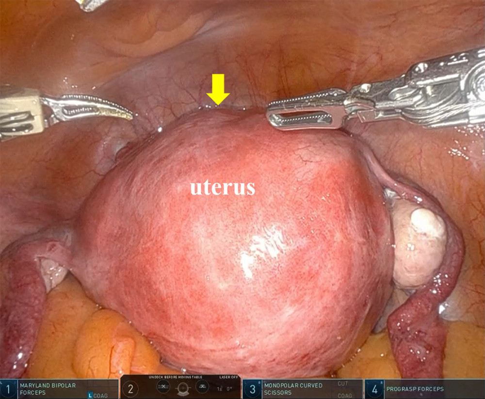 Figure 1. Intraoperative findings at the beginning of RASH. The enlarged uterus caused by adenomyosis was visible within the pelvic cavity (arrow). RASH, robot-assisted hysterectomy.
 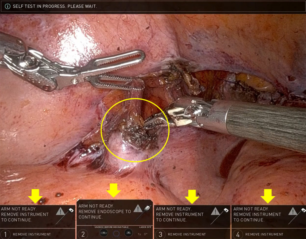 Figure 2. Emergency shutdown of the robotic system during RASH. After completion of vaginal cuff closure and intraperitoneal irrigation, an unrecoverable emergency shutdown of the da Vinci X system occurred during confirmation of hemostasis around the left infundibulopelvic ligament (circle). The console operation was suddenly disabled, making continuation of intracorporeal robotic manipulation impossible. The robotic system error messages displayed on the console monitor are shown (arrows). RASH, robot-assisted hysterectomy.
 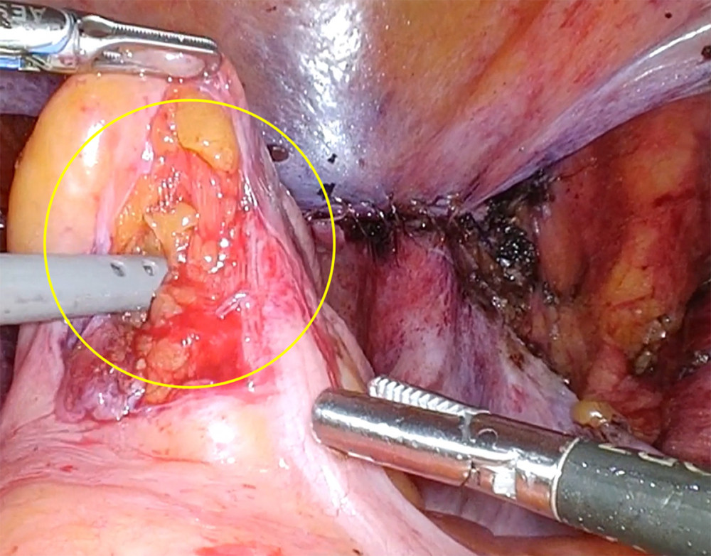 Figure 3. Intraoperative finding of rectal serosal injury (circle) that occurred during mobilization of the rectum using the prograsp forceps. The defect measured approximately 3 cm and was repaired laparoscopically using a single interrupted suture.
 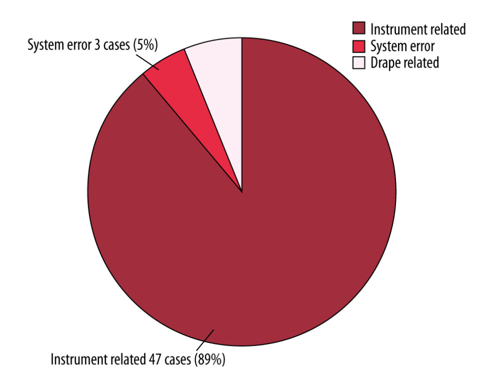 Figure 4. Classification of malfunctions during robot-assisted surgery at our hospital. Among 53 malfunction cases identified in 678 robot-assisted surgeries, instrument-related failures were the most common (47 cases, 89%). System errors occurred in 3 cases, including the unrecoverable malfunction in the present case, which required conversion to laparoscopic surgery.
 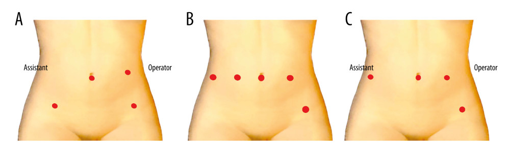 Figure 5. Comparison of port placement between standard laparoscopic surgery and robot-assisted surgery. (A) Port placement during standard laparoscopic surgery. (B) Port placement during robotic surgery. (C) Port placement after conversion from robotic surgery to laparoscopic surgery.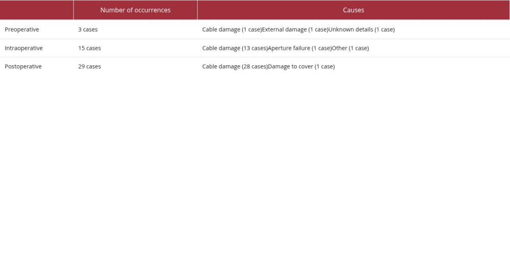
References
1. Surico D, Aquino CI, Vigone A, Robot-assisted hysterectomy for endometrial cancer: Is there a correlation between uterine diameters, pelvic volume, and surgical outcomes?: J Robot Surg, 2025; 19(1); 661
2. Chen C-C, Ou Y-C, Yang C-K, Malfunction of the da Vinci robotic system in urology: Int J Urol, 2012; 19; 736-40
3. Lanfranco AR, Castellanos AE, Desai JP, Meyers WC, Robotic surgery: A current perspective: Ann Surg, 2004; 239(1); 14-21
4. Kim WT, Ham WS, Jeong W, Failure and malfunction of da VINCI surgical systems during various robotic surgeries: Experience from six departments at a single institute: Urology, 2009; 74(6); 1234-37
5. Borden LS, Kozlowski PM, Porter CR, Corman JM, Mechanical failure rate of da VINCI robotic systems: Can J Urol, 2007; 14(2); 3499-501
6. Rajih E, Tholomier C, Cormier B, Error reporting from the da Vinci Surgical System un robotic surgery: A Canadian multispecialty experience at a single academic centre: Can J Urol, 2017; 11(5); 197-202
7. Jones N, Fleming ND, Nick AM, Conversion from robotic surgery to laparotomy: A case-control study evaluating risk factors for conversion: Gynecol Oncol, 2014; 134(2); 238-42
8. Flynn J, Larach JT, Kong JCH, The learning curve in robotic colorectal surgery compared with laparoscopic colorectal surgery: A systematic review: Colorectal Dis, 2021; 23(11); 2806-20
9. Vasudevan V, Reusche R, Wallace H, Kaza S, Clinical outcomes and cost-benefit analysis comparing laparoscopic and robotic colorectal surgeries: Surg Endosc, 2016; 30(12); 5490-93
Figures
Figure 1. Intraoperative findings at the beginning of RASH. The enlarged uterus caused by adenomyosis was visible within the pelvic cavity (arrow). RASH, robot-assisted hysterectomy.Figure 2. Emergency shutdown of the robotic system during RASH. After completion of vaginal cuff closure and intraperitoneal irrigation, an unrecoverable emergency shutdown of the da Vinci X system occurred during confirmation of hemostasis around the left infundibulopelvic ligament (circle). The console operation was suddenly disabled, making continuation of intracorporeal robotic manipulation impossible. The robotic system error messages displayed on the console monitor are shown (arrows). RASH, robot-assisted hysterectomy.Figure 3. Intraoperative finding of rectal serosal injury (circle) that occurred during mobilization of the rectum using the prograsp forceps. The defect measured approximately 3 cm and was repaired laparoscopically using a single interrupted suture.Figure 4. Classification of malfunctions during robot-assisted surgery at our hospital. Among 53 malfunction cases identified in 678 robot-assisted surgeries, instrument-related failures were the most common (47 cases, 89%). System errors occurred in 3 cases, including the unrecoverable malfunction in the present case, which required conversion to laparoscopic surgery.Figure 5. Comparison of port placement between standard laparoscopic surgery and robot-assisted surgery. (A) Port placement during standard laparoscopic surgery. (B) Port placement during robotic surgery. (C) Port placement after conversion from robotic surgery to laparoscopic surgery. In Press
Case report 
Am J Case Rep In Press; DOI: 10.12659/AJCR.953317
Case report 
Am J Case Rep In Press; DOI: 10.12659/AJCR.954643
Case report 
Am J Case Rep In Press; DOI: 10.12659/AJCR.954074
Case report 
Am J Case Rep In Press; DOI: 10.12659/AJCR.953592
Most Viewed Current Articles
07 Dec 2021 : Case report
28,712,429
DOI :10.12659/AJCR.934347
Am J Case Rep 2021; 22:e934347
06 Dec 2021 : Case report  180,530
DOI :10.12659/AJCR.934406
Am J Case Rep 2021; 22:e934406
21 Jun 2024 : Case report
124,276
DOI :10.12659/AJCR.944371
Am J Case Rep 2024; 25:e944371
08 May 2026 : Case report
95,045
DOI :10.12659/AJCR.952158
Am J Case Rep 2026; 27:e952158