


22 August 2026: Articles 
Werner Syndrome Masquerading as Type 2 Diabetes: A Diagnostic Odyssey Leading to Precision Medicine
Challenging differential diagnosis, Unusual or unexpected effect of treatment, Rare disease
Aibibai Yusufu AEFG 1,2*, Asiguli Adili EF 3,2, Sheng Jiang AE 1,2, Yihan Wei E 1,2, Hailin Ma E 1,2DOI: 10.12659/AJCR.952718
Am J Case Rep 2026; 27:e952718


Abstract
BACKGROUND: Werner syndrome (WS), also called adult progeria, is a rare hereditary progeroid disorder characterized by highly heterogeneous clinical manifestations, often leading to delayed diagnosis or misdiagnosis. Diabetes mellitus affects ~ 55% of patients with WS but is frequently misclassified as type 1 or type 2 diabetes due to overlapping metabolic features. This report aims to highlight the diagnostic challenges of WS-associated diabetes, emphasize the role of genetic testing in establishing the correct etiological diagnosis, and demonstrate how precision treatment concerning the underlying genetic defect can improve glycemic control and patient outcomes.
CASE REPORT: A 50-year-old man was admitted to the First Affiliated Hospital of Xinjiang Medical University in September 2024 with a history of poorly controlled diabetes. He exhibited diabetes mellitus, hyperlipidemia, hoarseness, and atrophy of subcutaneous fat and muscles in the face and limbs. His parents were consanguineous. Genetic sequencing revealed a homozygous mutation in the WRN gene (c.1846G>C, p.Ala616Pro), confirming the diagnosis of WS. His diabetes was subsequently reclassified as a specific type of monogenic diabetes rather than type 2 diabetes, and an appropriate precision treatment plan was developed.
CONCLUSIONS: This case underscores the importance of recognizing the association between WS and diabetes mellitus, particularly in patients with signs of premature aging. Early diagnosis and individualized management strategies are crucial for improving patient outcomes. Genetic testing plays a key role in confirming the diagnosis of WS and preventing misdiagnosis as type 1 or type 2 diabetes. This case also highlights the importance of identifying and managing rare diseases in clinical practice.
Keywords: Werner Syndrome, Progeria, Adult, genetic testing, Case Reports
Introduction
Werner syndrome (WS), also called adult progeria, is a rare autosomal recessive genetic disorder first described by Otto Werner in 1904 [1]. It has an extremely low estimated incidence, approximately 1 in 10 million to 1 in 1 million individuals. Relatively higher incidence rates have been reported in regions such as Japan, Sardinia, India, and Pakistan. The primary characteristics of WS include premature aging and highly heterogeneous clinical manifestations. The disease is mainly caused by mutations in the
This report describes the diagnostic and therapeutic course of a patient with WS who exhibited hyperglycemia and hyperlipidemia. The case highlights the critical importance of recognizing the distinctive clinical features and genetic basis of WS, particularly in the context of diabetes management. Through detailed clinical evaluation, genetic testing, and individualized therapeutic interventions, we aim to improve awareness of this rare form of diabetes associated with a genetic disorder. This case also underscores the importance of early diagnosis and personalized management strategies.
Case Report
A 50-year-old man presented with polydipsia, polyuria, polyphagia, and weight loss beginning in 2013. He was admitted to our hospital, where his random blood glucose level was 19.5 mmol/L and triglyceride level was 19.53 mmol/L. He was initially diagnosed with type 2 diabetes, hyperlipidemia, osteopenia, and lacunar infarction. His treatment regimen included subcutaneous injections of biphasic insulin aspart 30 (18 units before breakfast and 16 units before dinner), oral metformin (0.5 g, 3 times daily), and fenofibrate (0.2 g once daily). Despite these interventions, glycemic control remained poor, and he was repeatedly admitted to tertiary hospitals over the following years. He received multiple insulin regimens, including premixed insulin, basal insulin, and basal-bolus insulin therapy, along with oral antidiabetic agents such as biguanides, sulfonylureas, and α-glucosidase inhibitors. However, glycemic control remained suboptimal.
In 2018, the patient developed fatigue, progressive hair graying, alopecia, and reduced hair density; he reported feeling physically weaker than his peers. In 2019, he exhibited a symmetrical reduction in subcutaneous fat in the limbs and muscle atrophy, particularly involving the palms and soles. The following year, he experienced numbness in the limbs and blurred vision in both eyes. In 2022, after evaluation in our department, he was diagnosed with familial-partial-lipodystrophy-associated diabetes, diabetic peripheral neuropathy, and nonproliferative diabetic retinopathy. His treatment regimen was adjusted to include insulin aspart (16 units before each meal) and insulin degludec (38 units at bedtime), whereas his oral antidiabetic medications remained unchanged.
In 2023, the patient noticed a pronounced reduction in subcutaneous fat in the limbs, accompanied by facial fat atrophy and sunken cheeks. Genetic testing revealed a homozygous missense mutation in the
The patient had achieved normal developmental milestones during infancy, including head control at 3 months, rolling over at 6 months, crawling at 8 months, and independent standing at 12 months. During childhood, his growth rate was comparable to that of his peers; however, he did not experience a pubertal growth spurt, resulting in short stature (final height: 170 cm). He was diagnosed with hypertension in 2013 (peak blood pressure: 160/110 mm Hg) but was nonadherent to antihypertensive therapy. Bilateral high-frequency sensorineural hearing loss was diagnosed in 2015 but remained untreated. In 2020, he was diagnosed with an anxiety disorder and treated with lorazepam, which was subsequently discontinued. He also reported poor sleep quality. Cataracts were diagnosed in 2024, and no surgical intervention had been performed. His surgical history included an appendectomy in February 2002 and cholecystectomy in 2016, both with uneventful recoveries.
On physical examination, the patient was 50 years old chronologically but appeared to be in his sixties due to premature aging features characteristic of WS. He had a high-pitched, hoarse voice. His height was 170 cm, weight was 62 kg, body mass index was 21 kg/m2, and blood pressure was 145/94 mm Hg. He was conscious, alert, and fully oriented, with a Glasgow Coma Scale score of 15. His hair was sparse and gray-white. Mild bilateral proptosis was present, and subcutaneous fat in the cheeks was greatly reduced. He had a pointed nose, resulting in a characteristic bird-like facial appearance. His limbs were slender; the skin of both feet appeared mildly erythematous, taut, and thin. Pronounced atrophy of subcutaneous fat was observed in the buttocks and limbs, accompanied by substantial muscle atrophy over the lateral gluteal regions (Figure 1). No acanthosis nigricans was observed.
Laboratory investigations revealed hypertriglyceridemia, insulin resistance with elevated blood glucose and glycated hemoglobin A1c levels, normal ketone body and plasma lactic acid levels, and negative diabetes-related autoantibodies (Table 1).
Whole-body magnetic resonance imaging demonstrated symmetrical atrophy of subcutaneous fat throughout the body, except in the chest region, as well as mild hepatic steatosis. Ultrasound examination of the neck and lower-extremity arteries revealed atherosclerosis with multiple plaques. Electromyography showed bilateral motor and sensory nerve damage in the lower limbs. Fundus photography confirmed diabetic retinopathy. Bone mineral density assessment demonstrated osteopenia, with a lowest T-score of -1.6. Electronic laryngoscopy revealed a raised lesion on the left side of the posterior nasopharyngeal wall with a generally smooth surface. Genetic testing by whole-exome sequencing identified a homozygous
The treatment regimen was adjusted to include subcutaneous insulin degludec at 30 IU at bedtime and subcutaneous insulin lispro at 14 IU before each meal. Oral medications included metformin 0.5 g, 4 times daily; pioglitazone 45 mg once daily; dapagliflozin 10 mg once daily; fenofibrate 0.2 g once daily; acipimox 0.25 g twice daily; and atorvastatin 10 mg at bedtime, along with additional symptomatic treatments as needed.
Discussion
STRENGTHS AND LIMITATIONS OF THIS CASE:
This case report contributes several unique aspects to the existing literature on WS-associated diabetes. First, this case is among few reported cases of WS originating from Central Asia (Xinjiang, China), thereby expanding the geographic and ethnic spectrum of documented
Second, the patient experienced a prolonged diagnostic delay of 11 years (2013–2024), during which his diabetes was misclassified as type 2 diabetes. This aspect highlights the persistent challenge of recognizing WS in clinical practice, particularly when features of premature aging emerge gradually and are attributed to unrelated conditions.
Third, we identified a homozygous missense mutation, c.1846G>C (p.Ala616Pro), in the WRN gene. To our knowledge, this specific mutation has not been previously reported, thus expanding the mutational spectrum of WRN. The only structurally similar mutation reported within the same exon region is the nonsense variant c.1111G>T (p.Glu371*) described in a Lebanese family by Jaafar et al [17], which differs in both location and functional consequence (nonsense versus missense).
Fourth, this case demonstrates successful precision therapy guided by genetic diagnosis. The treatment strategy was modified from insulin monotherapy (86 U/day with poor glycemic control) to a combination of basal-bolus insulin, metformin, pioglitazone, and a sodium-glucose cotransporter 2 inhibitor, resulting in reduced insulin requirements and improved glycemic stability. This approach is consistent with emerging evidence that insulin sensitizers may be more effective than insulin alone in managing insulin resistance associated with WS.
This report has several limitations. First, as a single-case report, its findings require validation in larger cohorts; however, the rarity of WS inherently limits the feasibility of large case series.
Second, follow-up after genetic diagnosis was relatively short. Although glycemic stability was observed during the 3-month period after treatment modification, longer-term outcomes (eg, cardiovascular events, malignancy risk, and survival) remain unknown.
Third, functional studies were not performed to validate the effect of the c.1846G>C mutation on
Fourth, family screening was incomplete because only 3 first-degree relatives underwent genetic testing, and extended pedigree analysis was limited by geographic dispersion.
Finally, patient-reported outcomes were not assessed via standardized quality-of-life instruments.
Conclusions
This case highlights the importance of recognizing the diverse clinical manifestations of WS and the critical role of genetic testing in establishing an accurate diagnosis. Identification of the
Figures
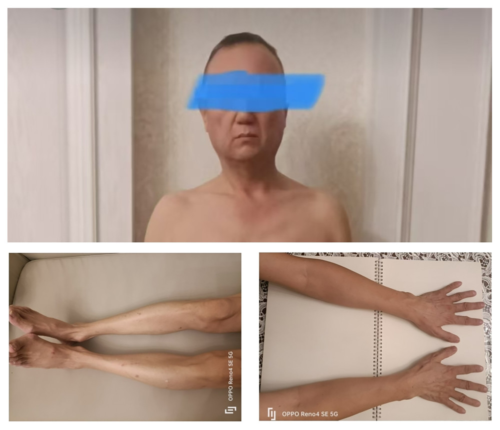 Figure 1. Atrophy of subcutaneous fat in the buttocks and limbs, accompanied by pronounced muscle atrophy of the lateral gluteal regions.
 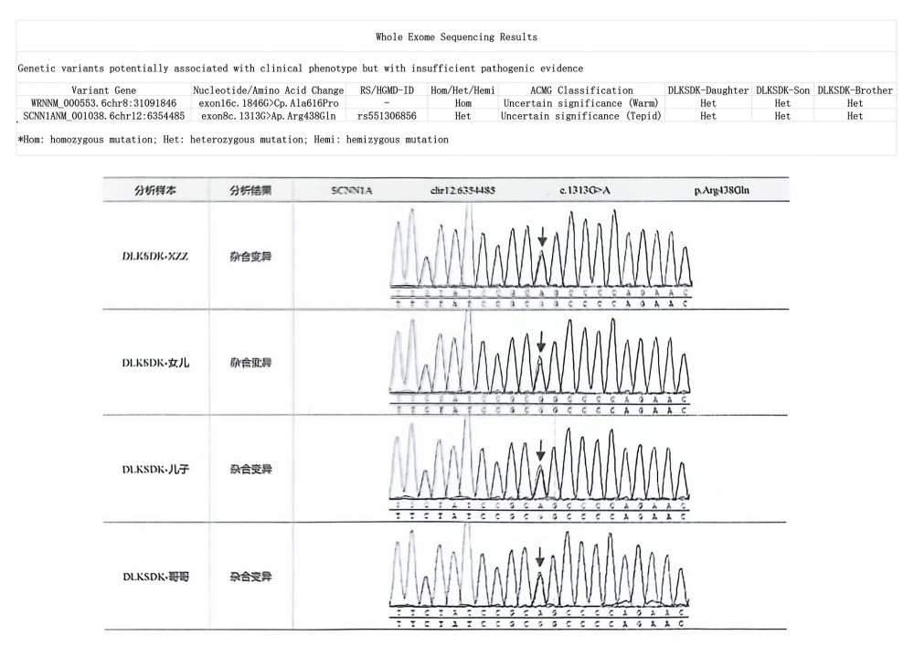 Figure 2. Whole-exome sequencing revealed a homozygous WRN gene mutation, c.1846G>C (p.Ala616Pro). The patient’s daughter, son, and 1 brother carried the mutation in the heterozygous state.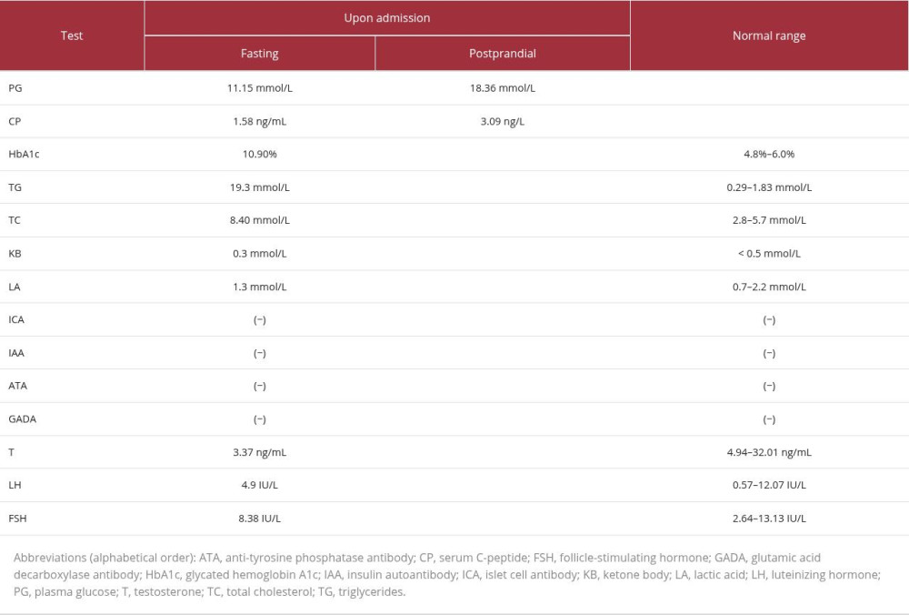
References
1. Oshima J, Sidorova JM, Monnat RJ, Werner syndrome: Clinical features, pathogenesis and potential therapeutic interventions: Ageing Res Rev, 2017; 33; 105-14
2. Oshima J, Hisama FM, Search and insights into novel genetic alterations leading to classical and atypical Werner syndrome: Gerontology, 2014; 60(3); 239-46
3. Saito T, Shimoda Y, Tagaya Y, A case of Werner syndrome with type 1 diabetes showing insulin resistance: Folia Endocrinol Jpn, 2014; 90; 345
4. Furuya M, Kurokawa R, Fujita N, A case of Werner syndrome diagnosed during treatment of juvenile diabetes: Folia Endocrinol Jpn, 2011; 87; 351
5. Kaylan KB, Philipson LH, Werner syndrome and diabetes: Opportunities for precision medicine: Diabetes Care, 2024; 47(5); 785-86
6. Bilgiç Ö, Do you know this syndrome? Werner syndrome: An Bras Dermatol, 2017; 92(2); 271-72
7. Lauper JM, Krause A, Vaughan TL, Spectrum and risk of neoplasia in Werner syndrome: A systematic review: PLoS One, 2013; 8(4); e59709
8. Tian Y, Lautrup S, Law P, WRN loss accelerates abnormal adipocyte metabolism in Werner syndrome: Cell Biosci, 2024; 14(1); 7
9. Yokote K, Werner syndrome and metabolic syndrome: Nihon Rinsho, 2006; 64(Suppl 9); 742-46
10. Tsukamoto K, Takemoto M, Kubota Y, Management guideline for Werner syndrome 2020 1. Dyslipidemia and fatty liver associated with Werner syndrome: Geriatr Gerontol Int, 2021; 21(2); 133-38
11. American Diabetes Association, 2. Classification and diagnosis of diabetes: Diabetes Care, 2015; 38(Suppl); S8-16
12. Yokote K, Honjo S, Kobayashi K, Metabolic improvement and abdominal fat redistribution in Werner syndrome by pioglitazone: J Am Geriatr Soc, 2004; 20; 1582-83
13. Hattori S, Kasai M, Namatame T, Pioglitazone treatment of insulin resistance in a patient with Werner’s syndrome: Diabetes Care, 2004; 27; 3021-22
14. Kitamoto T, Takemoto M, Fujimoto M, Sitagliptin successfully ameliorates glycemic control in Werner syndrome with diabetes: Diabetes Care, 2012; 35; e83
15. Ide S, Yamamoto M, Takemoto M, Improved glycemic control and vascular function and reduction of abdominal fat accumulation with liraglutide in a case of Werner syndrome with diabetes mellitus: J Am Geriatr Soc, 2016; 64; 687-88
16. Yamamoto R, Akasaki K, Horita M, Evaluation of glucose tolerance and effect of dietary management on increased visceral fat in a patient with Werner syndrome: Endocr J, 2020; 67(12); 1239-46
17. Jaafar B, Nasrallah M, Sievers B, Werner syndrome in a Lebanese family: Am J Med Genet A, 2022; 188(5); 1630-34
Figures
Figure 1. Atrophy of subcutaneous fat in the buttocks and limbs, accompanied by pronounced muscle atrophy of the lateral gluteal regions.Figure 2. Whole-exome sequencing revealed a homozygous WRN gene mutation, c.1846G>C (p.Ala616Pro). The patient’s daughter, son, and 1 brother carried the mutation in the heterozygous state. In Press
Case report 
Am J Case Rep In Press; DOI: 10.12659/AJCR.953543
Case report 
Am J Case Rep In Press; DOI: 10.12659/AJCR.954216
Case report
Am J Case Rep In Press; DOI: 10.12659/AJCR.954932
Case report 
Am J Case Rep In Press; DOI: 10.12659/AJCR.954146
Most Viewed Current Articles
07 Dec 2021 : Case report
28,712,695
DOI :10.12659/AJCR.934347
Am J Case Rep 2021; 22:e934347
06 Dec 2021 : Case report  180,721
DOI :10.12659/AJCR.934406
Am J Case Rep 2021; 22:e934406
21 Jun 2024 : Case report
124,419
DOI :10.12659/AJCR.944371
Am J Case Rep 2024; 25:e944371
08 May 2026 : Case report
95,462
DOI :10.12659/AJCR.952158
Am J Case Rep 2026; 27:e952158