


24 August 2026: Articles 
Ovarian Lymphoma and Bone Metastasis Presenting With Paraparesis in an Adolescent: A Case Report
Challenging differential diagnosis, Rare disease
Ryan Haryana Darajatun CDEF 1*, Aditiyono AB 2, Siti Salima ABCDEFG 1
DOI: 10.12659/AJCR.952834
Am J Case Rep 2026; 27:e952834


Abstract
BACKGROUND: Lymphoma comprises a heterogeneous group of lymphoid malignancies broadly categorized as Hodgkin lymphoma and non-Hodgkin lymphoma. Ovarian involvement is exceedingly rare, particularly in adolescents, and is most commonly associated with diffuse large B-cell lymphoma (DLBCL) as part of systemic disease. Its clinical and radiological resemblance to primary ovarian malignancies often leads to diagnostic challenges.
CASE REPORT: We describe the case of a 17-year-old nulligravid female adolescent who presented with a 2-month history of progressive bilateral lower extremity weakness accompanied by lower abdominal pain. Radiological evaluation revealed large bilateral ovarian masses with extensive pelvic organ infiltration, vertebral metastases, epidural extension causing spinal cord compression, and pleural effusion. The patient underwent exploratory laparotomy with suboptimal tumor debulking. Histopathological examination, supported by immunohistochemical analysis, demonstrated diffuse cluster of differentiation (CD)20 positivity, CD3 negativity, and a high Ki-67 proliferation index (approximately 80%), consistent with high-grade DLBCL. Despite postoperative supportive care and planned initiation of systemic chemotherapy, the patient’s condition rapidly deteriorated, resulting in death within 3 months of symptom onset.
CONCLUSIONS: Ovarian involvement by DLBCL in adolescents is a rare and highly aggressive clinical entity that can closely mimic advanced epithelial ovarian malignancy. The presence of atypical features, including neurological deficits secondary to spinal involvement, may further complicate diagnosis. Definitive diagnosis relies on histopathological and immunohistochemical confirmation. Early recognition and timely initiation of systemic therapy are essential; however, prognosis remains poor in cases with advanced dissemination.
Keywords: Adolescent, Case Reports, Lymphoma, Large B-Cell, Diffuse, Lymphoma, Non-Hodgkin, Ovarian Neoplasms
Introduction
Lymphoma, a group of malignancies originating from the lymphoid system, is broadly classified as Hodgkin lymphoma or non-Hodgkin lymphoma (NHL). The American Cancer Society reports that approximately 4% of all cancer cases are NHL, making it the most common hematologic malignancy after leukemia [1]. Extranodal involvement is observed in approximately 30% of NHL cases; it most commonly affects the gastrointestinal tract, head and neck region, skin and soft tissues, and central nervous system [2]. Ovarian lymphoma constitutes approximately 0.5% of NHL cases and 1.5% of all ovarian tumors; most cases represent secondary involvement, whereas primary ovarian lymphoma is exceedingly rare [3].
Most cases of ovarian lymphoma occur between 40 and 60 years of age, whereas its occurrence in children and adolescents is extremely rare [4]. In younger patients, ovarian involvement is generally part of systemic disease or diffuse large B-cell lymphoma (DLBCL) with multiorgan dissemination [3]. Clinical and radiological manifestations often resemble those of primary ovarian malignancy. Diagnosis is frequently established after exploratory surgery, followed by histopathological and immunohistochemical evaluation [5]. Here, we present the case of a female adolescent initially suspected to have ovarian carcinoma but subsequently diagnosed with DLBCL based on further diagnostic investigations. Given its rarity, this case highlights the diagnostic challenges and management considerations associated with ovarian DLBCL in adolescents.
Case Report
A 17-year-old nulligravid (G0P0A0), unmarried female adolescent presented to the emergency department with a 2-month history of progressive bilateral lower-extremity weakness that had worsened to the point that movement became difficult. The weakness was accompanied by bilateral lower-limb numbness. Symptoms were constant and progressively worsened over time. There was no history of trauma. The patient also reported a sensation of abdominal fullness for 1 month, accompanied by lower abdominal pain. Symptoms were not aggravated by weight-bearing or movement; they were relieved by rest. She denied any history of abnormal vaginal bleeding outside the menstrual cycle or urinary and fecal disturbances. Menstrual history was unremarkable, with menarche at 13 years of age, regular cycles every 28 to 30 days, and no severe dysmenorrhea. The patient denied weight loss and was not sexually active.
Physical examination revealed normal vital signs. The patient weighed 49 kg and was 159 cm tall, with a body mass index of 19.4 kg/m2. Neurological examination demonstrated motor strength of 1/5 in both lower extremities. Abdominal examination showed distension with suprapubic tenderness but no guarding. Shifting dullness and flank dullness were present. A solid abdominal mass measuring approximately 8 × 10 cm was palpable, with indistinct margins and limited mobility. No palpable lymphadenopathy was detected. A pelvic examination was not performed because of the patient’s unmarried status (in accordance with local cultural practices).
Laboratory evaluation revealed a hemoglobin level of 11.1 g/dL (reference range: 10.9–14.9 g/dL); all other parameters were within normal limits. A pregnancy test (beta-human chorionic gonadotropin) was not performed. Contrast-enhanced abdominal computed tomography (CT) demonstrated a heterogeneous solid pelvic mass measuring approximately 10.4 × 10.9 × 10.4 cm, which had infiltrated the uterus, bilateral adnexa, ileocecal region, sigmoid colon, and rectum. Minimal ascites and mesenteric thickening suggestive of peritoneal seeding were also observed. Additionally, bone metastasis involving the T11 vertebra was identified, along with a right paravertebral mass extending from T9 to L3, left pleural effusion, and compressive atelectasis of the left lower lobe (Figure 1).
Contrast-enhanced thoracic CT demonstrated infiltration of the T7 to T12 vertebrae and epidural space, resulting in severe spinal canal stenosis with compressive myelopathy, which supported metastatic involvement (Figure 1). Contrast-enhanced lumbosacral magnetic resonance imaging revealed altered signal intensity in the L1 vertebra and a paravertebral mass extending from L1 to L3, infiltrating the right longissimus dorsi, right iliocostalis lumborum, and right psoas muscles, as well as the proximal right ureter, resulting in right-sided hydronephrosis (Figures 2, 3). Furthermore, solid masses with cystic components were identified in the right adnexa, measuring 8.6 × 7.4 cm, and in the left adnexa, measuring 7.6 × 6.0 cm, with infiltration of adjacent intestinal structures. Contrast-enhanced chest CT demonstrated compression of the T12 vertebra with a paravertebral mass extending from T11 to L3, suggestive of metastatic disease. Left-sided pleural effusion with compressive atelectasis involving segments 6 and 10 of the left lung was observed. A solid lesion measuring 3.9 × 1.9 cm was noted in the anterior mediastinum, likely representing thymic tissue.
The patient subsequently underwent exploratory laparotomy. Intraoperative findings included minimal ascites and a uterus measuring 7 × 5 × 5 cm with adhesions involving the ovaries, cecum, pouch of Douglas, urinary bladder, and rectosigmoid colon. The left ovary appeared as a partially solid cystic mass measuring 8 × 8 × 8 cm, adherent to the uterus, rectosigmoid colon, pouch of Douglas, and omentum. The right ovary contained a partially solid cystic mass measuring 10 × 8 × 8 cm with similar adhesions (Figure 4). Peritoneal seeding was observed.
Frozen-section examination of the right adnexa revealed malignant features; therefore, debulking surgery was performed, including supravaginal hysterectomy, bilateral salpingo-oophorectomy, peritoneal lavage, omentectomy, and peritoneal biopsies from the bilateral pelvic peritoneum, bilateral paracolic gutters, pouch of Douglas, and prevesical peritoneum. Intraoperative blood loss was approximately 1000 mL. Total hysterectomy was not performed because of dense adhesions involving the cervix, pelvic wall, and rectosigmoid colon; thus, residual tumor exceeded 1 cm. Postoperatively, the patient was diagnosed as P0A0, status post suboptimal debulking for clinically staged IVB ovarian cancer with bone metastases, pleural effusion, and grade III to IV adhesions involving the rectosigmoid colon, pouch of Douglas, and omentum.
Histopathological examination revealed malignant lymphoma; adult granulosa cell tumor was considered in the differential diagnosis. The tumor involved the right ovary, with metastatic spread to the uterus, left ovary, peritoneum, pelvic lymph nodes, bilateral paracolic peritoneum, prevesical peritoneum, pouch of Douglas, and omentum. Immunohistochemical analysis demonstrated negative cluster of differentiation (CD)3 staining, diffuse CD20 positivity, and a Ki-67 proliferation index of approximately 80%, consistent with a diagnosis of high-grade DLBCL (Figure 5).
Postoperatively, the patient was admitted to the intensive care unit for hemodynamic monitoring. She received transfusions of 3 units of packed red blood cells and was discharged on postoperative day 5. During follow-up at the gynecologic oncology outpatient clinic, she was readmitted to improve her general condition due to poor oral intake and anemia. The patient was subsequently referred to the hematology-oncology department for further optimization and was scheduled to receive the first cycle of chemotherapy 78 days after initial presentation to the emergency department, pending clinical improvement. On the fifth day of hospitalization, the patient’s condition deteriorated, with suspected obstructive ileus, prompting emergency exploratory laparotomy by the general surgery team. Intraoperatively, partial obstructive ileus secondary to grade IV small-bowel adhesions was identified, and sharp adhesiolysis was performed. Postoperatively, the patient remained intubated and received supportive treatment in the intensive care unit. She died 71 days after initial presentation to the emergency department.
Discussion
Lymphoma, a hematologic malignancy originating from lymphoid cells, represents approximately 3% to 4% of all malignancies [1]. Ovarian tumors may arise from various tissues, including epithelial, germ cell, and gonadal stromal origins. Sex cord-stromal cell tumors are rare, comprising approximately 5% to 8% of all ovarian malignancies, with peak incidence in middle-aged and older women [6]. In contrast, NHL is a heterogeneous lymphoproliferative neoplasm that can involve both lymphoid and extranodal organs, including the ovaries, and may mimic other types of ovarian cancer. NHL risk is increased in immunocompromised individuals and those with certain viral infections, including human immunodeficiency virus, Epstein-Barr virus, and hepatitis C virus infection. Environmental exposures, including pesticides, benzene, and herbicides, have also been associated with the development of DLBCL [7,8]. The ovary is the most common gynecologic site of NHL involvement [9].
Lymphoma may involve the ovary as either a primary or secondary disease. In primary ovarian lymphoma, the neoplasm originates in the ovary; spread is initially limited to adjacent lymph nodes or structures. More distant lesions typically become evident several months later. In secondary ovarian lymphoma, ovarian involvement occurs through hematogenous or lymphatic dissemination of systemic disease. One pathogenic theory proposes that ovarian lymphoma arises from lymphocytes located within vascular structures of the ovarian hilum and corpus luteum [10]. The most common sites of lymphoma metastasis include the lymph nodes, liver, bone marrow, lungs, and gastrointestinal tract [3]. In the present case, the pattern of multiorgan involvement suggested hematogenous and infiltrative spread, consistent with systemic lymphoma rather than ovarian carcinoma. The presence of an extensive paravertebral mass infiltrating the epidural space, vertebral bodies, and paraspinal muscles, along with bilateral adnexal involvement and the absence of predominant ascites or extensive peritoneal implantation, supports a lymphoid neoplastic process disseminating via the bloodstream and regional lymphoid tissues [6].
Establishing a diagnosis of ovarian lymphoma is challenging because primary ovarian lymphoma is often misdiagnosed as epithelial ovarian malignancy. Primary ovarian lymphoma represents approximately 0.5% of all NHL cases and 1.5% of ovarian neoplasms. Patients with NHL commonly display fever, weight loss, or night sweats, collectively known as B symptoms. More than two-thirds of patients exhibit painless peripheral lymphadenopathy. Clinical manifestations vary according to disease location and subtype [9,11]. Ovarian lymphoma often presents with bilateral involvement, rapid growth, and systemic symptoms such as fever, weight loss, and night sweats. Another characteristic feature is the involvement of extranodal organs (eg, bone marrow, vertebrae, or paravertebral tissues), which is rarely observed in sex cord-stromal tumors [7,8]. In the present case, early imaging studies revealed vertebral involvement, which helped distinguish ovarian lymphoma from sex cord-stromal tumor.
The diagnostic criteria for primary ovarian lymphoma were proposed by Fox and Langley. First, the lymphoma must be clinically confined to the ovary at the time of diagnosis, without evidence of lymphoma elsewhere on comprehensive evaluation. Involvement of immediately adjacent lymph nodes is still considered consistent with primary ovarian lymphoma. Second, peripheral blood and bone marrow should not contain abnormal lymphoid cells. Third, lesions at sites remote from the ovary should not appear until several months after the detection of ovarian and adjacent extraovarian lesions [12]. Based on these criteria, it is difficult to diagnose primary ovarian lymphoma, particularly in the absence of histopathological examination [9]. Our patient presented with progressive lower-extremity weakness, an atypical presentation that did not initially suggest ovarian lymphoma and thus further complicated the diagnostic process. These symptoms were most likely caused by lymphomatous dissemination involving the vertebrae, which was only identified after radiological evaluation.
Histopathological and immunohistochemical examinations are essential for establishing the diagnosis; lymphoid markers such as CD5, CD19, CD20, CD22, CD30, CD45, CD79a, BCL-2, and BCL-6 frequently exhibit positivity in DLBCL [11]. CD3 is a crucial immunohistochemical marker for the diagnosis and classification of lymphomas, particularly in distinguishing T-cell lymphomas from B-cell lymphomas [13]. The CD3 molecule is a transmembrane protein complex that forms an integral component of the T-cell receptor and plays a critical role in T-cell activation and signal transduction. In contrast, B-cell lymphomas typically display CD3 negativity but show positivity for B-cell markers such as CD19, CD20, CD79a, and PAX5 [11].
Ki-67 is a cellular proliferation marker widely used in the histopathological evaluation of lymphomas to assess tumor proliferative activity. High-grade lymphomas, such as DLBCL, typically exhibit a high Ki-67 index (> 80% to 95%). A high Ki-67 index is generally correlated with rapid disease progression and a favorable initial response to chemotherapy; however, it is also associated with increased relapse risk and lower survival rates in aggressive lymphoma subtypes [11]. Patients with a Ki-67 index of at least 80% have lower 5-year overall survival rates, averaging approximately 40% to 50%, relative to patients with a Ki-67 index below 80%, whose survival rates may reach 70% to 80% [14]. These findings are consistent with observations in our patient, who demonstrated negative CD3 staining, diffuse CD20 positivity, and a Ki-67 proliferation index of 80%, supporting a diagnosis of high-grade DLBCL. The poor clinical outcome in the present case is also consistent with the unfavorable prognosis linked to a high Ki-67 index [14].
Primary treatment for suspected ovarian, fallopian tube, or primary peritoneal cancer generally consists of appropriate surgical staging, with subsequent systemic chemotherapy. Extranodal NHL is typically diagnosed by excisional biopsy or fine-needle aspiration. Because ovarian malignancies are commonly diagnosed and staged surgically, primary ovarian lymphoma is often initially managed via surgery followed by chemotherapy. Surgical management generally includes hysterectomy and bilateral salpingo-oophorectomy, along with comprehensive surgical staging and tumor debulking when indicated. Primary debulking surgery is recommended for advanced-stage disease (stages III and IV), as in the present case. However, surgical morbidity can delay the initiation of chemotherapy and adversely affect outcomes [15,16].
Neoadjuvant chemotherapy followed by interval debulking surgery (IDS) should be considered in patients with advanced-stage ovarian cancer who are poor candidates for primary debulking surgery due to advanced age, poor physical condition, poor performance status, comorbidities, or disease unlikely to be optimally cytoreduced. In most cases, IDS is performed after 2 to 4 cycles of neoadjuvant chemotherapy. A prolonged interval may selectively eliminate chemosensitive tumor cells while allowing chemoresistant clones to persist. Tangjitgamol et al reported no significant difference in survival between patients with advanced ovarian cancer treated via IDS and those undergoing primary surgery. IDS may be particularly beneficial for patients with less extensive tumor burden [15,17].
In contrast, surgery is not considered curative for ovarian lymphoma given the systemic nature of the disease. Diagnosis is established by tissue biopsy, as in the present case; the mainstay of treatment is systemic chemotherapy, most commonly the R-CHOP regimen (rituximab, cyclophosphamide, doxorubicin, vincristine, and prednisone). This regimen has been associated with favorable outcomes, including 5-year survival rates of approximately 70%. Complete remission after treatment has also been reported. In stage I to II disease, R-CHOP may be administered for 3 to 6 cycles, with or without radiotherapy. In stage III to IV disease, 6 cycles of R-CHOP are generally recommended [7,8,18]. Among patients with ovarian lymphoma, fertility preservation remains challenging due to the gonadotoxic effects of chemotherapy regimens. Potential fertility-preserving strategies focus on pre-chemotherapy interventions, including oocyte or embryo cryopreservation [6,19].
Despite available treatments and advances in diagnostic methods, ovarian lymphoma is associated with a poor prognosis, particularly in cases with advanced dissemination. Favorable prognostic factors include unilateral ovarian involvement, focal ovarian disease, and early-stage diagnosis. Conversely, rapid pelvic mass growth, severe systemic symptoms, and non-B-cell lymphoma subtypes are associated with worse outcomes [9,20]. The present case shares similarities with a previously reported case involving a 20-year-old patient. As in that report, the greatest diagnostic challenge was the broad differential diagnosis. Definitive diagnosis is typically established through immunohistochemical evaluation [16]. The unfavorable outcome in our patient was likely related to extensive disease dissemination.
Conclusions
DLBCL presenting as an ovarian tumor is a rare entity, particularly in adolescents. The clinical and radiological features of ovarian lymphoma can closely resemble those of epithelial ovarian cancer. Histopathological examination with immunohistochemical confirmation is essential to establish the diagnosis and guide appropriate management. Clinicians should maintain suspicion for lymphoma when an ovarian mass is accompanied by systemic manifestations or multiorgan involvement. Prompt diagnosis and timely initiation of systemic therapy are critical to improve patient outcomes.
Figures
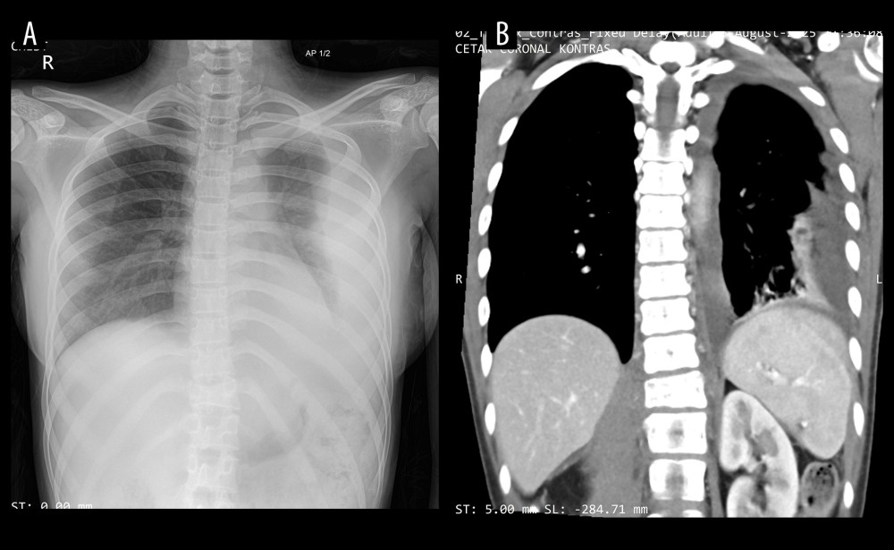 Figure 1. (A) Anteroposterior chest radiograph demonstrating left pleural effusion and compressive atelectasis of the left lower lung. (B) Contrast-enhanced thoracic computed tomography scan demonstrating infiltration of the T7–T12 vertebrae and epidural space.
 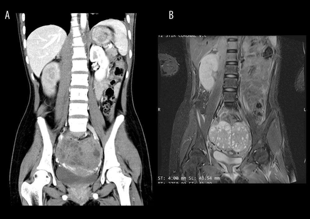 Figure 2. (A) Contrast-enhanced abdominal and lumbosacral computed tomography scan demonstrating compression of the T12 vertebra and a paravertebral mass extending from T11 to L3. (B) Contrast-enhanced abdominal and lumbosacral magnetic resonance image.
 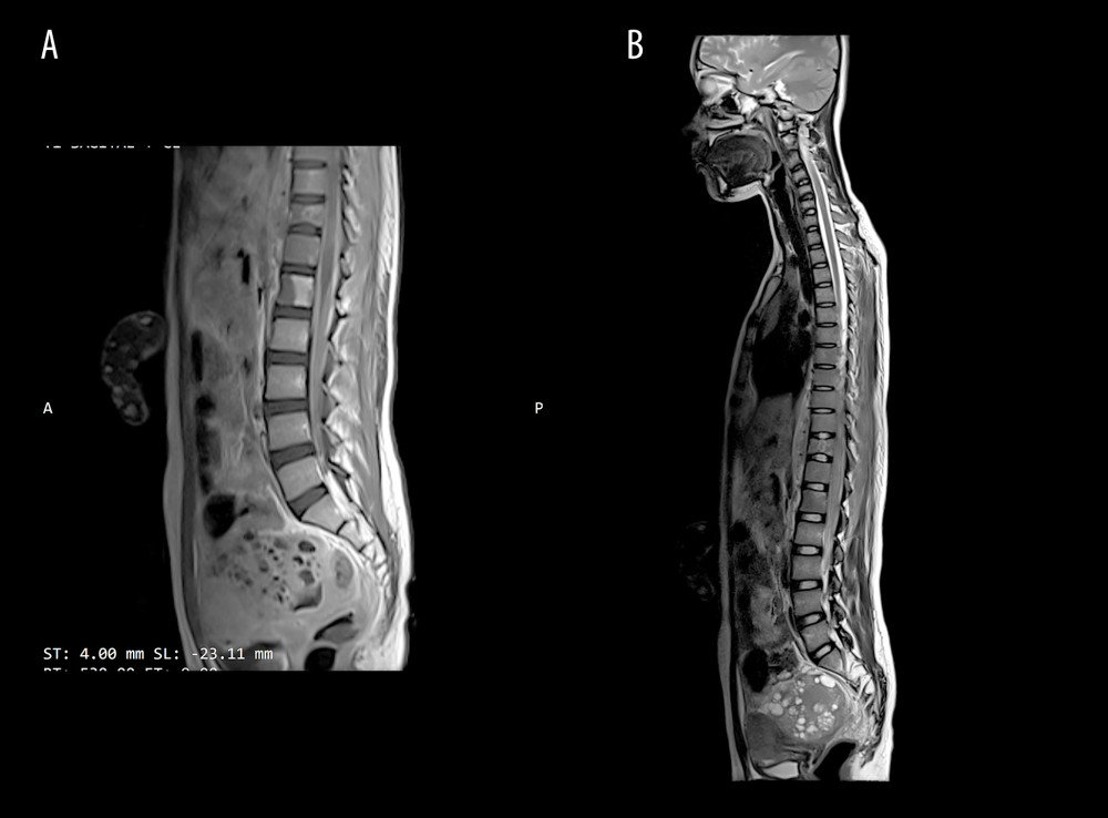 Figure 3. (A) Contrast-enhanced lumbar magnetic resonance image (sagittal view) demonstrating altered signal intensity of the L1 vertebra with a paravertebral mass extending from L1 to L3. (B) Contrast-enhanced thoracic magnetic resonance image demonstrating left-sided pleural effusion, compressive atelectasis involving segments 6 and 10 of the left lung, an anterior mediastinal lesion, a paravertebral mass, and bilateral adnexal masses.
 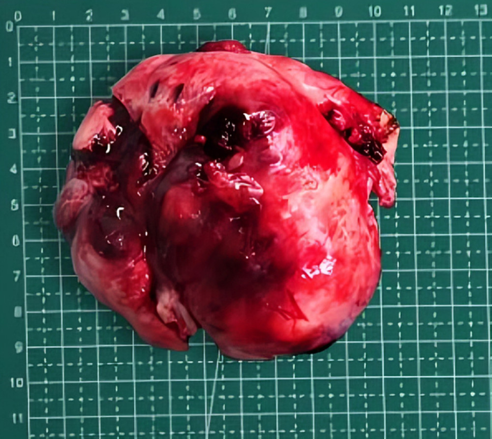 Figure 4. Right ovarian mass.
 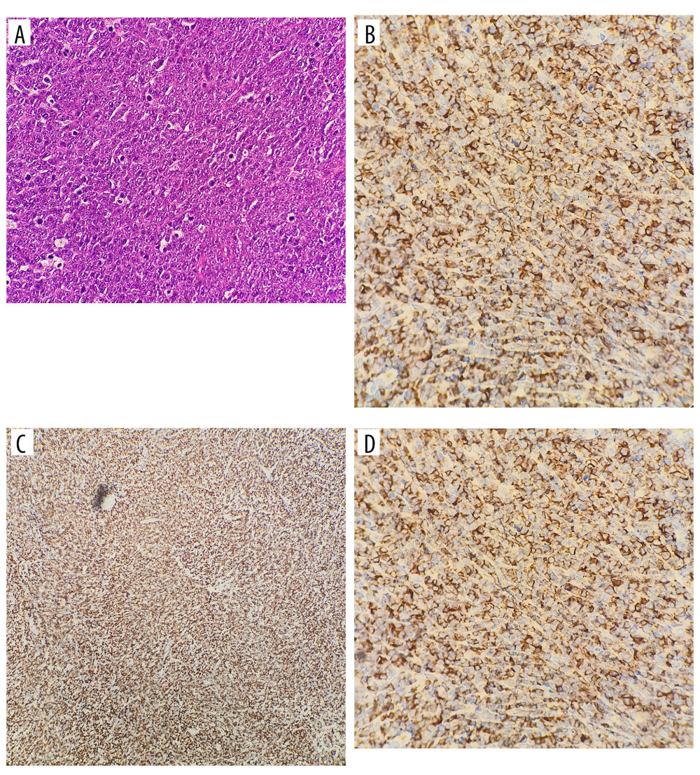 Figure 5. Immunohistochemical examination. (A) Hematoxylin and eosin staining demonstrating diffuse proliferation of large atypical lymphoid cells, consistent with diffuse large B-cell lymphoma. (B) CD20 immunohistochemical staining showing strong diffuse membranous positivity in neoplastic lymphoid cells, confirming B-cell lineage. (C) Ki-67 immunohistochemical staining demonstrating a high proliferative index (approximately 80%) within the tumor cell population. (D) CD3 immunohistochemical staining showing negative staining in neoplastic cells, supporting exclusion of T-cell lymphoma.
 References
1. Siegel RL, Kratzer TB, Giaquinto AN, Cancer statistics, 2025: Cancer J Clin, 2025; 75; 10-45
2. Yang H, Xun Y, Ke C, Tateishi K, You H, Extranodal lymphoma: Pathogenesis, diagnosis and treatment: Mol Biomed, 2023; 4; 29
3. Bhartiya R, Kumari N, Malik M, Singh RVN, Primary non-Hodgkin’s lymphoma of the ovary: A case report: J Clin Diagn Res, 2016; 10(5); ED10-11
4. Monterroso V, Jaffe ES, Merino MJ, Medeiros LJ, Malignant lymphomas involving the ovary: Am J Surg Pathol, 1993; 17; 154-70
5. Campo E, Swerdlow SH, Harris NL, The 2008 WHO classification of lymphoid neoplasms and beyond: Evolving concepts and practical applications: Blood, 2011; 117; 5019-32
6. Horta M, Cunha TM, Sex cord-stromal tumors of the ovary: A comprehensive review and update for radiologists: Diagn Interv Radiol, 2015; 21; 277-86
7. Shankland KR, Armitage JO, Hancock BW, Non-Hodgkin lymphoma: Lancet, 2012; 380; 848-57
8. Kachuri L, Beane Freeman LE, Spinelli JJ, Insecticide use and risk of non-Hodgkin lymphoma subtypes: A subset meta-analysis of the North American Pooled Project: Int J Cancer, 2020; 147; 3370-83
9. Perlman S, Ben-Arie A, Feldberg E, Hagay Z, Non-Hodgkin’s lymphoma presenting as advanced ovarian cancer: A case report and review of literature: Int J Gynecol Cancer, 2005; 15; 554-57
10. Crawshaw J, Sohaib SA, Wotherspoon A, Shepherd JH, Primary non-Hodgkin’s lymphoma of the ovaries: Imaging findings: Br J Radiol, 2007; 80; e155-58
11. Sapkota S, Shaikh H, Non-Hodgkin lymphoma: StatPearls [Internet], 2023, Treasure Island (FL), StatPearls Publishing [cited 2026 Jan 7]. Available from: https://www.ncbi.nlm.nih.gov/books/NBK559328/
12. Fox H, Langley FA, Govan ADT, Malignant lymphoma presenting as an ovarian tumour: A clinicopathological analysis of 34 cases: BJOG, 1988; 95; 386-90
13. Li S, Young KH, Medeiros LJ, Diffuse large B-cell lymphoma: Pathology, 2018; 50; 74-87
14. Huber F, Zwickl-Traxler E, Pecherstorfer M, Singer J, Evaluation of Ki-67 as a prognostic marker in diffuse large B-cell lymphoma—A single-center retrospective cohort study: Curr Oncol, 2021; 28; 4521-29
15. Armstrong DK, Alvarez RD, Bakkum-Gamez JN, Ovarian cancer, version 2.2020, NCCN clinical practice guidelines in oncology: J Natl Compr Canc Netw, 2021; 19; 191-226
16. Gerrity C, Mercadel A, Alghamdi A, Huang M, Primary ovarian lymphoma: A case report: Gynecol Oncol Rep, 2023; 47; 101212
17. Tangjitgamol S, Manusirivithaya S, Laopaiboon M, Interval debulking surgery for advanced epithelial ovarian cancer: Cochrane Database Syst Rev, 2013(4); CD006014
18. Colantonio MA, Dionne C, A rare presentation of ovarian diffuse large B-cell lymphoma: A case report: Radiol Case Rep, 2025; 20; 3409-13
19. Silber S, Ovarian tissue cryopreservation and transplantation: Scientific implications: J Assist Reprod Genet, 2016; 33; 1595-603
20. Zhao XY, Hong XN, Cao JN, Clinical features and treatment outcomes of 14 cases of primary ovarian non-Hodgkin’s lymphoma: A single-center experience: Med Oncol, 2011; 28(4); 1559-64
Figures
Figure 1. (A) Anteroposterior chest radiograph demonstrating left pleural effusion and compressive atelectasis of the left lower lung. (B) Contrast-enhanced thoracic computed tomography scan demonstrating infiltration of the T7–T12 vertebrae and epidural space.Figure 2. (A) Contrast-enhanced abdominal and lumbosacral computed tomography scan demonstrating compression of the T12 vertebra and a paravertebral mass extending from T11 to L3. (B) Contrast-enhanced abdominal and lumbosacral magnetic resonance image.Figure 3. (A) Contrast-enhanced lumbar magnetic resonance image (sagittal view) demonstrating altered signal intensity of the L1 vertebra with a paravertebral mass extending from L1 to L3. (B) Contrast-enhanced thoracic magnetic resonance image demonstrating left-sided pleural effusion, compressive atelectasis involving segments 6 and 10 of the left lung, an anterior mediastinal lesion, a paravertebral mass, and bilateral adnexal masses.Figure 4. Right ovarian mass.Figure 5. Immunohistochemical examination. (A) Hematoxylin and eosin staining demonstrating diffuse proliferation of large atypical lymphoid cells, consistent with diffuse large B-cell lymphoma. (B) CD20 immunohistochemical staining showing strong diffuse membranous positivity in neoplastic lymphoid cells, confirming B-cell lineage. (C) Ki-67 immunohistochemical staining demonstrating a high proliferative index (approximately 80%) within the tumor cell population. (D) CD3 immunohistochemical staining showing negative staining in neoplastic cells, supporting exclusion of T-cell lymphoma. In Press
Case report 
Am J Case Rep In Press; DOI: 10.12659/AJCR.953543
Case report 
Am J Case Rep In Press; DOI: 10.12659/AJCR.954216
Case report 
Am J Case Rep In Press; DOI: 10.12659/AJCR.954932
Case report 
Am J Case Rep In Press; DOI: 10.12659/AJCR.954146
Most Viewed Current Articles
07 Dec 2021 : Case report
28,712,695
DOI :10.12659/AJCR.934347
Am J Case Rep 2021; 22:e934347
06 Dec 2021 : Case report  180,721
DOI :10.12659/AJCR.934406
Am J Case Rep 2021; 22:e934406
21 Jun 2024 : Case report
124,419
DOI :10.12659/AJCR.944371
Am J Case Rep 2024; 25:e944371
08 May 2026 : Case report
95,462
DOI :10.12659/AJCR.952158
Am J Case Rep 2026; 27:e952158