


03 September 2026: Articles 
When Grief Strains the Heart: A Case of Reverse Takotsubo Cardiomyopathy Triggered by Mourning and Respiratory Failure
Rare disease
Peter Cwalina ABCDEF 1*, Ibrahim Rahmatullah DE 2, Ayesha Samad ABCDEF 2, Adam S. Budzikowski CDE 3DOI: 10.12659/AJCR.952896
Am J Case Rep 2026; 27:e952896


Abstract
BACKGROUND: Takotsubo cardiomyopathy is a transient stress-induced cardiomyopathy that can mimic acute coronary syndrome (ACS) but typically occurs without obstructive coronary disease. Reverse takotsubo is an uncommon variant characterized by basal hypokinesis with preserved apical contraction. We report an atypical case in which acute respiratory failure, rather than an abrupt emotional stressor, appeared to be the primary trigger.
CASE REPORT: A 69-year-old woman with coronary artery disease status after prior left anterior descending artery stent, hypertension, hyperlipidemia, and chronic obstructive pulmonary disease (COPD) presented with severe respiratory distress and became unresponsive, requiring emergency intubation. She tested positive for coronavirus OC43 and was managed in the intensive care unit (ICU) for COPD exacerbation with ventilator-dependent respiratory failure. High-sensitivity troponin rose from 56 ng/L to 937 ng/L initially, later peaking at 1928 ng/L after catheterization with ST-T changes on EKG, prompting cardiac evaluation. Transthoracic echocardiography revealed new segmental wall motion abnormalities consistent with stress cardiomyopathy and an estimated ejection fraction of 40%. Urgent coronary angiography demonstrated no obstructive coronary disease with a patent LAD stent; left ventriculography showed severe basal hypokinesis with preserved apical contraction, confirming reverse takotsubo. Further history-taking revealed ongoing grief after her sister’s death 3 months earlier.
CONCLUSIONS: Reverse takotsubo should be considered in patients with ACS-like presentations during acute critical illness, including respiratory failure, even when emotional stressors are chronic rather than sudden. Recognizing basal hypokinesis with preserved apical function can help avoid misdiagnosis and guide appropriate supportive management and follow-up imaging.
Keywords: Cardiology, Case Reports, Echocardiography, Grief, Respiratory Insufficiency, takotsubo cardiomyopathy
Introduction
Takotsubo cardiomyopathy, originally described in 1990 in Japan, refers to a transient reversible cardiomyopathy characterized by an apical ballooning of the left ventricle, resembling that of an octopus trap [1,2]. Classically, takotsubo cardiomyopathy occurs in older women due to a period of emotional or physical stress leading to catecholamine release resulting in symptoms that can mimic myocardial infarction (typically due to plaque rupture). However, takotsubo typically occurs without a culprit obstructive coronary lesion and is characterized by regional wall motion abnormalities extending beyond a single coronary vascular territory. Takotsubo is an acute coronary syndrome mimic with transient left ventricular systolic dysfunction and no culprit obstructive coronary lesion, although concomitant CAD can still be present [3]. Classically, this presents as a hyperdynamic base and akinetic apex, but several other variants have been reported. Takotsubo syndrome includes several morphologic variants, including apical, midventricular, basal (reverse), and focal forms. Reverse takotsubo is an atypical subtype characterized by basal hypokinesis or akinesis with relative apical sparing or hyperkinesis [2,4,5]. We present a case of reverse takotsubo cardiomyopathy triggered by acute respiratory failure rather than sudden emotional distress, underscoring an atypical anatomical and clinical presentation. This case is especially important because it demonstrates a non-classical anatomical pattern (basal hypokinesis with preserved apex), and a non-classical trigger (acute illness rather than sudden grief), highlighting the variability in takotsubo presentation.
Case Report
A 69-year-old woman presented by emergency medical service (EMS) to the emergency department for worsening respiratory distress. She has a significant past medical history of hyperlipidemia, hypertension, coronary artery disease status after stent to the left anterior descending artery 6 years ago (with follow-up cardiac catheterization 2 years ago revealing a patent stent), and chronic obstructive pulmonary disease (COPD) not on home oxygen. On presentation, she was in severe respiratory distress with wheezing and subsequently became unresponsive (Glasgow coma scale 3), requiring emergency intubation. Vital signs were respiratory rate of 21, blood pressure of 149/78, and initial EKG revealed sinus tachycardia (HR 120 bpm). Arterial blood gas analysis revealed pH 7.295, pCO2 50.9, bicarb 24.2, pO2 361, and O2 saturation 99.6% while on an FiO2 of 100%. Viral testing was positive for coronavirus OC43 and she was ultimately admitted to the ICU to be medically managed for COPD exacerbation complicated by acute ventilator-dependent respiratory failure.
During ICU evaluation, high-sensitivity troponin I rose from 56 ng/L and trended upwards to 937 ng/L, prompting a cardiology consultation (Table 1). Initial EKG (Figure 1) on admission showed a sinus tachycardia with ST-T wave changes and old anteroseptal MI. Transthoracic echocardiography (Figure 2) demonstrated severe hypokinesis of the proximal 2/3 of the septal wall anterior lateral and posterior with apical sparing consistent with stress cardiomyopathy (takotsubo) with a new reduction in left ventricular ejection fraction to 40% compared with previously normal systolic function. Following this result, the patient underwent urgent cardiac catheterization, which revealed no culprit obstruction in the left main, circumflex, right coronary artery, and a patent stent in the LAD with her troponin peaking at 1928ng/L after catheterization. Left ventriculography demonstrated preserved apical contraction with severe hypokinesis of the base, confirming a reverse takotsubo variant (Figure 3), highlighting the involvement of the base rather than the typical apex.
Following catheterization, she remained hemodynamically stable and in normal sinus rhythm. Because takotsubo syndrome is often clinically indistinguishable from ACS at presentation, the patient was initially managed with standard ACS directed evaluation while receiving ventilatory support for acute respiratory failure. After coronary angiography excluded an obstructive culprit lesion and ventriculography demonstrated a reverse takotsubo pattern, management was primarily supportive and focused on treatment of the precipitating respiratory illness. The patient received aspirin, heparin, and rosuvastatin. Regarding GDMT, beta-blocker therapy was initially deferred because of active bronchospasm and recent respiratory arrest. Sacubitril-valsartan and spironolactone were started instead. Repeat echocardiography was planned at approximately 90 days to assess for recovery of left ventricular systolic function. Upon further questioning, it was revealed that she had been experiencing significant grief due to the death of her sister 3 months prior, but this was a chronic stressor rather than sudden. Her acute critical illness was determined to be the primary precipitating factor for stress cardiomyopathy.
Ultimately, her clinical status improved with medical management, resulting in her extubation and metoprolol succinate was added. Following further improvement, she was discharged to a subacute rehabilitation facility and regained much of her functional status. Follow-up echocardiography 4 months later revealed an improved LVEF of 60% while continuing her GDMT regimen of metoprolol succinate, sacubitril-valsartan, and spironolactone.
Discussion
Epidemiologically, an estimated 1% to 2% of patients with suspected acute coronary syndrome are diagnosed with takotsubo, but the incidence is likely underestimated due to lack of consideration of pre-hospitalization sudden cardiac death [6]. Interestingly, in a study of 1750 patients with takotsubo, 81.7% had the classic form, while 2.2% were of the inverse variety, with the other varieties making up the remaining total [7]. Further stratifying for sex, we see a predominantly postmenopausal women being affected, with 1 registry reporting a 91% female and 9% male sex distribution [6]. Paradoxically, a study suggested that the reverse variant is actually associated with a younger age of onset (36 years) [5]. This makes our case particularly atypical given the patient’s older age and reverse anatomic pattern. Therefore, we present a case of a rare takotsubo variant given the patient’s demographic factors.
Much of the pathophysiology of takotsubo is still rather unclear, with the prevailing theory revolving around sudden surges of either endogenous or exogenous catecholamines during periods of stress [6]. Additionally, histological changes in takotsubo resemble those seen in human catecholamine toxicity, with recent evidence suggesting that the pathophysiology is related to changes in β-adrenergic receptor signaling. Other proposed pathophysiologies include metabolic stunning of the myocardium, coronary microvascular impairment, and multivessel epicardial coronary artery spasm [2]. Nevertheless, our patient’s main risk factors seemed to be pre-existing CAD and the excessive physiological and emotional stress she had experienced recently.
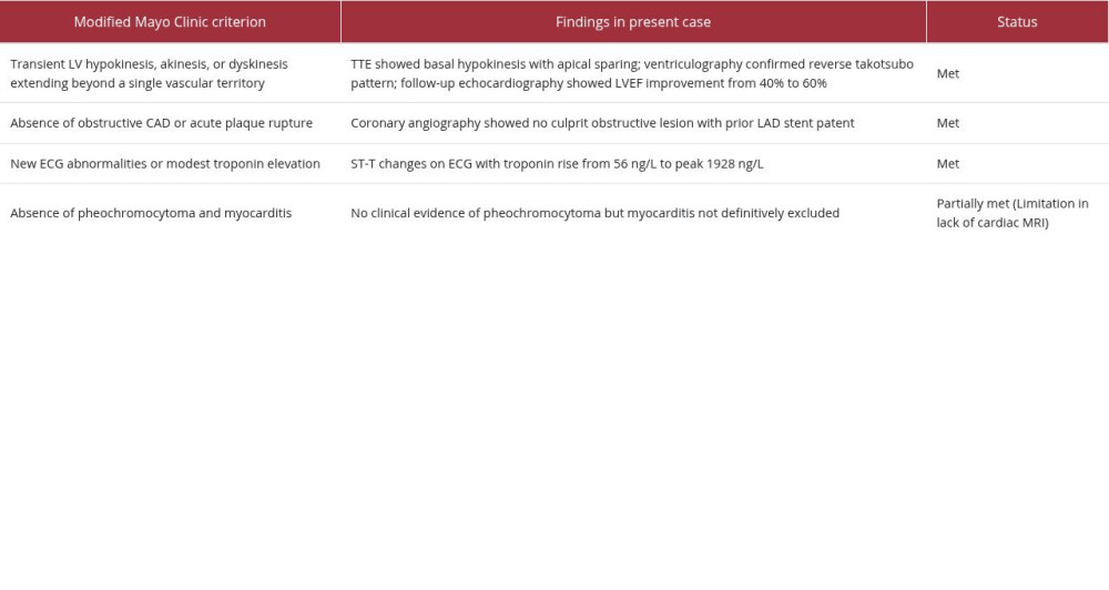
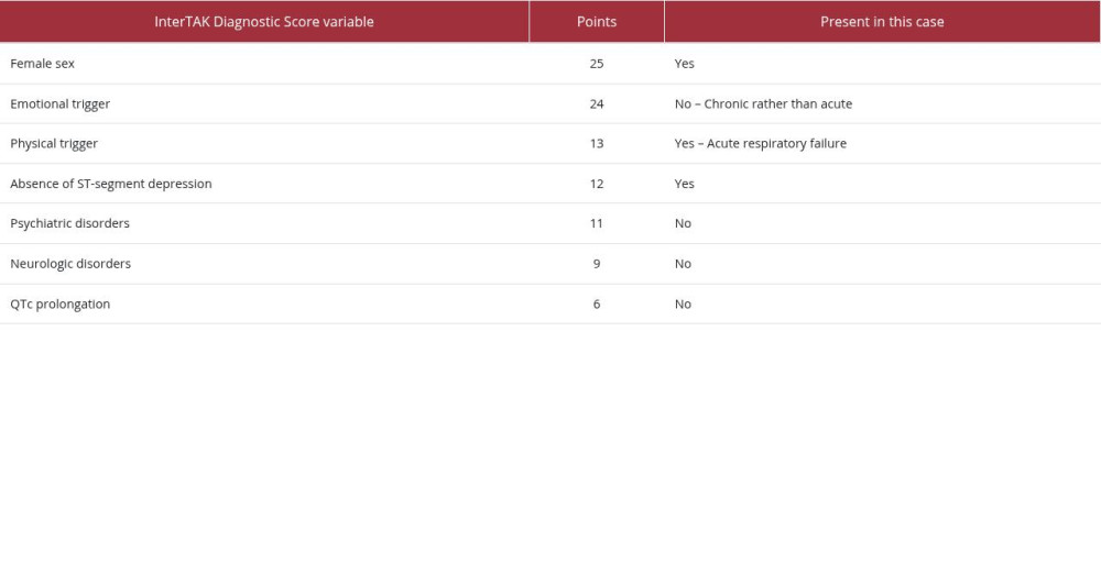
This case fits contemporary diagnostic frameworks for takotsubo syndrome. The patient had a reverse (basal) pattern of left ventricular dysfunction, new ECG abnormalities, elevated cardiac biomarkers, and no obstructive culprit lesion on urgent coronary angiography. These findings are consistent with the modified Mayo Clinic criteria meeting criteria for transient LV hypokinesis, absence of obstructive CAD, and positive for ECG changes with troponin elevation (Table 2). Using the InterTAK Diagnostic Score, the patient accrued 25 points for female sex, 13 points for a physical trigger, and 12 points for absence of ST-segment depression outside of lead aVR, for a total score of 50, indicating intermediate risk (Table 3). In addition, the clinical trigger was predominantly physical (acute respiratory failure requiring intubation), with chronic bereavement likely serving as a background stressor rather than the sole acute precipitant. However, cardiac MRI was not performed, and this should be acknowledged as a limitation because the modified Mayo Clinic criteria recommend excluding myocarditis when clinically relevant [4,8].
Presentation of takotsubo cardiomyopathy can be variable and a distinction between primary takotsubo presenting with cardiac symptoms and secondary takotsubo occurring during evaluation or treatment of another illness such as with our patient. An analysis of US Medicare data showed a 3-fold increase in the discharge diagnosis of takotsubo cardiomyopathy, much of which belongs to the secondary group [9]. Our patient’s presentation was most consistent with secondary takotsubo triggered by acute illness, with prior grief acting as a chronic, rather than sudden, stressor.
The present case is distinctive for several reasons. First, it demonstrates a reverse or basal takotsubo pattern rather than the classic apical form. Second, although atypical forms tend to occur at a younger age, our patient was an older woman, making the anatomic pattern less expected. Third, the likely dominant trigger was acute respiratory failure during critical illness rather than an abrupt emotional event, while chronic grief may have lowered the threshold for stress cardiomyopathy. Finally, the presence of known coronary artery disease with a prior LAD stent made urgent angiographic exclusion of a culprit obstructive lesion essential before attributing the presentation to takotsubo syndrome [3,4,7].
Although once considered relatively benign, takotsubo syndrome is now recognized to carry substantial hospital complications and meaningful long-term risk, particularly in patients with physical triggers or secondary takotsubo occurring during acute medical illness. There is additionally a reported higher rate of serious disease and comorbidities in takotsubo than that of patients with AMI/CAD/ACS [3,4,10]. This highlights the need for randomized clinical trials supporting a specific treatment for takotsubo. Management is currently based on established regimens for other cardiovascular diseases such as CAD, AMI, and HF [2–5,7,10].
Conclusions
This case paves the way for future investigation to standardize therapy specifically targeting takotsubo cardiomyopathy. It also emphasizes the need for clinicians to recognize atypical takotsubo patterns, including basal hypokinesis with preserved apex, to avoid misdiagnosis. Recognition of these non-classical anatomical patterns and non-classical stressors is essential for accurate diagnosis and appropriate management. Most importantly, this case reinforces the importance of acknowledging a vigilant differential diagnosis for takotsubo syndrome in patients presenting with ACS-like symptoms, while also highlighting the need for monitoring and follow-up in high-stress clinical settings to reduce the risk of reverse takotsubo.
Figures
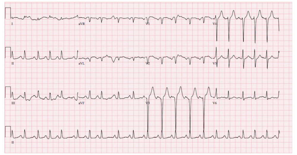 Figure 1. Initial electroencephalogram showing sinus tachycardia with ST-T wave changes and old anteroseptal myocardial infarction.
 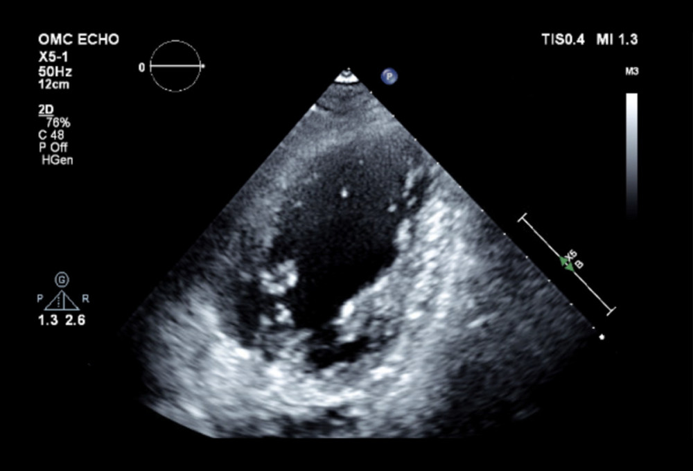 Figure 2. 2D echocardiogram demonstrated severe hypokinesis of the proximal 2/3 of the septal wall anterior lateral and posterior with apical sparing consistent with reverse takotsubo cardiomyopathy and new left ventricle ejection fraction of 40%.
 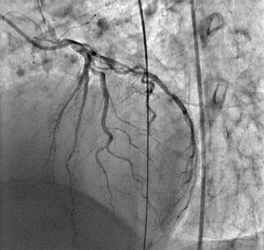 Figure 3. Cardiac catheterization with no new obstructive culprit lesions suggesting takotsubo cardiomyopathy.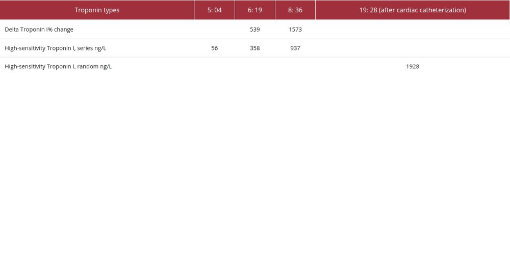
References
1. Dote K, Sato H, Tateishi HMyocardial stunning due to simultaneous multivessel coronary spasms: A review of 5 cases: J Cardiol, 1991; 21(2); 203-14 [in Japanese]
2. Ono R, Falcão LM, Takotsubo cardiomyopathy systematic review: Pathophysiologic process, clinical presentation and diagnostic approach to takotsubo cardiomyopathy: Int J Cardiol, 2016; 209; 196-205
3. Ghadri JR, Wittstein IS, Prasad A, International Expert Consensus Document on Takotsubo Syndrome (Part I): Clinical characteristics, diagnostic criteria, and pathophysiology: Eur Heart J, 2018; 39(22); 2032-46
4. Ghadri JR, Cammann VL, Napp LC, Differences in the clinical profile and outcomes of typical and atypical Takotsubo syndrome: JAMA Cardiology, 2016; 1(3); 335
5. Ramaraj R, Movahed MR, Reverse or inverted takotsubo cardiomyopathy (reverse left ventricular apical ballooning syndrome) presents at a younger age compared with the mid or apical variant and is always associated with triggering stress: Congestive Heart Failure, 2010; 16(6); 284-86
6. Lyon AR, Bossone E, Schneider B, Current state of knowledge on Takotsubo syndrome: a Position statement from the Taskforce on Takotsubo Syndrome of the Heart Failure Association of the European Society of Cardiology: Eur J Heart Fail, 2016; 18(1); 8-27
7. Templin C, Ghadri JR, Diekmann J, Clinical features and outcomes of takotsubo (stress) cardiomyopathy: N Engl J Med, 2015; 373(10); 929-38
8. Madhavan M, Prasad A, Proposed Mayo Clinic criteria for the diagnosis of Tako-Tsubo cardiomyopathy and long-term prognosis: Herz, 2010; 35(4); 240-44
9. Sharkey SW, A clinical perspective of the Takotsubo syndrome: Heart Fail Clin, 2016; 12(4); 507-20
10. Madias JE, Takotsubo cardiomyopathy: Current treatment: J Clin Med, 2021; 10(15); 3440
Figures
Figure 1. Initial electroencephalogram showing sinus tachycardia with ST-T wave changes and old anteroseptal myocardial infarction.Figure 2. 2D echocardiogram demonstrated severe hypokinesis of the proximal 2/3 of the septal wall anterior lateral and posterior with apical sparing consistent with reverse takotsubo cardiomyopathy and new left ventricle ejection fraction of 40%.Figure 3. Cardiac catheterization with no new obstructive culprit lesions suggesting takotsubo cardiomyopathy. Tables
In Press
Case report
Am J Case Rep In Press; DOI: 10.12659/AJCR.954146
Case report 
Am J Case Rep In Press; DOI: 10.12659/AJCR.953555
Case report
Am J Case Rep In Press; DOI: 10.12659/AJCR.953169
Case report 
Am J Case Rep In Press; DOI: 10.12659/AJCR.954027
Most Viewed Current Articles
07 Dec 2021 : Case report
28,713,109
DOI :10.12659/AJCR.934347
Am J Case Rep 2021; 22:e934347
06 Dec 2021 : Case report  180,916
DOI :10.12659/AJCR.934406
Am J Case Rep 2021; 22:e934406
21 Jun 2024 : Case report
124,581
DOI :10.12659/AJCR.944371
Am J Case Rep 2024; 25:e944371
08 May 2026 : Case report
95,842
DOI :10.12659/AJCR.952158
Am J Case Rep 2026; 27:e952158