


13 August 2026: Articles 
Delayed-Onset Giant Bleb Formation and Persistent Intraocular Pressure Elevation in a Patient With COVID-19 and a Long-Term Ahmed Glaucoma Valve
Unusual clinical course, Diagnostic / therapeutic accidents
Elizabeth Sun ABDEF 1, Andrew G. Iwach DE 2,3, Shan C. Lin ABDE 2,3*
DOI: 10.12659/AJCR.953273
Am J Case Rep 2026; 27:e953273


Abstract
BACKGROUND: Glaucoma is a progressive optic neuropathy often associated with elevated intraocular pressure (IOP). Implantation of an Ahmed glaucoma valve (AGV), a silicone or polypropylene shunt tube, can regulate aqueous humor drainage through a filtering bleb and lower IOP. A rare postoperative complication of AGV implantation is giant bleb formation, which typically occurs early in the postoperative period and can require surgical revision if IOP becomes uncontrolled or if the bleb causes discomfort/cosmetic concerns. We describe a case of bilateral refractory steroid-induced glaucoma and delayed-onset giant bleb formation after AGV implantation, temporally associated with coronavirus disease 2019 (COVID-19).
CASE REPORT: A 39-year-old man with bilateral steroid-induced glaucoma presented for an unscheduled visit 3 years after AGV implantation in the left eye with complaints of photophobia and foreign body sensation. He also reported recent COVID-19 onset. Tonometry revealed elevated IOP in the left eye (50 mm Hg). Examination showed an enlarged superotemporal bleb overlying the Ahmed valve plate. Treatment comprising topical glaucoma medications and oral acetazolamide improved bleb appearance and stabilized IOP within 3 months. At the last follow-up, the patient remained stable, with well-controlled IOP in both eyes on 4 topical glaucoma agents (6 medications total) and oral acetazolamide.
CONCLUSIONS: This report describes delayed-onset giant bleb formation and pronounced IOP elevation coincident with COVID-19 onset several years after AGV implantation. Conservative medical management improved bleb appearance and IOP; long-term pressure control required maximal medical therapy. Clinicians should remain vigilant for persistent IOP elevation despite favorable bleb appearance, including after COVID-19 onset.
Keywords: COVID-19, Glaucoma Drainage Implants, Intraocular Pressure, Postoperative Complications
Introduction
Glaucoma, characterized by the gradual loss of retinal ganglion cells and optic nerve axons, is the leading cause of irreversible blindness worldwide [1]. Elevated intraocular pressure (IOP), the primary physiologic risk factor for open-angle glaucoma, typically results from impaired aqueous humor drainage through the trabecular meshwork at the anterior chamber angle [2]. Steroid-induced glaucoma is a form of secondary open-angle glaucoma caused by elevated IOP after topical or systemic corticosteroid use [3]. Diagnosis of glaucoma relies on clinical history, dilated slit-lamp examination, tonometry (IOP measurement), visual field testing, and optic nerve imaging. The primary goals of glaucoma management are to mitigate risk factors and lower IOP to prevent further optic nerve damage. These goals can be achieved through topical medications, laser treatments, and surgical procedures [2].
Glaucoma drainage devices, including the Ahmed glaucoma valve (AGV), are widely used in the treatment of glaucoma. The AGV consists of a silicone or polypropylene plate and drainage tube with a self-regulating 1-way valve mechanism that controls aqueous humor outflow. The tube drains aqueous humor into a subconjunctival reservoir overlying the plate, known as a filtering bleb, where fluid is subsequently reabsorbed into the systemic circulation [4]. Successful AGV implantation typically results in sustained IOP control with a reduced need for glaucoma medications [5]. Early postoperative complications are well described, but bleb-related complications and IOP elevation occurring years after implantation are uncommon [5,6]. Although rare, symptomatic giant bleb formation can occur and often requires surgical revision [7,8]. We report a case of bilateral refractory steroid-induced glaucoma and delayed-onset giant bleb formation with persistent IOP elevation that occurred 3 years after AGV implantation in the context of a recent coronavirus disease 2019 (COVID-19) diagnosis.
Case Report
A 39-year-old man with bilateral steroid-induced glaucoma following penetrating keratoplasty (PKP) underwent AGV implantation (FP7 model, New World Medical, Rancho Cucamonga, CA, USA) in the left eye (OS) in January 2020. The early postoperative course was complicated by low IOP and mild hyphema, which were managed with cycloplegics and corticosteroids. The patient subsequently developed progressive corneal edema, requiring repeat PKP in the OS. IOP in the OS remained stable (< 14 mm Hg) throughout recovery, and all glaucoma medications for that eye were discontinued.
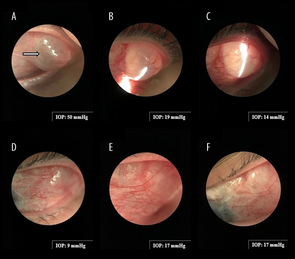
Three years after AGV implantation, the patient presented for an unscheduled visit with complaints of photophobia and a persistent sensation of “rubbing” against his eyelid in the OS. At presentation, visual acuity with current spectacle correction was 20/25 in the right eye (OD) and 20/50 OS; baseline measurements obtained 6 weeks earlier were 20/25 OD and 20/80 OS. Tonometry (rebound tonometer, IC200 model, iCare USA, Raleigh, NC, USA) revealed IOP values of 12 mm Hg OD and 50 mm Hg OS; baseline measurements were 12.5 mm Hg OD and 10.4 mm Hg OS. In the OD, the visual field index was 94%, pattern standard deviation was 1.99 dB, and mean deviation was −6.52 dB. In the OS, the visual field index was 85%, pattern standard deviation was 5.32 dB, and mean deviation was −9.62 dB. Medications in the OD included brimonidine tartrate 0.15% 3 times daily, a combination of dorzolamide HCl and timolol maleate 22.3/6.8 mg/mL twice daily, and fixed-combination netarsudil and latanoprost 0.02%/0.005% once nightly. Medications in the OS consisted only of prednisolone acetate 1% once daily in the morning and preservative-free artificial tears as needed. The patient’s medical history was notable for well-controlled type 2 diabetes mellitus and recent COVID-19 onset, self-diagnosed at home by a positive antigen nasal swab test without confirmatory laboratory testing approximately 1 to 2 weeks before presentation. Systemic medications included empagliflozin, atorvastatin, and metformin. Slit-lamp examination revealed a newly enlarged superotemporal bleb overlying the Ahmed plate in the OS (Figure 1A). Mild haze was present in the PKP graft; the tip of the AGV tube was positioned near, but not touching, the edge of the graft. There was no evidence of overt ocular inflammation, including cells, flare, discharge, or hyperemia. Given the pronounced IOP elevation in the OS, the patient was treated immediately with topical glaucoma medications and oral acetazolamide 500 mg, then closely monitored in the clinic throughout the day. Repeat tonometry after treatment showed progressive IOP reductions in the OS to 44 mm Hg at 1 hour and 40 mm Hg at 2.5 hours. The patient was instructed to use his current OD glaucoma regimen in both eyes and to take acetazolamide 500 mg by mouth twice daily.
At the next-day follow up, visual acuity values were 20/20 OD and 20/30 OS; tonometry demonstrated substantial decreases in IOP (10 mm Hg OD and 19 mm Hg OS). Slit-lamp examination revealed that the bleb was less prominent (Figure 1B). The anterior chamber was deep and quiet, without evidence of inflammation. Follow-up examinations showed that IOP in the OS initially decreased and then stabilized as the bleb progressively decreased in size during medical management (Figure 1A–1F). The PKP graft remained clear throughout follow-up. No further surgical intervention was required. After normalization of IOP, the patient was monitored every 3 months. At the last follow-up, the patient remained stable, with well-controlled IOP in both eyes (13.9 mm Hg OD and 12 mm Hg OS) and overall stable visual field results (OD: visual field index 94%, pattern standard deviation 2.29 dB, mean deviation −5.43 dB; OS: visual field index 89%, pattern standard deviation 4.86 dB, mean deviation −8.01 dB) while receiving 4 topical glaucoma agents (6 medications total) in both eyes and oral acetazolamide 500 mg twice daily by mouth. The topical regimen included pilocarpine 1% 3 times daily, brimonidine tartrate 0.15% 3 times daily, a combination of dorzolamide HCl and timolol maleate 22.3/6.8 mg/mL twice daily, and fixed-combination netarsudil and latanoprost 0.02%/0.005% once nightly.
Discussion
We present an unusual case of giant bleb formation accompanied by sustained IOP elevation occurring 3 years after AGV implantation in the context of recent COVID-19 onset, which highlights the importance of maintaining clinical vigilance for rare late postoperative complications. Although bleb size and appearance improved upon initiation of medical therapy, the patient continued to require both topical and oral glaucoma medications to maintain adequate IOP control.
AGV implantation typically provides long-term IOP control with a reduced need for glaucoma medications [5]. Giant bleb formation secondary to AGV implantation is a rare complication that has been documented in only a small number of case reports. Ugarte et al described a patient with anterior enlargement of the superotemporal bleb overlying the Ahmed plate who presented with foreign body sensation in the absence of IOP compromise; surgical revision was ultimately required [8]. Reports by Manabe et al. and Jeon et al. identified variability in ocular symptoms, timing of presentation, and need for surgical intervention [7,9]. Although our case shares several features with previously reported cases, there are important differences. First, giant bleb formation in the present case occurred several years after AGV implantation; prior cases developed within the first few postoperative months. Second, our patient developed a persistent hypertensive phase requiring maximal topical glaucoma therapy and oral acetazolamide, without spontaneous resolution. This finding differs from results in prior reports and deviates from the typical postoperative course after successful implantation. Notably, the bleb showed substantial improvement in appearance, reduction in size, and decreased IOP following conservative medical therapy, suggesting that some cases of giant bleb formation do not require surgical intervention.
Due to the rarity of giant bleb formation, no established guidelines exist for its diagnosis and management. Diagnosis in the present case was complicated by the unusual timing and clinical presentation. Bleb encapsulation, a common complication after AGV implantation, was considered in the differential diagnosis; however, the delayed presentation and absence of fibrotic features made this diagnosis unlikely [10]. Prior case reports have described the use of orbital magnetic resonance imaging to aid diagnosis [9]. Although this modality was not available at our center, careful slit-lamp examination revealed a large cystic, fluid-filled elevation in the superotemporal region without evidence of fibrosis or overt ocular inflammation, raising suspicion for atypical giant bleb formation. Empirical treatment with glaucoma medications was sufficient to improve bleb appearance and IOP, consistent with some previous reports of giant bleb formation [11].
Intriguingly, our patient’s bleb complication was temporally associated with recent COVID-19 onset, implying that systemic immune responses to viral infection could influence postoperative bleb architecture and aqueous outflow dynamics. Multiple case reports have linked COVID-19 onset to various ocular manifestations, including conjunctivitis, dry eye, photophobia, iritis, and uveitis [12]. It has been hypothesized that ocular inflammation can occur secondary to the systemic release of proinflammatory cytokines in response to COVID-19 onset [13]. Although systemic corticosteroids were not used to manage COVID-19 in our patient, clinicians should be aware that these agents can independently contribute to IOP elevation [14]. It is important to emphasize that current evidence is insufficient to support a causal relationship between viral infection and bleb dysfunction. The mechanism underlying giant bleb formation remains poorly understood, and further studies are needed to determine whether immune-mediated factors contribute to its development.
Conclusions
Giant bleb formation can present with variable clinical manifestations and have diverse management outcomes. In the present case of delayed-onset giant bleb formation, our patient’s ocular symptoms and visual acuity improved shortly after initiation of glaucoma therapy; however, the clinical course was notable for a persistent hypertensive phase requiring maximal topical and oral glaucoma therapy. Ophthalmologists managing patients with long-term glaucoma drainage devices should be aware of late bleb changes, including giant bleb formation—particularly following COVID-19 onset—and recognize that prominent bleb morphology does not necessarily indicate adequate filtration. A stepwise approach to conservative and surgical management may be warranted in cases of persistent IOP elevation.
References
1. Quigley HA, Broman AT, The number of people with glaucoma worldwide in 2010 and 2020: Br J Ophthalmol, 2006; 90(3); 262-67
2. Dietze J, Blair K, Zeppieri M, Havens SJ, Glaucoma: StatPearls [Internet], 2026, Treasure Island (FL), StatPearls Publishing
3. Feroze KB, Zeppieri M, Khazaeni L, Steroid-induced glaucoma: StatPearls [Internet], 2026, Treasure Island (FL), StatPearls Publishing
4. Riva I, Roberti G, Oddone F, Ahmed glaucoma valve implant: Surgical technique and complications: Clin Ophthalmol, 2017; 11; 357-67
5. Radhakrishnan S, Kots-Gotlib N, Pickering TD, Outcomes of Ahmed glaucoma valve implantation with subsequent trans-scleral diode cyclophotocoagulation as the main intervention if IOP remained medically uncontrolled: Clin Ophthalmol, 2024; 18; 3825-36
6. Lee CK, Ma KT, Hong YJ, Kim CY, Long-term clinical outcomes of Ahmed valve implantation in patients with refractory glaucoma: PLoS One, 2017; 12(11); e0187533
7. Manabe K, Ikeda Y, Fujihara E, Tanito M, Bleb wall recession technique to repair giant bleb formation after Ahmed glaucoma valve implantation: A case report: J Med Case Rep, 2019; 13(1); 211
8. Ugarte R, Sugihara K, Sano I, Two different types of giant bleb formation following Ahmed glaucoma valve implantation: Am J Ophthalmol Case Rep, 2024; 33; 102008
9. Jeon TY, Kim HJ, Kim ST, MR imaging features of giant reservoir formation in the orbit: An unusual complication of Ahmed glaucoma valve implantation: Am J Neuroradiol, 2007; 28(8); 1565-66
10. Bae K, Suh W, Kee C, Comparative study of encapsulated blebs following Ahmed glaucoma valve implantation and trabeculectomy with mitomycin-C: Korean J Ophthalmol, 2012; 26(4); 265-70
11. Younger JR, Kooner KS, Upper eyelid extension of a filtering bleb following glaucoma shunt surgery: Ophthalmic Surg Lasers Imaging, 2006; 37(4); 324-26
12. Huang D, Xuan W, Li Z, Impact of COVID-19 on ocular surface health: infection mechanisms, immune modulation, and inflammatory responses: Viruses, 2025; 17(1); 68
13. Douglas KAA, Douglas VP, Moschos MM, Ocular manifestations of COVID-19 (SARS-CoV-2): A critical review of current literature: In Vivo, 2020; 34(3 Suppl); 1619-28
14. Zhang Z, Wu T, Xu W, The hidden ophthalmic dangers of steroids in COVID-19 treatment: A case report: Transl Pediatr, 2025; 14(3); 516-21
In Press
Case report 
Am J Case Rep In Press; DOI: 10.12659/AJCR.953543
Case report 
Am J Case Rep In Press; DOI: 10.12659/AJCR.954216
Case report 
Am J Case Rep In Press; DOI: 10.12659/AJCR.954932
Case report
Am J Case Rep In Press; DOI: 10.12659/AJCR.954146
Most Viewed Current Articles
07 Dec 2021 : Case report
28,712,695
DOI :10.12659/AJCR.934347
Am J Case Rep 2021; 22:e934347
06 Dec 2021 : Case report  180,721
DOI :10.12659/AJCR.934406
Am J Case Rep 2021; 22:e934406
21 Jun 2024 : Case report
124,419
DOI :10.12659/AJCR.944371
Am J Case Rep 2024; 25:e944371
08 May 2026 : Case report
95,462
DOI :10.12659/AJCR.952158
Am J Case Rep 2026; 27:e952158