


28 September 2026: Articles 
Coexisting Postural Orthostatic Tachycardia Syndrome on Autonomic Reflex Testing in Functional Neurologic Disorder
Unusual clinical course, Challenging differential diagnosis
Sushmita Das AEF 1, Sri Rama Narayana Yerraballa AEF 1, Priyanka Shekhawat AEF 1*
DOI: 10.12659/AJCR.953323
Am J Case Rep 2026; 27:e953323


Abstract
BACKGROUND: Functional neurological disorder (FND) is characterized by neurological symptoms identified through positive clinical signs that are incongruent with recognized structural neurological disease. Postural orthostatic tachycardia syndrome (POTS), in contrast, is defined by excessive orthostatic tachycardia and is associated with a range of multisystem autonomic symptoms. Shared clinical features can blur diagnostic boundaries. As a result, new or evolving symptoms in patients with a prior functional diagnosis may be overlooked, increasing the risk of diagnostic overshadowing.
CASE REPORT: An 18-year-old woman with a prior diagnosis of FND presented with fluctuating lower-extremity weakness, gait disturbance, and recurrent drop attacks. She also reported persistent orthostatic intolerance and symptom improvement with recumbency. These symptoms had previously been attributed to the functional diagnosis. Neurological examination demonstrated positive signs of functional weakness, consistent with the prior FND diagnosis. The Composite Autonomic Symptom Score (COMPASS-31) was markedly elevated. Head-up tilt table testing demonstrated sustained sinus tachycardia, with reproduction of typical orthostatic symptoms; other autonomic responses were largely preserved. Generalized joint hypermobility (Beighton score 4/9 with positive historical hypermobility) met the 2017 criteria for hypermobile Ehlers-Danlos syndrome based on family history and chronic musculoskeletal pain. Over several months, functional status improved from wheelchair dependence to independent standing, with reduced symptoms during multidisciplinary care; these findings are reported as a temporal association rather than causal inference.
CONCLUSIONS: This case demonstrates the coexistence of POTS in a patient with established FND and highlights the importance of systematic autonomic evaluation in individuals with persistent orthostatic symptoms. A prior functional diagnosis should not preclude investigation for coexisting physiological disorders.
Keywords: Orthostatic Intolerance, Conversion Disorder, Psychogenic Nonepileptic Seizures, postural orthostatic tachycardia syndrome, Autonomic Nervous System Diseases
Introduction
Functional neurologic disorder (FND) is a disorder of nervous system functioning characterized by neurological symptoms that are internally inconsistent with recognized disease and are diagnosed by positive clinical signs rather than exclusion alone. Presentations include functional weakness, abnormal movements, sensory disturbances, and nonepileptic attacks. Psychogenic nonepileptic seizures are a common category of FND in which individuals exhibit episodes of abnormal limb movements with apparent loss of consciousness resembling epileptic seizures but without any changes in cortical activity [1,2]. FND is common in neurology practice and is among the leading causes of disability in young adults.
Postural orthostatic tachycardia syndrome (POTS) is a disorder of orthostatic intolerance characterized by a heart rate increase of 30 or more beats/min (≥ 40 beats/min in ages 12–19) within 10 minutes of standing, in the absence of orthostatic hypotension, accompanied by symptoms of lightheadedness, palpitations, weakness, blurred vision, and fatigue. Patients with joint hypermobility disorders have a significantly higher prevalence of orthostatic intolerance and POTS [3,4].
Patients with FND frequently report paroxysmal events, fatigue, cognitive dysfunction, and sleep disturbance. In contrast, POTS is characterized by orthostatic tachycardia with lightheadedness, palpitations, presyncope, exercise intolerance, gastrointestinal dysmotility, and cognitive impairment related to cerebral hypoperfusion [3,5,6]. Despite these distinctions, both conditions can present with some overlapping symptoms such as fatigue, cognitive dysfunction, and transient loss of consciousness or presyncope. FND is not associated with a primary hemodynamic abnormality, whereas POTS is defined by objective orthostatic tachycardia. The clinical similarity occurs despite important physiologic differences. This overlap creates a risk of diagnostic overshadowing, whereby potentially treatable autonomic dysfunction may be misattributed to a functional diagnosis.
Both disorders have been associated in prior studies with alterations in central autonomic network function and heightened interoceptive awareness. These observations have been proposed as potential mechanisms contributing to overlapping symptom profiles, although their relevance to individual cases remains uncertain.
Although FND and POTS are individually well described, several clinically relevant uncertainties remain [7]. In particular, the prevalence of objectively confirmed autonomic dysfunction in patients with established FND is not well defined, and the frequency with which orthostatic symptoms are misattributed to a functional diagnosis (diagnostic overshadowing) is unclear. Diagnostic overshadowing may be further compounded when objective orthostatic assessment is limited or not systematically performed, reinforcing attribution of symptoms to a functional etiology.
There is also limited guidance on when persistent orthostatic symptoms in patients with FND should prompt escalation to structured autonomic testing. This gap has direct implications for clinical decision-making, as reliance on single time-point bedside measurements—or the absence of standardized orthostatic assessment—may fail to detect physiologic orthostatic abnormalities, particularly in patients with limited upright tolerance [8].
We present a case of a patient with established FND in whom persistent orthostatic symptoms and nondiagnostic bedside evaluation were followed by diagnostic confirmation of POTS on formal autonomic reflex testing. This case illustrates a practical clinical question: When should clinicians pursue formal autonomic evaluation in patients with established FND who present with persistent orthostatic symptoms, to avoid diagnostic overshadowing and identify coexisting physiologic pathology?
Case Report
An 18-year-old female patient was referred for evaluation of longstanding episodic lower-extremity weakness, paroxysmal “drop attacks”, and dizziness.
Her neurologic symptoms began at age 14 years in 2022 and were characterized by intermittent inability to support weight in the legs, fluctuating gait difficulty, and recurrent episodes of behavioral arrest with loss of postural tone described as “zoning out”. These events occurred multiple times per week, were often precipitated by fatigue or prolonged standing, and were followed by rapid recovery without postictal confusion. She also had a history of motor and vocal tics beginning in childhood. Episodes were associated with lightheadedness and worsening symptoms with upright posture, heat exposure, and visually complex environments, with improvement on recumbency. There was no history of tongue biting, incontinence, or prolonged loss of consciousness, and injury was not reported.
She had been diagnosed with functional neurologic disorder (FND) based on the variability of her motor symptoms and prior documentation of examination findings consistent with functional weakness. She demonstrated good insight into this diagnosis and reported that symptom severity increased during periods of environmental transition or heightened stress. At that time, she had also been seen for new persistent daily headache and vertigo. Brain magnetic resonance imaging and laboratory studies were normal. Amitriptyline was ineffective as a preventive therapy, and headache-related disability progressed, limiting school participation. There was no history of structural neurologic disease, inflammatory disorder, or known cardiac condition. A diagnosis of psychogenic nonepileptic seizures had also been made based on clinical evaluation and video-electroencephalographic (vEEG) confirmation.
Throughout this time period, she endorsed persistent orthostatic symptoms that had not been formally evaluated. These included daily lightheadedness, chronic fatigue, “brain fog”, exercise intolerance, and worsening of both dizziness and leg weakness with prolonged standing, heat exposure, and busy visual environments. Symptoms improved with recumbency. These symptoms had largely been attributed to anxiety, deconditioning, or her functional neurologic diagnosis. At age 14 years, she had a cardiology evaluation for episodic orthostatic lightheadedness. Orthostatic vital signs obtained at that time did not meet criteria for POTS or orthostatic hypotension; however, the duration of upright posture was limited. Conservative measures, including increased fluid and salt intake and gradual positional changes, were recommended.
Psychiatric comorbidities were managed primarily by outside providers, including the patient’s treating provider at the therapeutic school. Review of the available records demonstrated that, over several years, the patient underwent multiple psychopharmacologic trials, including bupropion, duloxetine, quetiapine, hydroxyzine, dextroamphetamine-amphetamine and lamotrigine, with adjustments in therapy over time. Because much of the psychiatric care occurred outside our institution, the exact chronology, indications, and duration of individual medication trials could not be reliably reconstructed from the available records.
Notably, she experienced a marked exacerbation in gait impairment and frequency of paroxysmal events after transitioning to a full-time therapeutic school program in September 2024. She had participated in physical therapy earlier in her course with substantial but temporary improvement in mobility.
On neurological examination in our clinic, mental status and cranial nerves were normal. Motor testing demonstrated fluctuating, give-way weakness in the lower extremities with inconsistent effort, alongside preservation of automatic movements during distraction, consistent with positive functional motor signs. Gait assessment revealed variability in base and stride with inconsistency across tasks and improvement during distraction, further supporting a functional etiology rather than structural neurologic disease. These findings were consistent with previously documented functional motor signs in this patient. The patient presented to clinic using a wheelchair due to activity-limiting lower-extremity weakness and orthostatic symptoms. Sensory testing and coordination were normal. When moving from supine to standing, she developed marked lightheadedness, subjective leg heaviness, and visible tachycardia. During the visit, she also exhibited episodes of abnormal limb movements with apparent unresponsiveness, consistent with her prior diagnosis of psychogenic nonepileptic seizures.
Given the chronic and pervasive nature of her orthostatic symptoms, autonomic symptom burden was quantified using the Composite Autonomic Symptom Score (COMPASS-31), which demonstrated an elevated total score of 63.4/100 indicating significant multidomain autonomic involvement. The largest contribution to the total score derived from the orthostatic intolerance domain, driven by frequent and severe lightheadedness with standing and worsening symptoms over time. Substantial contributions were also observed in the secretomotor domain due to increased sweating and persistent sicca symptoms, and in the gastrointestinal domain, primarily related to severe, constant constipation. Additional points accrued from vasomotor symptoms, including color changes in feet, and pupillomotor symptoms such as photophobia and difficulty focusing. This supported clinically meaningful autonomic involvement that had not previously been formally evaluated.
Examination also demonstrated generalized joint hypermobility (Beighton score 4/9) (Table 1). The Beighton score included bilateral fifth finger hyperextension over 90° and thumb apposition to the forearm. Criterion 2 was met based on positive family history of hypermobility and chronic musculoskeletal pain and joint instability. Alternative heritable connective tissue disorders were excluded based on clinical evaluation and absence of syndromic features. The patient met the 2017 diagnostic criteria for hypermobile Ehlers-Danlos syndrome, supported by historical photographs demonstrating longstanding joint hypermobility and related phenotypic features since childhood.
Given the prominent orthostatic symptoms, she underwent formal autonomic reflex testing. Autonomic reflex testing was performed under standard laboratory preparation protocols, including medication review, avoidance of caffeine and other stimulants before testing, hydration instructions, and continuous electrocardiogram monitoring during tilt. Menstrual phase was not standardized as part of the testing protocol.
After 5 minutes of supine rest, her baseline heart rate was 95 beats/min and blood pressure was 130/80 mm Hg. During head-up tilt to 70°, heart rate increased to 115 beats/min at 1 minute, 118 beats/min at 3 minutes, 130 beats/min at 5 minutes, and 144 beats/min at 10 minutes, representing a sustained increase of over 40 beats/min without a fall in systolic or diastolic blood pressure meeting criteria for orthostatic hypotension. Blood pressure remained elevated (160/120 to 175/130 mm Hg). During head-up tilt, the patient developed progressive orthostatic symptoms, including heaviness of the head and legs at 2 minutes and diffuse headache with generalized heaviness by 5 minutes, accompanied by pallor and slowed speech. Symptoms improved shortly after return to the supine position, with residual fatigue. Continuous monitoring demonstrated sustained sinus tachycardia without arrhythmia. Blood pressure did not demonstrate a sustained fall meeting criteria for orthostatic hypotension. Findings were consistent with POTS (Figure 1). Testing of other autonomic responses was largely preserved. During the Valsalva maneuver, blood pressure and heart rate responses were overall within normal limits, although the phase IV blood pressure overshoot was slightly delayed, suggesting mild slowing of adrenergic baroreflex recovery without evidence of significant adrenergic failure (Figure 2). Deep breathing testing demonstrated reduced heart rate variability compared with age-adjusted norms, indicating mildly reduced parasympathetic (cardiovagal) function, but without features of generalized autonomic failure.
Laboratory evaluation did not identify secondary causes of tachycardia or orthostatic intolerance. Screening studies including complete blood count and iron studies, thyroid function testing, and basic metabolic profile were within reference ranges. Clinical evaluation did not suggest acute infection, dehydration, or medication-induced tachycardia. Pregnancy was not applicable. Orthostatic hypotension was excluded based on absence of sustained blood pressure reduction during tilt. Vasovagal syncope was considered less likely given the sustained tachycardic response without hypotension or bradycardia. Inappropriate sinus tachycardia was considered but was less consistent with the orthostatic pattern of symptoms and heart rate increase observed during tilt.
The patient had prior cardiology evaluation including ambulatory patch monitoring, which demonstrated sinus tachycardia without evidence of arrhythmia, including during episodes of heart rate up to 202 beats/min. These findings argued against supraventricular tachycardia or other primary arrhythmias. Conservative management strategies, including hydration, salt intake, and physical conditioning, were recommended.
Together, findings supported a diagnosis of POTS. At follow-up over several months, the patient reported improvement in orthostatic tolerance, with reduction in frequency and severity of presyncope and paroxysmal symptoms compared with baseline daily symptoms associated with functional limitation. At baseline, symptoms limited ambulation and required wheelchair use for mobility; at follow-up, the patient was able to stand independently for an extended period of time and participate more fully in daily activities. Management included participation in a structured POTS rehabilitation program incorporating fluid and sodium optimization, compression strategies, and graded recumbent exercise progressing toward upright activity, alongside reintroduction of physical therapy. Low-dose propranolol (twice daily) was initiated for management of POTS. The patient also continued care in a specialized FND clinic, which addressed symptom awareness and functional neurological symptoms during the same interval. Her psychiatric medication regimen remained stable throughout follow-up, including bupropion, dextroamphetamine-amphetamine (Adderall), doxylamine, and duloxetine. Improvement occurred in the context of multidisciplinary care and is therefore described as temporal association rather than causal inference in a single case. Table 2 outlines the clinical timeline, diagnostic evaluation, and management, highlighting the recognition of coexisting autonomic dysfunction in a patient with established FND.
Over the course of longitudinal follow-up, the patient’s broader clinical phenotype became apparent. Subsequent evaluations led to diagnoses of POTS, hypermobile Ehlers-Danlos syndrome, and chronic migraine. Management was refined accordingly to include nonpharmacologic and pharmacologic measures for orthostatic intolerance and onabotulinumtoxinA for chronic migraine. Approximately 6 months after the diagnosis of POTS and initiation of low-dose propranolol (twice daily), the COMPASS-31 questionnaire was repeated, demonstrating an improvement in the total score from 63.4 to 30, and she no longer required a wheelchair, reporting substantially improved mobility and daily functioning.
Discussion
LIMITATIONS:
This report should be interpreted in the context of a few limitations. As a single-patient case, causal inferences regarding the relationship between autonomic findings and clinical improvement are limited, and observed changes may reflect the combined effects of multidisciplinary management, including physical therapy and FND-directed interventions. The characterization of paroxysmal events is informed by prior vEEG evaluation at an outside institution, during which typical events were captured and a diagnosis of psychogenic nonepileptic seizures was established; however, repeat vEEG was not performed during the current evaluation. Finally, although autonomic testing demonstrated objective orthostatic tachycardia, the extent to which these physiologic findings directly account for all reported symptoms or paroxysmal events cannot be definitively established.
Conclusions
Diagnostic overshadowing is an important consideration in complex neurologic presentations. A prior diagnosis of FND should not preclude continued evaluation for coexisting, objectively measurable conditions when symptoms are physiologically plausible. In patients with FND who report orthostatic intolerance or multisystem autonomic symptoms, systematic orthostatic assessment and, when indicated, formal autonomic testing may enable timely recognition of treatable autonomic disorders. Maintaining diagnostic vigilance is essential to optimize clinical outcomes and to avoid misattribution of reversible physiologic pathology.
Figures
![Continuous heart rate and blood pressure during head-up tilt table test. Heart rate increases from 95 beats/min at baseline to 144 beats/min within 10 minutes of upright posture without fall in blood pressure, consistent with postural orthostatic tachycardia syndrome (excessive orthostatic tachycardia defined as > 40 beats/min for ages 12–19 years) [17].](https://jours.isi-science.com/imageXml.php?i=amjcaserep-27-e953323-g001.jpg&idArt=953323&w=1000) Figure 1. Continuous heart rate and blood pressure during head-up tilt table test. Heart rate increases from 95 beats/min at baseline to 144 beats/min within 10 minutes of upright posture without fall in blood pressure, consistent with postural orthostatic tachycardia syndrome (excessive orthostatic tachycardia defined as > 40 beats/min for ages 12–19 years) [17].
Figure 1. Continuous heart rate and blood pressure during head-up tilt table test. Heart rate increases from 95 beats/min at baseline to 144 beats/min within 10 minutes of upright posture without fall in blood pressure, consistent with postural orthostatic tachycardia syndrome (excessive orthostatic tachycardia defined as > 40 beats/min for ages 12–19 years) [17]. 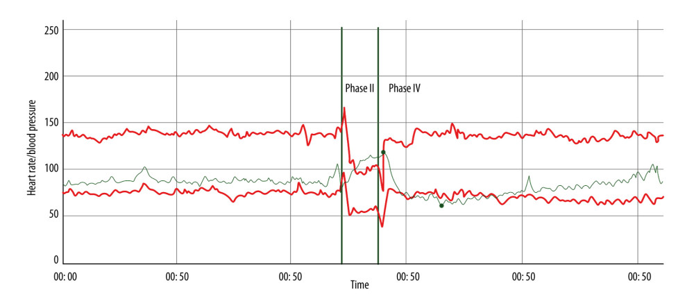 Figure 2. Beat-to-beat heart rate and blood pressure during the Valsalva maneuver demonstrating normal late phase II blood pressure recovery, indicating preserved sympathetic vasoconstrictor function. Phase IV blood pressure overshoot is mildly delayed, suggesting subtle slowing of adrenergic baroreflex recovery without evidence of significant adrenergic failure. The Valsalva ratio is 2.64 (5th percentile 1.46), consistent with preserved cardiovagal function. Red tracings denote systolic and diastolic blood pressure, and the green tracing denotes heart rate. Vertical green lines indicate onset and release of strain during the Valsalva maneuver.
 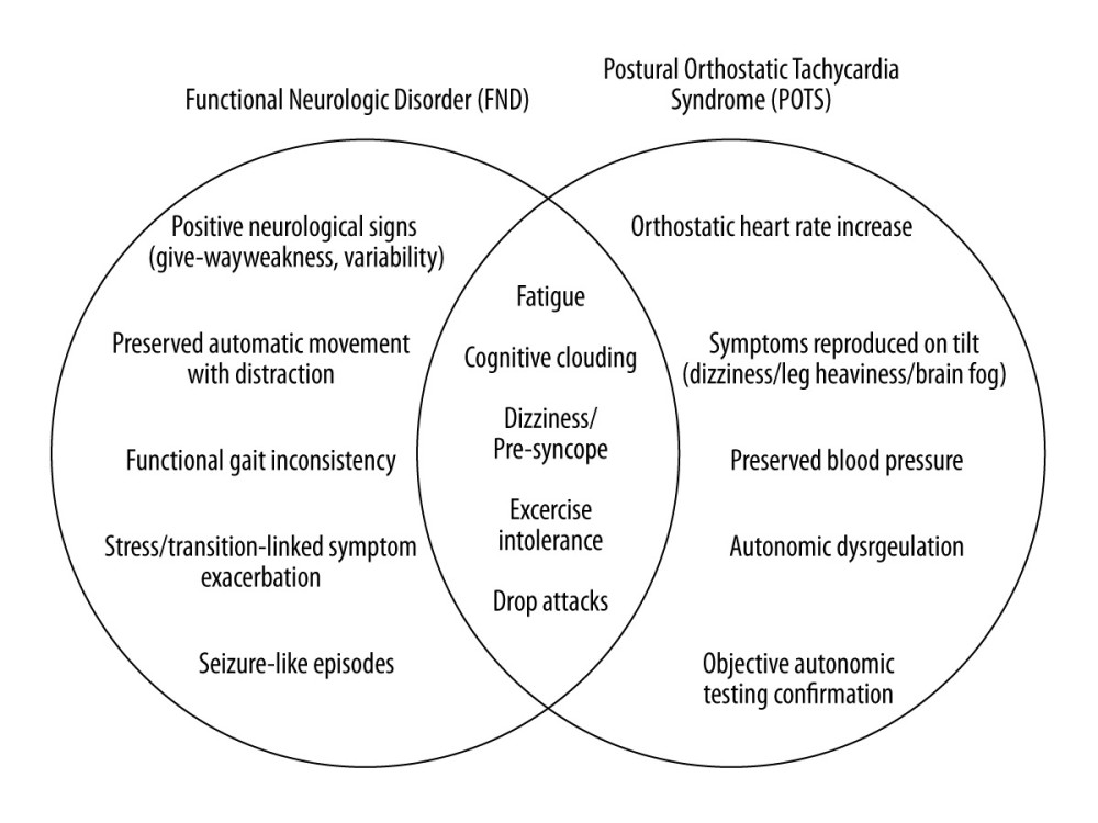 Figure 3. Conceptual diagram illustrating overlapping symptoms and distinguishing diagnostic features of functional neurologic disorder (FND) and postural orthostatic tachycardia syndrome (POTS). The central overlap highlights shared clinical manifestations while the outer sections depict condition-specific diagnostic signs and pathophysiologic mechanisms.
 Tables
Table 1. Diagnostic criteria for hypermobile Ehlers-Danlos syndrome (hEDS), adapted from the 2017 international classification.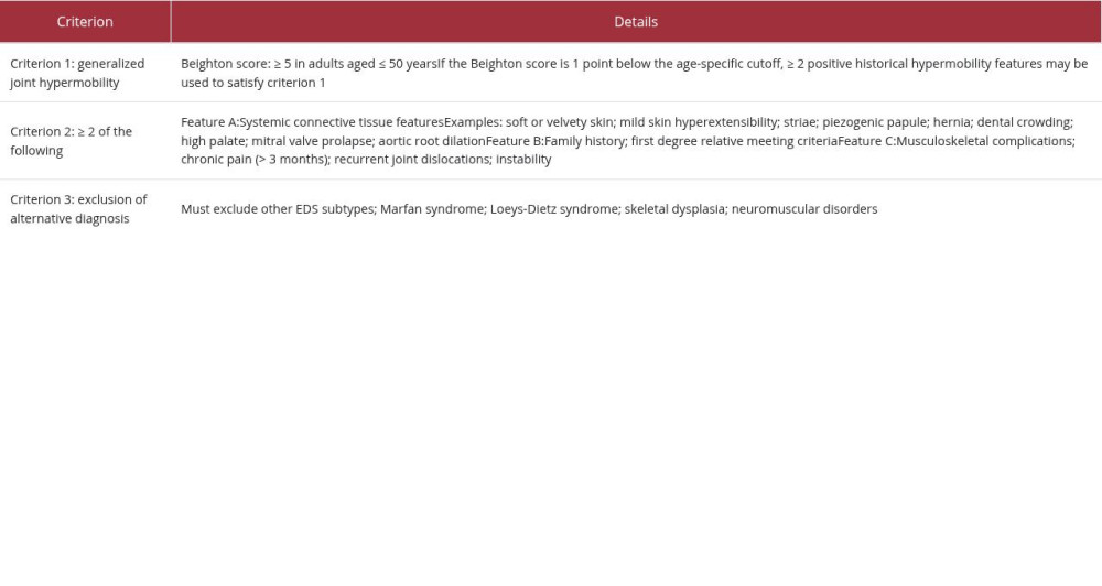 Table 2. Clinical timeline, diagnostic evaluation, and management highlighting the stepwise recognition of coexisting autonomic dysfunction in a patient with established functional neurological disorder (FND).
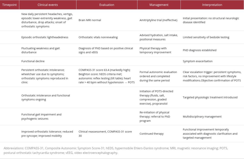 Table 3. Stepwise approach to orthostatic symptoms in patients with established functional neurological disorder (FND).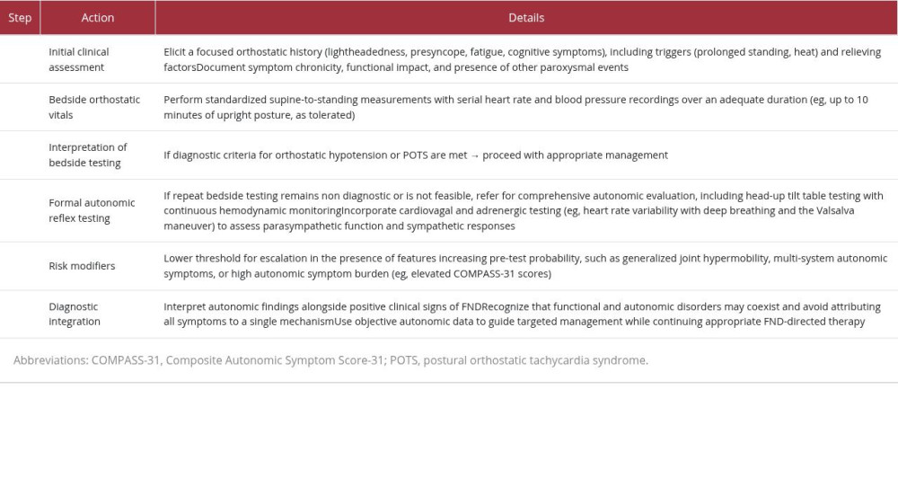
References
1. American Psychiatric Association: Diagnostic and statistical manual of mental disorders, 2013, Arlington (VA), American Psychiatric Association
2. Perez DL, LaFrance WC, Nonepileptic seizures: An updated review: CNS Spectr, 2016; 21(3); 239-46
3. Bryarly M, Phillips LT, Fu Q, Postural orthostatic tachycardia syndrome: JACC focus seminar: J Am Coll Cardiol, 2019; 73(10); 1207-28
4. Roma M, Marden CL, De Wandele I, Postural tachycardia syndrome and other forms of orthostatic intolerance in Ehlers-Danlos syndrome: Auton Neurosci, 2018; 215; 89-96
5. Fedorowski A, Postural orthostatic tachycardia syndrome: Clinical presentation, aetiology and management: J Intern Med, 2019; 285(4); 352-66
6. Hallett M, Aybek S, Dworetzky BA, Functional neurological disorder: New subtypes and shared mechanisms: Lancet Neurol, 2022; 21(6); 537-50
7. Varma-Doyle A, Robbins N, Autonomic disorders and functional neurological disorder: Semin Neurol, 2026; 46(2); 189-95
8. Wieling W, Kaufmann H, Claydon VE, Diagnosis and treatment of orthostatic hypotension: Lancet Neurol, 2022; 21(8); 735-46
9. Lazris A, Roth A, Haskell H, James J, Diagnostic overshadowing: When cognitive biases can harm patients: Am Fam Physician, 2023; 107(3); 244-45
10. Hallyburton A, Diagnostic overshadowing: An evolutionary concept analysis on the misattribution of physical symptoms to pre-existing psychological illnesses: Int J Ment Health Nurs, 2022; 31(5); 1105-16
11. Perez DL, Aybek S, Popkirov S, A review and expert opinion on the neuropsychiatric assessment of motor functional neurological disorders: J Neuropsychiatry Clin Neurosci, 2021; 33(1); 14-26
12. Walzl D, Solomon AJ, Stone J, Functional neurological disorder and multiple sclerosis: A systematic review of misdiagnosis and clinical overlap: J Neurol, 2022; 269(3); 1158-70
13. Shaw BH, Stiles LE, Bourne K, The face of postural tachycardia syndrome: Insights from a large cross-sectional online community-based survey: J Intern Med, 2019; 286(4); 438-48
14. Kesserwani H, Postural orthostatic tachycardia syndrome misdiagnosed as anxiety: A case report with a review of therapy and pathophysiology: Cureus, 2020; 12(10); e10829
15. Aziz Q, Harris LA, Goodman BP, AGA clinical practice update on gastrointestinal manifestations and autonomic or immune dysfunction in hypermobile Ehlers-Danlos syndrome: Expert review: Clin Gastroenterol Hepatol, 2025; 23(1); 15-27
16. Leger R, Dysautonomia-predominant functional neurological symptom disorder: Am J Psychiatry Resid J, 2021; 16(2); 6-8
17. Shekhawat P, Varma-Doyle A, Farhad K, How to interpret the head-up tilt table test: Teaching the 3 core orthostatic hemodynamic patterns: Neurol Educ, 2026; 5(2); e200315
Figures
Figure 1. Continuous heart rate and blood pressure during head-up tilt table test. Heart rate increases from 95 beats/min at baseline to 144 beats/min within 10 minutes of upright posture without fall in blood pressure, consistent with postural orthostatic tachycardia syndrome (excessive orthostatic tachycardia defined as > 40 beats/min for ages 12–19 years) [17].Figure 2. Beat-to-beat heart rate and blood pressure during the Valsalva maneuver demonstrating normal late phase II blood pressure recovery, indicating preserved sympathetic vasoconstrictor function. Phase IV blood pressure overshoot is mildly delayed, suggesting subtle slowing of adrenergic baroreflex recovery without evidence of significant adrenergic failure. The Valsalva ratio is 2.64 (5th percentile 1.46), consistent with preserved cardiovagal function. Red tracings denote systolic and diastolic blood pressure, and the green tracing denotes heart rate. Vertical green lines indicate onset and release of strain during the Valsalva maneuver.Figure 3. Conceptual diagram illustrating overlapping symptoms and distinguishing diagnostic features of functional neurologic disorder (FND) and postural orthostatic tachycardia syndrome (POTS). The central overlap highlights shared clinical manifestations while the outer sections depict condition-specific diagnostic signs and pathophysiologic mechanisms. Tables
Table 1. Diagnostic criteria for hypermobile Ehlers-Danlos syndrome (hEDS), adapted from the 2017 international classification.Table 2. Clinical timeline, diagnostic evaluation, and management highlighting the stepwise recognition of coexisting autonomic dysfunction in a patient with established functional neurological disorder (FND).Table 3. Stepwise approach to orthostatic symptoms in patients with established functional neurological disorder (FND).Table 1. Diagnostic criteria for hypermobile Ehlers-Danlos syndrome (hEDS), adapted from the 2017 international classification.Table 2. Clinical timeline, diagnostic evaluation, and management highlighting the stepwise recognition of coexisting autonomic dysfunction in a patient with established functional neurological disorder (FND).Table 3. Stepwise approach to orthostatic symptoms in patients with established functional neurological disorder (FND). In Press
Case report 
Am J Case Rep In Press; DOI: 10.12659/AJCR.953555
Case report
Am J Case Rep In Press; DOI: 10.12659/AJCR.953169
Case report 
Am J Case Rep In Press; DOI: 10.12659/AJCR.954027
Case report
Am J Case Rep In Press; DOI: 10.12659/AJCR.954380
Most Viewed Current Articles
07 Dec 2021 : Case report
28,713,109
DOI :10.12659/AJCR.934347
Am J Case Rep 2021; 22:e934347
06 Dec 2021 : Case report  180,916
DOI :10.12659/AJCR.934406
Am J Case Rep 2021; 22:e934406
21 Jun 2024 : Case report
124,581
DOI :10.12659/AJCR.944371
Am J Case Rep 2024; 25:e944371
08 May 2026 : Case report
95,842
DOI :10.12659/AJCR.952158
Am J Case Rep 2026; 27:e952158