


24 September 2026: Articles 
Giant Well-Differentiated Liposarcoma of the Spermatic Cord Associated With Inguinal Hernia: Clinical Insights From a Case Report
Challenging differential diagnosis, Rare disease
Kristian Chrz ABCDE 1,2*, David Hoskovec ABDE 1,2, Zdeněk Krška ADG 1,2, M.E. Dämmrich BDF 3, Pavol Klobušický ABCDEF 2,4
DOI: 10.12659/AJCR.953433
Am J Case Rep 2026; 27:e953433


Abstract
BACKGROUND: Spermatic cord liposarcoma is a rare adipocytic malignancy, with fewer than 200 cases described worldwide. Due to its rarity and nonspecific clinical presentation, it is frequently misdiagnosed as more common inguinoscrotal conditions such as hernia or hydrocele. Histologically, liposarcomas comprise several subtypes with varying prognoses. Accurate diagnosis is often challenging preoperatively and typically relies on imaging and definitive histopathological evaluation.
CASE REPORT: We present the case of an 87-year-old man with a progressively enlarging, painless mass in the left inguinoscrotal region, significantly impairing mobility. Computed tomography revealed a large, well-circumscribed mass originating from the spermatic cord, with both cystic and fatty components, along with a concurrent inguinal hernia and hydrocele. No metastatic spread was identified. The patient underwent complete surgical excision of the tumor with simultaneous orchiectomy and inguinal herniotomy. Histopathological analysis confirmed a well-differentiated liposarcoma (G1) with negative surgical margins (R0 resection). Given the patient’s advanced age and comorbidities, no adjuvant therapy was indicated. The postoperative course was uneventful.
CONCLUSIONS: Spermatic cord liposarcoma remains a diagnostic and therapeutic challenge due to its rarity and clinical resemblance to benign conditions. Radical surgical excision with negative margins is the cornerstone of treatment and offers favorable outcomes, particularly in well-differentiated, low-grade tumors. However, the high rate of local recurrence necessitates long-term follow-up. The role of adjuvant therapies such as radiotherapy or chemotherapy remains unclear and should be individualized based on tumor characteristics. Increased awareness and reporting of such cases are essential to improve diagnostic accuracy and optimize management strategies.
Keywords: Liposarcoma, Orchiectomy, Spermatic Cord
Introduction
Sarcoma is a complex form of malignant tumor that arises from mesenchymal tissue, which serves to support and connect tissues in the body. Mesenchymal tissue includes various cell types, including fat tissue (adipocytes), muscle tissue (myocytes), and connective tissue (fibroblasts). Sarcomas are less common than carcinomas, which originate from epithelial tissue, and often appear in muscle, fat, nerves, or other spongy tissues. The identification of over 137 different entities indicates that sarcomas can be categorized into a variety of subtypes, each with distinct characteristics, behaviors, and treatment options [1].
Liposarcoma is one of the most frequent soft tissue sarcomas, second only to malignant fibrous histiocytoma, and originates from adipocytic tissue. There are several different types of liposarcoma, differing in their aggressiveness and histological appearance, including de-differentiated, pleomorphic, and myxoid liposarcomas. One of the main challenges with sarcomas is their propensity for early hematogenous metastasis. This means that cancer cells can often spread through the bloodstream to other parts of the body, often before the disease is locally diagnosed. This can significantly affect the prognosis and usually requires a comprehensive multimodal treatment approach, including surgery, chemotherapy, and sometimes radiation therapy [1,2].
Liposarcoma arising from the spermatic cord is a rare malignant tumor, typically presenting as a painless inguinoscrotal mass with gradual progression. To date, fewer than 200 cases have been described [2]. Its clinical resemblance to benign entities, such as hydrocele or inguinal hernia, frequently leads to diagnostic uncertainty. We report a case in which definitive diagnosis was achieved only after histopathological evaluation.
In this case description, we detail the symptoms, the diagnostic challenges, and the measures undertaken. Additionally, we discuss the therapeutic options and the prognosis for patients with liposarcoma to raise awareness of this rare tumor type and emphasize the importance of accurate diagnosis. This case contributes to expanding the knowledge about liposarcomas of the spermatic cord and may assist other clinicians in the diagnosis and treatment of similar cases in the future.
Case Report
An 87-year-old patient presented to the Surgery Department of Helios St. Elisabeth Hospital Bad Kissingen, Germany in early October 2023 with a progressively painless thickening in the left groin area that extended into the scrotum. The patient reported that this swelling had increased rapidly in size since February 2023 and significantly impaired his ability to walk and engage in sports. His medical history included bladder emptying dysfunction and benign prostatic hyperplasia. To establish a differential diagnosis, we ordered a computed tomography scan of the abdomen and pelvis. The scan revealed a smooth-bordered mass in the spermatic cord measuring 20 × 13 × 12 cm, which was partially cystic and partially fat-equivalent, with a 2-cm nodule. Additionally, a small hydrocele and a medial inguinal hernia were visible on the left side (Figure 1). Both testicles appeared unremarkable. There was no evidence of affected regional lymph nodes or metastasis. Due to the patient’s pacemaker status, magnetic resonance imaging was not performed.
The indication for inguinal herniotomy with tumor excision was established. A complete scrotal tumor excision (Figure 2) was performed, with simultaneous orchiectomy and subsequent left-sided inguinal herniotomy according to Lichtenstein.
The postoperative course was unremarkable from a surgical perspective. A transient thrombocytopenia was detected in the laboratory tests. The tumor specimen was sent for histopathological examination. The microscopic analysis revealed the histology of a completely resected, well-differentiated liposarcoma of the left spermatic cord associated with an inguinal hernia (R0 G1) (Figure 3). Histopathology revealed a completely resected, well-differentiated liposarcoma (G1, ICDO 8851/3) of the left spermatic cord, associated with a concurrent inguinal hernia, measuring 24 cm and fully resected (R0) with a tumor-free spermatic cord and ductus deferens, alongside age-appropriate testicular parenchyma presenting with a hydrocele. The TNM Classification (2020) was pT4 pNx L0 V0 Pn0, R0; G1. During the case presentation at the interdisciplinary tumor conference, no further therapy was indicated based on age and comorbidity.
Discussion
Liposarcoma of the spermatic cord is considered an exceptionally rare malignancy, with fewer than 200 cases reported to date, as noted by Di Pilla et al [3]. It is a soft tissue malignancy derived from mesodermal tissue embryologically. It is the most common histological type (46%), followed by leiomyosarcoma (20%), histiocytoma (13%), and rhabdomyosarcoma (9%) [4]. Tumor size > 10 cm is considered “giant”, with only a few cases being described in the literature [1]. Chaker et al note that this entity accounts for 20% of adult paratesticular tumors and more frequently involves the right side [5].
Liposarcomas are predominantly found in the deep soft tissue of the lower extremities, with the thigh being particularly affected. Depending on their location, liposarcomas can reach considerable sizes and weights of several kilograms. Histopathological examination allows for the differentiation of several subtypes of liposarcoma, each exhibiting distinct prognoses.
The degree of differentiation of a liposarcoma indicates how morphologically distinct the
tumor tissue is from mature adipose tissue. The more immature and poorly differentiated the type, the worse the prognosis [1,4]. Nadeem and Jadoon demonstrate the utility of molecular techniques, specifically MDM2 gene amplification via fluorescence in situ hybridization, to confirm the diagnosis of de-differentiated subtypes [6].
The various subtypes of liposarcoma differ based on differentiation: well-differentiated or highly differentiated liposarcoma (WDL), myxoid/round cell liposarcoma, pleomorphic liposarcoma, and poorly differentiated or de-differentiated liposarcoma [1].
The discussion surrounding liposarcomas encompasses the challenges of diagnosis, particularly due to their rarity and the potential for confusion with other soft tissue tumors. Prognosis largely depends on the distribution probability of the various subtypes and their degree of differentiation. Early and precise diagnosis is crucial for achieving good treatment outcomes. Different therapeutic approaches, including surgery [7], chemotherapy, and radiation therapy, are evaluated based on the differentiation grade and location of the tumor. In the 2020 WHO classification of soft tissue tumors there are 5 reported types of malignant adipocytic tumors: well-differentiated, de-differentiated, myxoid, pleomorphic, and myxoid pleomorphic [8]. Well-differentiated liposarcoma and de-differentiated liposarcoma are a histological and behavioral spectrum of a single disease entity [3,9]. Most spermatic cord liposarcomas are well-differentiated low-grade malignancies with no or minimal tendency to metastasize. Liposarcomas are locally aggressive tumors with a high incidence of local recurrence [4]. Most reported cases occur in adults, with patient age spanning from 24 to 79 years and a mean age at presentation of 61 years. A majority of patients (57.9%) were over 60 years of age (22/38 cases). Symptom duration has been shown to vary widely, ranging from 1 week to several years [3]. The most common presentation of liposarcoma is a progressively enlarging, non-tender mass, usually arising in the groin or within the scrotum, often without associated pain [9]. In most cases, liposarcoma presents without pain, with only occasional reports of painful lesions [10–12]. Preoperative identification is often difficult, as the condition is commonly misdiagnosed as more prevalent inguinoscrotal pathologies, such as inguinal hernia, hydrocele, spermatocele, or tumors involving the testis or epididymis [1,9,10].
The clinical diagnosis of primary liposarcoma is challenging, as it can present similarly to an inguinoscrotal hernia or lipoma, often appearing as a painless left inguinoscrotal mass. Consequently, radiological imaging is essential for guiding the diagnosis. Ultrasound can identify solid, hyperechoic, and heterogeneous lesions; however, it cannot reliably differentiate between a lipoma and a small liposarcoma. Magnetic resonance imaging remains the gold standard for evaluating soft tissue tumors, as it assists in characterizing and defining the extent of local tumor spread, thereby aiding in staging [3].
Radiotherapy may have a role in the management of liposarcoma due to its partial radiosensitivity, particularly in reducing the likelihood of local recurrence [13]. Evidence regarding the optimal dosing of radiotherapy in liposarcoma remains limited. Its use is typically restricted to specific clinical scenarios, including higher-grade tumors or recurrent disease. In our previous case series, only 4 patients underwent radiotherapy, most often in the setting of de-differentiated subtypes or positive surgical margins [13]. The role of adjuvant chemotherapy in LSC remains uncertain. Local recurrence rates are relatively high (55%–70%), highlighting the need for long-term follow-up. In our analysis, recurrence was observed in 9 cases, with a mean time to recurrence of 3.3 years (range 1–6 years), including 1 patient with multiple relapses.
Despite this, prognosis is generally favorable, particularly after complete surgical resection with negative margins. Re-excision can improve outcomes even in cases of incomplete removal. Tumor size and absence of metastasis at diagnosis remain key prognostic factors. Only 1 disease-related death was reported, associated with rapid metastatic progression [1,13–15]. Panther et al reported that novel therapies targeting the MDM2 oncoprotein to restore p53 function show promise in preclinical trials for managing advanced disease [16]. Wetzel et al emphasize that de-differentiated variants exhibit aggressive behavior, often necessitating adjuvant radiotherapy when surgical margins are compromised [17]. Valverde Martínez et al underscored the importance of lifelong surveillance because local recurrence rates remain high, reaching up to 70% even in well-differentiated cases [18].
Conclusions
Spermatic cord liposarcoma is a rare malignancy that often resembles benign inguinoscrotal conditions, complicating diagnosis. Surgical excision remains the mainstay of treatment, but recurrence is not uncommon, and the role of adjuvant therapy remains unclear. Long-term surveillance is essential. Further research is needed to identify biomarkers that may improve prognostication and treatment strategies.
Figures
![CT scan of the pelvis [A] sagittal; [B] coronal; [C] transversal plane: a well-defined mass measuring approximately 20 × 13 × 12 cm originating from the spermatic cord, surrounded by a hydrocele.](https://jours.isi-science.com/imageXml.php?i=amjcaserep-27-e953433-g001.jpg&idArt=953433&w=1000) Figure 1. CT scan of the pelvis [A] sagittal; [B] coronal; [C] transversal plane: a well-defined mass measuring approximately 20 × 13 × 12 cm originating from the spermatic cord, surrounded by a hydrocele.
Figure 1. CT scan of the pelvis [A] sagittal; [B] coronal; [C] transversal plane: a well-defined mass measuring approximately 20 × 13 × 12 cm originating from the spermatic cord, surrounded by a hydrocele. 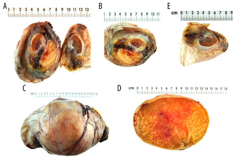 Figure 2. (A–E) Completely resected liposarcoma of the left spermatic cord measuring 20 × 13 × 12 cm, weighing approximately 3 kg.
 ![Histological specimen (H&E): In the upper section of the image, the normal adipose tissue of the well-differentiated liposarcoma can be seen, exhibiting a harmonious structure (R0 Grade 1 UICC). In the lower section, the neoplastic tissue is evident, characterized by numerous mitoses and nuclear polymorphisms, displaying pleomorphic cellular morphology. [A] Liposarcoma G1 Structure; [B] Neoplastic Lipoblasts and Lipocytes; [C] Analyzing immunohistochemistry (IHC): S100+, MDM2+, CDK4+.](https://jours.isi-science.com/imageXml.php?i=amjcaserep-27-e953433-g003.jpg&idArt=953433&w=1000) Figure 3. Histological specimen (H&E): In the upper section of the image, the normal adipose tissue of the well-differentiated liposarcoma can be seen, exhibiting a harmonious structure (R0 Grade 1 UICC). In the lower section, the neoplastic tissue is evident, characterized by numerous mitoses and nuclear polymorphisms, displaying pleomorphic cellular morphology. [A] Liposarcoma G1 Structure; [B] Neoplastic Lipoblasts and Lipocytes; [C] Analyzing immunohistochemistry (IHC): S100+, MDM2+, CDK4+.
Figure 3. Histological specimen (H&E): In the upper section of the image, the normal adipose tissue of the well-differentiated liposarcoma can be seen, exhibiting a harmonious structure (R0 Grade 1 UICC). In the lower section, the neoplastic tissue is evident, characterized by numerous mitoses and nuclear polymorphisms, displaying pleomorphic cellular morphology. [A] Liposarcoma G1 Structure; [B] Neoplastic Lipoblasts and Lipocytes; [C] Analyzing immunohistochemistry (IHC): S100+, MDM2+, CDK4+. References
1. Chan K, Odubanjo T, Swamy R, Hosny M, Giant paratesticular liposarcoma mimicking a left-sided groin hernia: A case report: Cureus, 2022; 14(9); e28856
2. Li F, Tian R, Yin C, Liposarcoma of the spermatic cord mimicking a left inguinal hernia: A case report and literature review: World J Surg Oncol, 2013; 11(1); 18
3. Di Pilla MA, Capuano MA, Rossi M, Liposarcoma of the spermatic cord: The state of art and our experience: Radiol Case Rep, 2023; 18(11); 3941-44
4. Vukmirovic F, Zejnilovic N, Ivovic J, Liposarcoma of the paratesticular tissue and spermatic cord: A case report: Vojnosanit Pregl, 2013; 70(7); 693-96
5. Chaker K, Ouanes Y, Zehani A, A case of liposarcoma of the spermatic cord: Urol Case Rep, 2024; 55; 102761
6. Nadeem MS, Jadoon A, Review of literature and case report of spermatic cord dedifferentiated liposarcoma: J Pak Med Assoc, 2024; 74(8); 1538-40
7. Chowdhry VK, Kane JM, Wang K, Testicular, spermatic cord, and scrotal soft tissue sarcomas: Treatment outcomes and patterns of failure: Sarcoma, 2021; 2021; 8824301
8. García Morúa A, Fermín Lozano Salinas J, Valdés Sepúlveda FLiposarcoma of the espermatic cord: Our experience and review of the literature: Actas Urol Esp, 2009; 33(7); 811-15 [in Spanish]
9. Noguchi H, Naomoto Y, Haisa M, Retroperitoneal liposarcoma presenting a indirect inguinal hernia: Acta Med Okayama, 2001; 55(1); 51-54
10. Hassan JM, Quisling SV, Melvin WV, Sharp KW, Liposarcoma of the spermatic cord masquerading as an incarcerated inguinal hernia: Am Surg, 2003; 69(2); 163-65
11. Bouropoulos C, Skopelitou A, Vaggos G, Papamichael C, Liposarcoma of the spermatic cord: Int Urol Nephrol, 2001; 33(2); 397-98
12. Hsu YF, Chou YY, Cheng YH, Spermatic cord myxoid liposarcoma presenting as an incarcerated inguinal hernia: Report of a case and review of literatures: Hernia, 2012; 16(6); 719-22
13. Rodríguez D, Barrisford GW, Sanchez A, Primary spermatic cord tumors: Disease characteristics, prognostic factors, and treatment outcomes: Urol Oncol Semin Orig Investig, 2014; 32(1); 52e19-e25
14. Dündar M, Erol H, Koçak İ, Kaçar F, Liposarcoma of the spermatic cord: Urol Int, 2001; 67(1); 102-3
15. Bestman TJR, Populaire J, Lauwers K, Molderez C, Liposarcoma of the spermatic cord: Report of 2 cases: Acta Chir Belg, 2007; 107(1); 58-59
16. Panther EJ, Lyons H, Shychuk AJ, Dedifferentiated liposarcoma of the spermatic cord: BMJ Case Rep, 2024; 17(4); e258954
17. Wetzel E, Adjamian N, Diaz G, Steen S, Hobbs J, Liposarcoma of the spermatic cord presenting as an inguinal hernia: Int J Surg Case Rep, 2020; 76(C); 274-77
18. Valverde Martínez S, Salcedo Mercado W, Grinard de León ESpermatic cord liposarcoma. Report of two cases and bibliographic review: Arch Esp Urol, 2018; 71(6); 549-54 [in Spanish]
Figures
Figure 1. CT scan of the pelvis [A] sagittal; [B] coronal; [C] transversal plane: a well-defined mass measuring approximately 20 × 13 × 12 cm originating from the spermatic cord, surrounded by a hydrocele.Figure 2. (A–E) Completely resected liposarcoma of the left spermatic cord measuring 20 × 13 × 12 cm, weighing approximately 3 kg.Figure 3. Histological specimen (H&E): In the upper section of the image, the normal adipose tissue of the well-differentiated liposarcoma can be seen, exhibiting a harmonious structure (R0 Grade 1 UICC). In the lower section, the neoplastic tissue is evident, characterized by numerous mitoses and nuclear polymorphisms, displaying pleomorphic cellular morphology. [A] Liposarcoma G1 Structure; [B] Neoplastic Lipoblasts and Lipocytes; [C] Analyzing immunohistochemistry (IHC): S100+, MDM2+, CDK4+. In Press
Case report 
Am J Case Rep In Press; DOI: 10.12659/AJCR.953543
Case report 
Am J Case Rep In Press; DOI: 10.12659/AJCR.954216
Case report 
Am J Case Rep In Press; DOI: 10.12659/AJCR.954932
Case report 
Am J Case Rep In Press; DOI: 10.12659/AJCR.954146
Most Viewed Current Articles
07 Dec 2021 : Case report
28,712,695
DOI :10.12659/AJCR.934347
Am J Case Rep 2021; 22:e934347
06 Dec 2021 : Case report  180,721
DOI :10.12659/AJCR.934406
Am J Case Rep 2021; 22:e934406
21 Jun 2024 : Case report
124,419
DOI :10.12659/AJCR.944371
Am J Case Rep 2024; 25:e944371
08 May 2026 : Case report
95,462
DOI :10.12659/AJCR.952158
Am J Case Rep 2026; 27:e952158