


18 September 2026: Articles 
A 16-Year-Old Male Patient With Severe Electrical Burns With Chronic and Recurrent Hospital-Acquired Opportunistic Infection With Carbapenem-Resistant Acinetobacter baumannii
Unusual clinical course
Jiawen Hong ABCDEF 1,2, Shengke Wang CE 2,3, Jimei Du AE 2, Qiongqian Pan D 1, Jiao Qian ADE 1*DOI: 10.12659/AJCR.953461
Am J Case Rep 2026; 27:e953461


Abstract
BACKGROUND: Acinetobacter baumannii is an opportunistic gram-negative bacterium found in health facilities, including intensive care units (ICUs), and for patients with burn injuries, it can cause severe wound infections and systemic infection. Electrical burn is a unique type of trauma. Burns and electrical injuries disrupt the integrity of skin and mucous membrane barriers, which results in impairment of the body’s defense mechanisms, thus significantly increasing the possibility of infection. This report describes the case of a 16-year-old male patient with severe electrical burns and chronic and recurrent ICU-acquired opportunistic infection with carbapenem-resistant A. baumannii (CRAB).
CASE REPORT: A 16-year-old male patient was admitted with 220-V electrical burns and was subsequently transferred to the ICU. On admission, he had impaired consciousness and recurrent fever, complicated by respiratory failure, gastrointestinal bleeding, nasal hemorrhage, hypoxic brain injury, and pulmonary infection. During hospitalization, cutaneous ulceration of the left lower limb, necrosis of the third to fifth toes of the left foot, and skin lesions involving the buttocks, sacrococcygeal region, and occiput developed sequentially. CRAB was successively isolated from sputum, blood, and multiple skin wound specimens. The patient underwent repeated debridement, wound expansion, and limb reconstructive surgery. He was discharged in stable condition after 156 days of hospitalization.
CONCLUSIONS: Early identification of CRAB colonization and invasive infection is essential. Strict infection control measures should be implemented in ICUs and burn wards. This case provides clinical reference for the management of recurrent CRAB infection in patients with severe electrical burns and for the prevention and control of nosocomial infection.
Keywords: Acinetobacter baumannii, Electric Injuries, Intensive Care Units, Bacterial Infections
Introduction
Electrical burn is a unique type of trauma. Long-term exposure to low-voltage alternating electric current can not only lead to the formation of burn wounds on the skin but can also induce arrhythmias, and in extreme conditions, even respiratory and cardiac arrest [9]. Burns and electrical injuries disrupt the integrity of skin and mucous membrane barriers, which results in impairment of the body’s defense mechanisms, thus significantly increasing the possibility of infection by pathogens. Studies have shown that the prevalence of
Case Report
CONSENT DECLARATIONS:
This study involving a human participant was approved by the institutional ethics committee. The study was conducted in accordance with local legislation and institutional requirements (approval No. KL20250113). Given the retrospective nature of this study, the ethics committee waived the requirement for additional written informed consent. The patient had previously provided informed consent for their clinical imaging examinations as part of routine medical care, and all identifiable personal information was anonymized to protect patient privacy.
Upon admission, a thorough examination and diagnostic testing were done. Physical examination results showed that there was an arrhythmia, a soft abdomen with bowel sounds heard thrice per minute, and non-compliance in the muscle strength test because of the patient’s altered mental status. Extracorporeal membrane oxygenation (ECMO) support was initiated. Additional clinical observations included muscle rigidity of the left lower limb, stiffness of the left ankle, mild active nasal bleeding, and aspiration of black fluid during gastrointestinal decompression.
Evaluation of the echocardiogram showed dysfunction of left ventricular systolic function in terms of the global reduction of amplitude of motion in the interventricular septum and the left ventricle’s posterior wall. The findings included mild regurgitation of the mitral valve, tricuspid valve, and pulmonary valve with an ejection fraction of 25% and fractional shortening of 11%.
Biochemical analysis yielded the following results: albumin level was 25.5 g/L (reference range: 40.0–55.0 g/L); alanine aminotransferase level was 231 U/L (reference range: 9–50 U/L); aspartate aminotransferase level was 861 U/L (reference range: 15–40 U/L); C-reactive protein (CRP) level was 2.1 mg/L (reference range: < 8.0 mg/L); and procalcitonin (PCT) level was 5.93 ng/mL (reference range: < 0.05 ng/mL).
Hematological evaluation revealed a white blood cell count of 13.1 × 10⁹/L (reference range: 3.9–9.5 × 10⁹/L); hemoglobin level of 95 g/L (reference range: 130–175 g/L); absolute neutrophil count of 11.79 × 10⁹/L (reference range: 1.8–6.3 × 10⁹/L); prothrombin time of 18.5 seconds (reference range: 11.0–14.5 seconds); activated partial thromboplastin time of 52 seconds (reference range: 28.0–42.0 seconds); and thrombin time exceeding 100 seconds (reference range: 14.0–21.0 seconds).
Admission diagnoses included electrical burn, cardiac arrest requiring CPR, shock, hypoxic encephalopathy, respiratory failure, gastrointestinal bleeding, acute renal insufficiency, mediastinal emphysema, left lower limb ischemia, and cardiac insufficiency.
After admission, the patient was maintained on ECMO, endotracheal intubation, and mechanical ventilation. He was unconscious and had compromised immune function. Acute renal failure developed on the second day of hospitalization, and blood purification therapy was initiated. The patient also presented with gastrointestinal bleeding and mild nasal bleeding. Mottled skin of the left lower limb and partial stiffness of the calf muscles suggested possible tissue necrosis. A large amount of sputum was suctioned through the endotracheal tube. Chest computed tomography (CT) revealed increased and thickened lung markings, indicating pulmonary infection. Intravenous meropenem was administered at 1.0 g every 8 hours for anti-infective treatment.
On hospital day 3, nucleic acid testing for pathogenic bacteria in sputum yielded a weakly positive result for
On hospital day 9, sputum pathogen nucleic acid testing was positive for
On hospital day 11, CRAB was isolated from the sputum culture. Susceptibility testing revealed the organism to be sensitive to tigecycline and polymyxin. As a result, the treatment protocol was modified to include the use of a combination of drugs, namely tigecycline (100 mg every 12 hours) along with cefoperazone (2.0 g every 8 hours). Laboratory test results showed elevated inflammatory markers: high-sensitivity CRP was 178.6 mg/L (reference range: < 8.0 mg/L), serum amyloid A protein exceeding 320 mg/L (reference range: 0–10 mg/L), and PCT level was 87.93 ng/mL (reference range: < 0.05 ng/mL).
On hospital day 14, the presence of CRAB was detected in the blood culture. On chest CT, there was evidence of pulmonary infection. Redness, swelling, and significant local hyperthermia were observed on the left lower extremity with skin ulceration and significant exudation on the toes of the left foot. As a result, the treatment strategy was changed to include tigecycline (100 mg every 12 hours) combined with meropenem (0.5 g every 8 hours).
On hospital day 23, the patient was conscious but apathetic. Neuroimaging revealed hypoxic cerebral alterations. In terms of clinical picture, there were episodes of uncontrollable tremors and confusion with complications such as otomastoiditis. There were observed skin ulcerations of the left lower limb, necrosis of the third to fifth toes of the left leg, lesions of the occipital skin, and sacral pressure ulcers. A temperature increase was recorded. CRAB pathogens were found repeatedly in samples collected from lesions of the skin of the left leg, the tip of the left subclavian catheter, and the gluteal area. Due to difficult expectoration and dyspnea, endotracheal intubation was done on day 25, then tracheostomy and bronchoscopy with the aspiration of sputum on day 27. Antibiotic therapy was escalated to tigecycline (75 mg every 12 hours) combined with polymyxin B injection (500 000 units every 12 hours).
On hospital day 37, the skin of the patient’s left lower extremity showed dryness with minimal tissue growth. Moist dressings helped remove eschars. The interdigital space of the left foot was cleaned and treated using iodophor gauze. Samples of secretions were taken for culturing. There was an improvement in the mental state of the patient, with stable hemodynamic parameters. No abnormalities in the lungs were found. Oxygen therapy replaced previous ventilation.
On hospital day 42, the patient had recurrent fever, and CRAB was detected in lower-limb secretions. However, at that point, the inflammatory biomarkers were still under control. Hemodynamics revealed stabilization of heart rate and blood pressure, and the levels of both brain natriuretic peptide and troponin were decreased. Magnetic resonance imaging (MRI) of the leg revealed bone infarction, significant edema, and hematoma formation in the muscles of the left calf. Because of a poor response to the previously used antibiotics, surgery became necessary. The patient had the revision of the left toe stump and debridement of the calf. Antibiotic therapy was adjusted to polymyxin B (500 000 units every 12 hours) combined with tigecycline (50 mg every 12 hours).
On hospital day 47, CRAB was detected in left foot secretions, and
On hospital days 49 and 58, the patient underwent 2 debridement procedures. His temperature remained well controlled, consciousness was clear, his condition was stable, and oxygen was administered via nasal cannula.
On hospital day 66, CRAB was detected in sputum culture, and
On hospital day 70, the patient experienced low-grade fever, a rapid heart rate (141 beats per minute), and rapid breathing (approximately 30 breaths per minute). Chest X-ray showed no pulmonary exudation. Due to the previous detection of
On hospital day 84,
On hospital day 93, partial necrosis of the left lower leg skin, necrosis of the third to fifth toes on the left foot, and a sacrococcygeal pressure injury were noted. CRAB was isolated from sacrococcygeal secretions. Cefoperazone-sulbactam sodium 1.0 g IV for 2 hours was started, while piperacillin-tazobactam was stopped. After that, the patient was stable. With supportive treatment and repeated surgeries to the limbs, the patient improved and was discharged after 156 days in the hospital.
The antibiotic duration, detected bacterial species, and maximum daily temperature are summarized in a time-series format in Figure 1. Chest X-ray findings are presented in Figure 2.
All clinical specimens were processed under sterile conditions by professional microbiology personnel according to standardized procedures. After obtaining pure colonies, bacterial identification was performed using matrix-assisted laser desorption/ionization time-of-flight mass spectrometry, confirming the isolated strain as
Since the bacteria, CRAB, were found at a high frequency in the hospitalization period, 2 CRAB isolates (WMTZ1 and WMTZ2) were selected for whole-genome sequencing (WGS) to evaluate their genetic relatedness. Isolate WMTZ1 was found in the blood sample collected on the 14th day after admission, while isolate WMTZ2 was identified in the bronchial fluid sample on the 84th day after admission.
Two purified colonies of
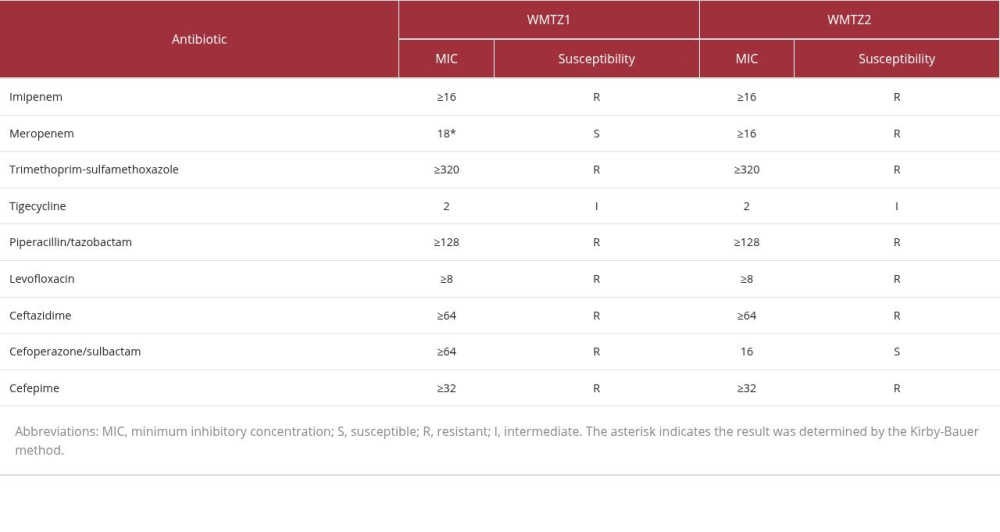
The sequencing results identified 135 single-nucleotide polymorphisms (SNPs) between the 2 CRAB isolates, suggesting they were not derived from the same clonal strain. WMTZ1 harbored 2 additional resistance genes, blaTEM-1 and aph(3’)-Ia, as well as the resistance-related insertion sequence IS26. WMTZ2 lacked these genes, consistent with its antibiotic-susceptibility phenotypes to cefoperazone/sulbactam and aminoglycosides. Resistance genes and insertion sequences detected in WMTZ1 and WMTZ2 are presented in Table 1. Drug susceptibility test results of WMTZ1 and WMTZ2 are shown in Table 2.
Discussion
This case illustrates that patients with severe electrical burns are susceptible to multisite invasive CRAB infections due to multiple organ damage and impaired immune function. Given the difficulty in distinguishing CRAB colonization from true infection, continuous monitoring of clinical manifestations and inflammatory biomarkers is essential; meanwhile, we should place greater emphasis on infection control in burn units.
Electrical burns often lead to multi-system trauma and are characterized by high morbidity and mortality rates [13]. Epidemiologic data suggest that electrical burns are the fourth most common cause of death at workplaces, where the number of yearly deaths is 500 to 1000 cases [3]. Electrical burns can lead to considerable tissue destruction and, consequently, to multi-system infection [13]. CRAB is one of the most common opportunistic pathogens in burn ICUs and has a considerable resistance to drying and disinfection techniques [1]. There is limited literature on the clinical features and pathogenesis of CRAB infections developing after electrical trauma.
The patient in this case was a 16-year-old male with no prior underlying disease. He developed cardiac arrest after a 220-V electrical injury and was revived by CPR. On admission, cardiac insufficiency, organ damage, shock, and hypoxic brain dysfunction increased the risk of CRAB colonization, invasion, and infection. An immune system disorder caused by the electrical injury further increased the likelihood of CRAB infection.
CRAB was isolated from a blood culture collected from the patient on day 13 of admission, indicating hematogenous spread and a CRAB bloodstream infection. Later studies repeatedly isolated CRAB from samples collected from the lower extremities, buttocks, sacrococcygeal area, and sputum. Thus, it was confirmed that the patient had sustained a severe electrical injury with a multisite invasion of CRAB infection. Continuous monitoring of inflammatory markers revealed persistently and markedly elevated levels of high-sensitivity CRP, serum amyloid A, and PCT, further supporting the diagnosis of severe, drug-resistant bacterial infection.
In this case, prompt culture of the bacteria was key to treating a patient with secondary infections after electrical burn. CRAB and other microbes were repeatedly isolated from the patient’s sputum, blood, and wound discharge. According to a 10-year surveillance study of microbial complications in high-voltage electrical burns by Váňa et al, infection of the burn wound was the most common infectious complication, followed by bloodstream infection, lower respiratory tract infection, and urinary tract infection [15]. Data show that the rate of resistant infections in burns is 56.82% [10]. The severity of damage, whether caused by low- or high-voltage electricity, depends on the length of time the individual is exposed to the current and the extent of contact with the electrical power supply [16]. In the present case, the patient developed shock and cardiac arrest immediately after electric shock and presented in critical condition, with recurrent fever, persistent pulmonary infection, and secondary wound infection. Serial bacterial cultures and antimicrobial susceptibility testing supported rational antibiotic administration throughout treatment.
Bacterial strains isolated from burn wards generally exhibit multidrug resistance and high genetic diversity, which facilitates cross-transmission within wards [12]. Shenoy et al noted that traditional epidemiological criteria for distinguishing community-acquired from nosocomial infections, based solely on onset setting, ward distribution, and time window, have clear limitations and are insufficient for tracing pathogen transmission. They performed WGS on CRAB strains isolated from patient specimens and environmental samples, highlighting the risk of burn patients for early-onset nosocomial infections [11]. WGS is now widely used for molecular tracing of nosocomial infection outbreaks and provides accurate molecular evidence to distinguish recurrent CRAB infection from new nosocomial CRAB reinfection [17]. In the present case, 2 representative CRAB strains, WMTZ1 and WMTZ2, isolated at different time points and from different infectious sites during hospitalization, were subjected to WGS. A total of 135 SNPs were detected between the 2 strains, along with differences in drug resistance genes and insertion sequences. These findings indicate that the 2 isolates likely belonged to distinct clones, suggesting that subsequent positive CRAB results were most likely due to the acquisition of a new strain rather than the recurrence of the original strain. This also indirectly reflects the high genetic diversity of CRAB strains in burn ICUs, where alternating colonization and infection by multiple clones can occur.
Therefore, in addition to anti-infective therapy, strict adherence to nosocomial isolation protocols, environmental disinfection, and aseptic procedures may help reduce the risk of cross-transmission and recurrent infections caused by drug-resistant bacteria. There is still the possibility of overestimating the actual SNP diversity, even with the small sample size, because information about the relevant historical strains was not available for further comparison. The WGS method can accurately describe genetic differences between strains, identify potential links to infections resistant to medications, and serve as a basis for infection tracing, but this technique is rarely used in nosocomial outbreak surveillance. WGS requires storing previous strains of the pathogen, and it is difficult to fully reconstruct the chain of transmission of drug-resistant bacteria without them [11].
Conclusions
Early identification of CRAB colonization and invasive infection is central to the clinical management of such cases. This case highlights the importance of standardized antimicrobial stewardship and rigorous infection prevention and control strategies in ICUs and burn wards. This case report has several limitations. Large-scale clinical studies focusing on individualized antimicrobial regimens for patients with electrical injuries complicated by CRAB infection remain scarce. Further large-scale clinical investigations are needed to optimize comprehensive diagnostic and treatment strategies for severe electrical injury complicated by recurrent CRAB infection.
Figures
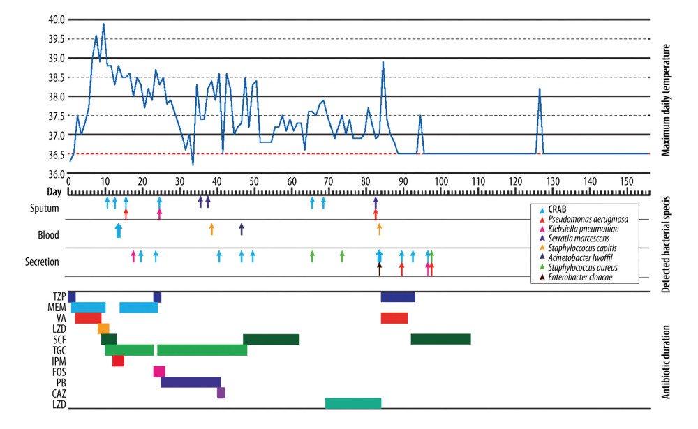 Figure 1. Antibiotic duration, detected bacterial species, and maximum daily temperatureThis figure shows the patient’s daily maximum body temperature during hospitalization, the sites, names, and timing of pathogen detection, as well as antibiotic usage. Abbreviations: CRAB, carbapenem-resistant Acinetobacter baumannii; TZP, piperacillin/tazobactam; MEM, meropenem; VA, vancomycin; LZD, linezolid; SCF, cefoperazone/sulbactam; TGC, tigecycline; IPM, imipenem; FOS, fosfomycin; PB, polymyxin B; CAZ, ceftazidime.
 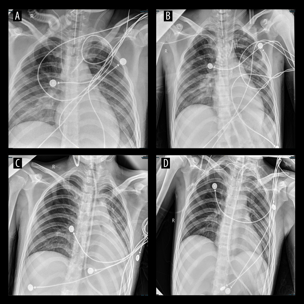 Figure 2. Chest X-ray findings of the patientChest radiographs obtained at different time points during hospitalization. On hospital day 3 (A), slightly increased and thickened pulmonary markings with patchy opacities and blurred margins were observed in both lower lung fields. On hospital day 7 (B), a patchy area of increased density with blurred margins was observed in the middle field of the left lung. On hospital day 24 (C), compared with previous findings, patchy opacities in the middle and lower lung fields were partially absorbed. A patchy area of increased density with relatively clear margins was observed in the upper field of the right lung, with local pleural adhesion at the right lung apex. On hospital day 34 (D), pulmonary markings were distinct, with no obvious abnormalities detected.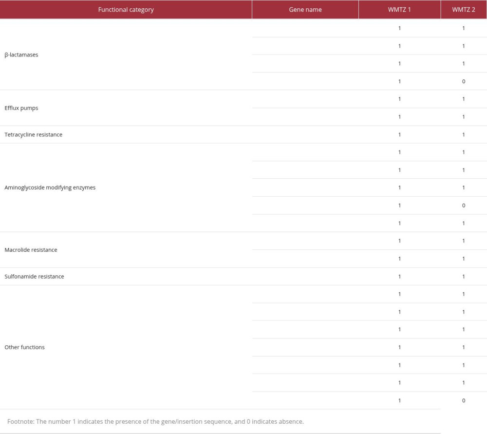
References
1. Bento D, Tobin EH, Acinetobacter. [Updated 2026 Mar 6]: StatPearls [Internet], 2026, Treasure Island (FL), StatPearls Publishing Available from: https://www.ncbi.nlm.nih.gov/books/NBK430784/
2. Bartal C, Rolston KVI, Nesher L: Infect Dis Ther, 2022; 11(2); 683-94
3. Lee CR, Lee JH, Park M: Front Cell Infect Microbiol, 2017; 7; 55
4. Lees-Miller RG, Iwashkiw JA, Scott NE: Mol Microbiol, 2013; 89(5); 816-30
5. Harding CM, Hennon SW, Feldman MF: Nat Rev Microbiol, 2018; 16(2); 91-102
6. Jiang Y, Ding Y, Wei Y, Jian C: Front Microbiol, 2022; 13; 1045206
7. Adeyemi FM, Akinlade EA, Yusuf-Omoloye NA: BMC Infect Dis, 2025; 25(1); 786
8. Sati H, Carrara E, Savoldi A, The WHO Bacterial Priority Pathogens List 2024: A prioritisation study to guide research, development, and public health strategies against antimicrobial resistance: Lancet Infect Dis, 2025; 25(9); 1033-43
9. Zemaitis MR, Guirguis M, Cindass R, Electrical injuries: StatPearls July 6, 2025, Treasure Island (FL), StatPearls Publishing
10. Maharjan S, Shrestha S, Acharya U, Prevalence and determinants of multi-drug resistance bacterial infection among burn patients in a tertiary care center in Nepal: Infect Drug Resist, 2026; 19; 559803
11. Shenoy ES, Pierce VM, Sater MRA: Infect Control Hosp Epidemiol, 2020; 41(5); 531-38
12. Gao J, Zhao X, Bao Y: Burns, 2014; 40(2); 295-99
13. Koumbourlis AC, Electrical injuries: Crit Care Med, 2002; 30(11 Suppl); S424-S30
14. Harding CM, Hennon SW, Feldman MF: Nat Rev Microbiol, 2018; 16(2); 91-102
15. Váňa V, Lipový B, Cvanová M, Results of microbiological surveillance in patients with high-voltage electrical injuries: A 10-year single center experience: Epidemiol Mikrobiol Imunol, 2025; 74(2); 97-106
16. Moukawam E, Aoun CB, Sfeir J, Electrical burns: A retrospective study at the lebanese burn center in Geitaoui Hospital, Lebanon (2011–2024): Int Wound J, 2025; 22(8); e70732
17. Jia H, Chen Y, Wang J: Int J Med Microbiol, 2019; 309(7); 151339
Figures
Figure 1. Antibiotic duration, detected bacterial species, and maximum daily temperatureThis figure shows the patient’s daily maximum body temperature during hospitalization, the sites, names, and timing of pathogen detection, as well as antibiotic usage. Abbreviations: CRAB, carbapenem-resistant Acinetobacter baumannii; TZP, piperacillin/tazobactam; MEM, meropenem; VA, vancomycin; LZD, linezolid; SCF, cefoperazone/sulbactam; TGC, tigecycline; IPM, imipenem; FOS, fosfomycin; PB, polymyxin B; CAZ, ceftazidime.Figure 2. Chest X-ray findings of the patientChest radiographs obtained at different time points during hospitalization. On hospital day 3 (A), slightly increased and thickened pulmonary markings with patchy opacities and blurred margins were observed in both lower lung fields. On hospital day 7 (B), a patchy area of increased density with blurred margins was observed in the middle field of the left lung. On hospital day 24 (C), compared with previous findings, patchy opacities in the middle and lower lung fields were partially absorbed. A patchy area of increased density with relatively clear margins was observed in the upper field of the right lung, with local pleural adhesion at the right lung apex. On hospital day 34 (D), pulmonary markings were distinct, with no obvious abnormalities detected. In Press
Case report
Am J Case Rep In Press; DOI: 10.12659/AJCR.953317
Case report 
Am J Case Rep In Press; DOI: 10.12659/AJCR.954643
Case report 
Am J Case Rep In Press; DOI: 10.12659/AJCR.954074
Case report 
Am J Case Rep In Press; DOI: 10.12659/AJCR.953592
Most Viewed Current Articles
07 Dec 2021 : Case report
28,712,429
DOI :10.12659/AJCR.934347
Am J Case Rep 2021; 22:e934347
06 Dec 2021 : Case report  180,530
DOI :10.12659/AJCR.934406
Am J Case Rep 2021; 22:e934406
21 Jun 2024 : Case report
124,276
DOI :10.12659/AJCR.944371
Am J Case Rep 2024; 25:e944371
08 May 2026 : Case report
95,045
DOI :10.12659/AJCR.952158
Am J Case Rep 2026; 27:e952158