


14 September 2026: Articles 
A 35-Year-Old Woman Presenting With Life-Threatening Fulminant Myocarditis After COVID-19: A Case Report
Management of emergency care, Rare disease, Educational Purpose (only if useful for a systematic review or synthesis)
Darejan Mdinaradze AF 1,2*, Ia Barabadze AF 1, Nikoloz Natsvlishvili AF 1DOI: 10.12659/AJCR.953506
Am J Case Rep 2026; 27:e953506


Abstract
BACKGROUND: Fulminant myocarditis is a rare but severe and life-threatening cardiovascular complication of severe acute respiratory syndrome coronavirus 2 (SARS-CoV-2) infection. Its sudden onset can result in severe heart failure, even in previously healthy adults. This report describes the case of a 35-year-old woman who presented with life-threatening fulminant myocarditis after coronavirus disease 2019 (COVID-19).
CASE REPORT: A 35-year-old previously healthy woman presented to the emergency department on the fourth day of acute constitutional symptoms with progressive dyspnea, severe chest pain, and cardiogenic shock. She had previously received a 2-dose primary series of the Sinovac COVID-19 vaccine. Initial evaluation showed pronounced elevation of cardiac biomarkers along with electrocardiographic abnormalities. Coronary angiography promptly excluded acute coronary occlusion. Following transient hemodynamic improvement with inotropic support, the patient developed rapid electrical deterioration 17 hours after admission, which manifested as polymorphic ventricular tachycardia characterized by a sine-wave-like QRS pattern that progressed to refractory ventricular fibrillation, cardiac arrest, and death. Echocardiographic findings, along with the hyperacute clinical course, were consistent with a diagnosis of probable fulminant COVID-19-associated myocarditis.
CONCLUSIONS: Fulminant myocarditis remains a rare but potentially fatal complication of SARS-CoV-2 infection, capable of causing rapid hemodynamic collapse and malignant electrical instability even in previously healthy individuals. Early recognition, supported by cardiac biomarkers and echocardiographic assessment, is essential to facilitate the timely initiation of aggressive supportive care and may improve clinical decision-making in acute settings.
Keywords: COVID-19, myocarditis, Arrhythmias, Cardiac, Ventricular Dysfunction, Left, Case Reports
Introduction
Since the emergence of coronavirus disease 2019 (COVID-19), numerous cardiac complications have been reported, including cardiomyopathies, arrhythmias, acute coronary syndromes, and heart failure [1]. Among these, fulminant myocarditis is the rarest and most severe form, characterized by rapid onset, extensive myocardial inflammation, and a high risk of cardiogenic shock, malignant arrhythmias, and multiorgan failure [2]. Fulminant myocarditis is estimated to comprise approximately 10% to 30% of acute myocarditis cases and typically presents with abrupt hemodynamic deterioration [3]. The pathophysiology is thought to be multifactorial, involving a hyperinflammatory response, direct myocardial injury mediated through angiotensin-converting enzyme 2 receptor interaction, and immune-mediated damage [4]. Although COVID-19-associated fulminant myocarditis has been increasingly reported, additional clinical descriptions remain important to improve understanding and optimize management strategies. Epidemiological data suggest that myocarditis secondary to COVID-19 occurs in approximately 0.2% to 0.7% of hospitalized patients [5]. Diagnosis requires integration of the clinical presentation, cardiac biomarker findings, and echocardiographic assessment. Management is primarily based on intensive supportive care, early initiation of inotropic therapy when indicated, and close monitoring in a critical care setting, with escalation to advanced circulatory support when necessary. Recent reviews emphasize that severe acute respiratory syndrome coronavirus 2 (SARS-CoV-2)-associated fulminant myocarditis often follows a particularly hyperacute clinical course, requiring prompt multidisciplinary intervention [6]. Here, we report the case of a 35-year-old woman with a fatal course of COVID-19-associated fulminant myocarditis, highlighting its aggressive nature, diagnostic challenges, and the critical need for early recognition and intervention to inform the management of future cases.
Case Report
A 35-year-old woman developed fever, tachycardia, and general malaise. On the second day after symptom onset, she underwent polymerase chain reaction testing at an outpatient clinic, which confirmed SARS-CoV-2 infection. Viral variant sequencing was unavailable; however, based on the timeline, the Omicron variant was the dominant circulating strain. Her condition deteriorated rapidly; on the fourth day after symptom onset, she presented to the emergency department with sudden onset of severe chest pain and rapid hemodynamic collapse. She had previously received a 2-dose primary series of the Sinovac COVID-19 vaccine and reported no history of cardiovascular disease or other clinically significant comorbidities. Targeted testing to exclude common respiratory viral infections, including influenza and respiratory syncytial virus, yielded negative results. SARS-CoV-2 infection was considered the most likely cause of the patient’s clinical presentation. On admission, vital signs were notable for hypotension (blood pressure 70/40 mm Hg), tachycardia (heart rate 129 bpm), respiratory rate of 24 breaths/min, SpO2 of 91%, and fever (38.5 °C). After initiation of inotropic support, her blood pressure improved to 130/70 mm Hg.
Laboratory evaluation revealed a near-normal pH with reduced pCO2 (pH 7.37, pCO2 30.9 mm Hg, pO2 35.4 mm Hg), elevated glucose (11.0 mmol/L), and normal electrolyte levels (K+ 4.02 mmol/L, Na+ 138.6 mmol/L). Cardiac biomarkers were substantially elevated (troponin I > 15.0 ng/mL, N-terminal pro-B-type natriuretic peptide 5256 pg/mL), along with elevated liver enzyme levels (aspartate aminotransferase 324.7 IU/L, alanine aminotransferase 225.9 IU/L) and inflammatory markers (C-reactive protein 18.9 mg/L, procalcitonin 0.17 ng/mL). Coagulation studies indicated hypocoagulation (prothrombin time 30.3 s, prothrombin index 43.9%, international normalized ratio 2.96, activated partial thromboplastin time 48.3 s, fibrinogen 182.2 mg/dL).
Electrocardiography demonstrated bidirectional ventricular tachycardia (Figure 1). Transthoracic echocardiography revealed severely reduced systolic function (ejection fraction 30%–35%) with diffuse left ventricular hypokinesia, a left ventricular end-diastolic volume of 83 mL, and normal left atrial size. Chest radiography showed well-aerated lungs with mild right basilar fibrotic changes, peribronchial thickening, and minimal pleural reaction (Figure 2). Abdominal ultrasonography and computed tomography demonstrated gallbladder wall thickening, biliary sludge, perivascular infiltration, and a small amount of free fluid. The pancreas was structurally normal, with mild peripancreatic inflammatory changes. Pulmonary embolism was excluded by computed tomography angiography, and acute surgical pathology was ruled out after surgical consultation. Urgent coronary angiography revealed no evidence of atherosclerotic lesions (Figure 3A, 3B).
Given the clinical presentation combined with electrocardiographic and echocardiographic findings, fulminant myocarditis was suspected. According to the European Society of Cardiology criteria [7], a diagnosis of probable fulminant myocarditis was established based on the clinical presentation, substantially elevated cardiac biomarkers, severe global left ventricular systolic dysfunction, and exclusion of obstructive coronary artery disease by coronary angiography. Cardiac magnetic resonance imaging and endomyocardial biopsy could not be performed due to the patient’s hemodynamic instability.
The patient was admitted to the intensive care unit, where she received phenylephrine and norepinephrine, oxygen therapy, intravenous fluids, correction of metabolic acidosis and electrolyte abnormalities, nonsteroidal anti-inflammatory drugs, antiarrhythmic therapy (amiodarone and lidocaine), and anticoagulation (later discontinued due to progressive coagulopathy). Supportive care was provided in accordance with institutional protocols. Specific antiviral and anti-inflammatory therapies, including remdesivir and corticosteroids, were not administered because the patient’s rapid and profound hemodynamic collapse on admission precluded a therapeutic window in which these agents could achieve clinical benefit. Nonsteroidal anti-inflammatory drugs were primarily used for symptomatic relief of acute chest pain and systemic distress. However, their role in the management of fulminant viral myocarditis remains a focus of clinical debate.
Seventeen hours after admission, the patient experienced rapid electrical deterioration, presenting with polymorphic ventricular tachycardia characterized by a wide, sine-wave-like QRS morphology that quickly degenerated into refractory ventricular fibrillation (Figure 4). Despite repeated electrical defibrillation (200–360 J; 6 attempts), cardiopulmonary resuscitation according to protocol, rapid orotracheal intubation, intravenous atropine and epinephrine, continued fluid resuscitation, and correction of metabolic acidosis, the rhythm progressed to asystole; effective cardiac contractility could not be restored. Resuscitation efforts were terminated, and death was pronounced.
Discussion
This case illustrates the rapid progression of COVID-19-associated fulminant myocarditis in a previously healthy young woman. After the onset of nonspecific symptoms, the patient developed severe left ventricular dysfunction, cardiogenic shock, and malignant ventricular arrhythmias within a few days, ultimately resulting in a fatal outcome.
Fulminant myocarditis is an uncommon complication of SARS-CoV-2 infection, with reported incidence rates ranging from 0.2% to 0.7% among hospitalized patients. Although most cases of myocarditis are mild, fulminant myocarditis represents a particularly severe presentation associated with substantial morbidity and mortality. Previous studies have shown that myocardial involvement can occur even in individuals without known cardiovascular disease [8].
In the present case, SARS-CoV-2 infection was confirmed 2 days before hospital admission. During this period, there was no evidence of cardiac involvement. At presentation, however, the patient demonstrated substantially elevated cardiac biomarkers, severe left ventricular systolic dysfunction, cardiogenic shock, and ventricular arrhythmias, indicating extensive myocardial injury. The short interval between COVID-19 diagnosis and cardiovascular collapse highlights the potential for rapid clinical deterioration in affected patients.
Similar cases have been described in the literature. Dao et al reported a patient who developed fulminant myocarditis after COVID-19 onset but survived after receiving mechanical circulatory support [9]. In contrast, Mirza et al described a fatal case associated with concurrent SARS-CoV-2 and influenza B infection [10]. Unlike the case reported by Mirza et al, tests for influenza and respiratory syncytial virus showed negative results in our patient, making SARS-CoV-2 infection the most likely cause of the observed myocardial involvement.
Extracorporeal membrane oxygenation (ECMO) has become an important therapeutic option for patients with fulminant myocarditis complicated by refractory cardiogenic shock, providing temporary circulatory support while myocardial recovery occurs. Previous studies have demonstrated improved survival when ECMO is initiated early in appropriately selected patients [11]. In the present case, ECMO could not be implemented because of the patient’s rapidly progressive hemodynamic deterioration and the lack of immediate access to advanced mechanical circulatory support. This clinical course underscores the importance of early recognition of fulminant myocarditis and timely consideration of ECMO prior to onset of irreversible circulatory collapse. The establishment of referral pathways and standby ECMO protocols may facilitate earlier intervention in similar cases and potentially improve outcomes [12].
According to current European Society of Cardiology guidelines on the management of myocarditis, cardiac magnetic resonance imaging is recommended as the noninvasive gold standard for diagnosing suspected myocarditis; however, its use may be limited in patients with hemodynamic instability or cardiogenic shock [7].
Although endomyocardial biopsy remains the reference standard for the definitive diagnosis of myocarditis [13], it was not performed in this case due to the patient’s profound hemodynamic instability and rapidly progressive shock. Consequently, the diagnosis was based on the clinical presentation, substantially elevated cardiac biomarkers, electrocardiographic abnormalities, severe left ventricular systolic dysfunction on echocardiography, and exclusion of obstructive coronary artery disease and other alternative diagnoses. Under these circumstances, immediate stabilization and supportive treatment took precedence over invasive diagnostic procedures.
Conclusions
This case demonstrates a fatal course of COVID-19-associated fulminant myocarditis in a previously healthy young woman. Rapidly progressive cardiogenic shock and severe myocardial injury warranted intensive supportive care. Early consideration of mechanical circulatory support, including ECMO, may be lifesaving in selected patients with rapidly progressive fulminant myocarditis. Although endomyocardial biopsy remains the diagnostic gold standard, its use may be limited in critically ill patients, in whom immediate hemodynamic stabilization is the highest priority.
Figures
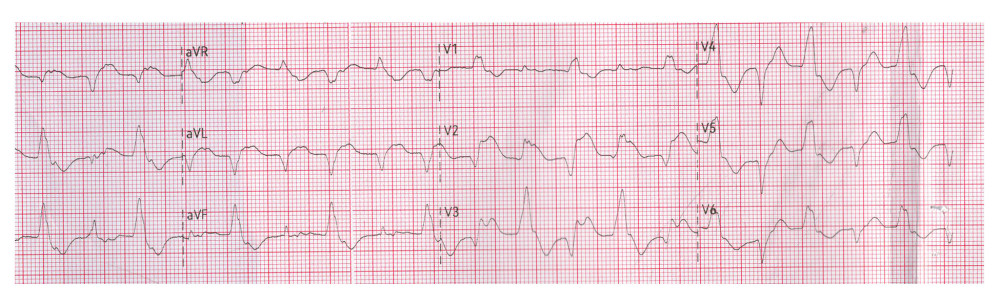 Figure 1. Electrocardiogram obtained at presentation demonstrating bidirectional ventricular tachycardia. The rhythm is characterized by a ventricular rate of approximately 125 to 130 bpm, widened QRS complexes (140–160 ms), and beat-to-beat alternation of the QRS axis, consistent with bidirectional ventricular tachycardia.
 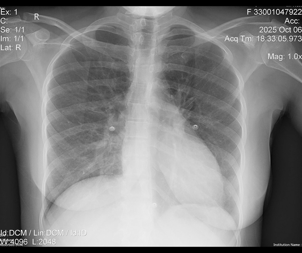 Figure 2. Anteroposterior chest radiograph. The image demonstrates well-aerated lungs without clinically significant loss of aeration. Structural findings include mild right basilar fibrotic changes, localized peribronchial thickening, and minimal pleural reaction. The hila appear prominent without focal abnormality, and the pulmonary vasculature is mildly prominent. The costophrenic angles and mediastinal contours are within normal limits.
 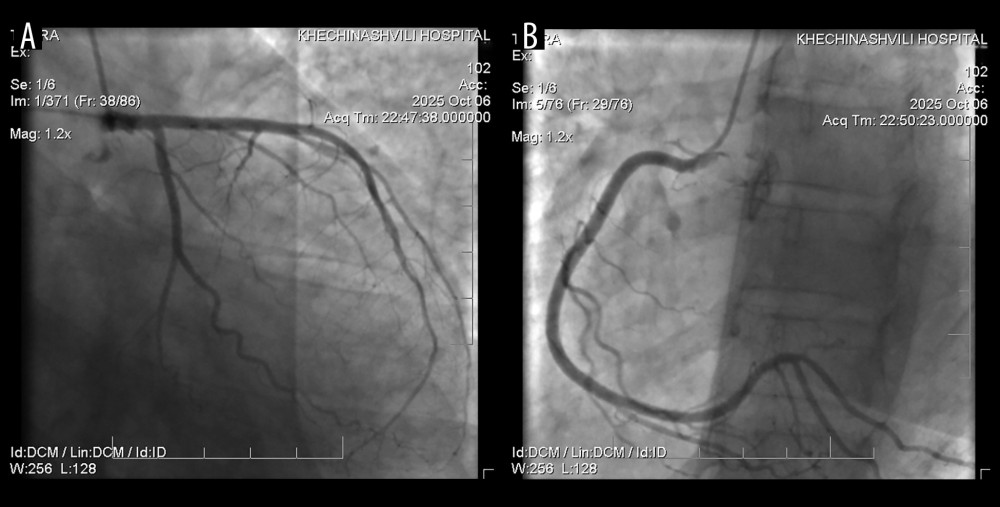 Figure 3. (A) Left coronary angiogram demonstrating a normal left main coronary artery bifurcating into the left anterior descending and left circumflex arteries, without evidence of atherosclerotic disease. (B) Right coronary angiogram showing a normally originating right coronary artery with smooth vessel walls and no evidence of atherosclerotic disease.
 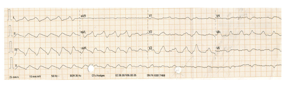 Figure 4. Electrocardiogram rhythm strip demonstrating polymorphic ventricular tachycardia. The single-lead rhythm strip documents the onset of polymorphic ventricular tachycardia with a ventricular rate of approximately 150 bpm, characterized by substantially widened (> 240 ms) and deformed QRS complexes approaching a sine-wave pattern, accompanied by severe repolarization abnormalities secondary to global myocardial electrical instability. This rhythm carries a high risk of degeneration into ventricular fibrillation, which subsequently developed.
 References
1. Denegri A, Dall’Ospedale V, Covani M, Cardiovascular complications of COVID-19 disease: A narrative review: Diseases, 2025; 13(8); 252
2. Guglin ME, Etuk A, Shah C, Ilonze OJ, Fulminant myocarditis and cardiogenic shock following COVID-19 infection versus COVID-19 vaccination: A systematic literature review: J Clin Med, 2023; 12(5); 1849
3. Veronese G, Ammirati E, Cipriani M, Frigerio M, Fulminant myocarditis: Characteristics, treatment, and outcomes: Anatol J Cardiol, 2018; 19(4); 279-86
4. Irabien Ortiz Á, Carreras Mora J, Sionis A, Fulminant myocarditis due to COVID-19: Rev Esp Cardiol (Engl Ed), 2020; 73(6); 503-4
5. Ammirati E, Lupi L, Palazzini M, Prevalence, characteristics, and outcomes of COVID-19-associated acute myocarditis: Circulation, 2022; 145(15); 1123-39
6. Ono R, Iwahana T, Aoki K, Fulminant myocarditis with SARS-CoV-2 infection: A narrative review from the case studies: Can J Infect Dis Med Microbiol, 2024; 2024; 9000598
7. Schulz-Menger J, Collini V, Gröschel J, 2025 ESC guidelines for the management of myocarditis and pericarditis: Eur Heart J, 2025; 46(1); 12-118
8. Zhang T, Pan N, Wang J, Understanding COVID-19-related myocarditis: Pathophysiology, diagnosis, and management: Cardiol Plus, 2023; 8(2); 72-81
9. Dao T, Barrow S, Lee R, Fulminant myocarditis after recovery from COVID-19 infection: A case report: J Am Coll Cardiol, 2022; 79(9 Suppl); 2344
10. Mirza N, Mirza M, Rayad MN, A rare case of fulminant myocarditis caused by COVID-19 and influenza B co-infection: Cureus, 2023; 15(6); e39905
11. Asaumi Y, Yasuda S, Morii I, Favourable clinical outcome in patients with cardiogenic shock due to fulminant myocarditis supported by percutaneous extracorporeal membrane oxygenation: Eur Heart J, 2005; 26(20); 2185-92
12. Lorusso R, Combes A, Coco VL, ECMO for COVID-19-associated severe acute respiratory distress syndrome and acute cardiomyopathy: ELSO guidelines and consensus statement: ASAIO J, 2021; 67(11); 1195-205
13. Kociol RD, Cooper LT, Fang JC, Recognition and initial management of fulminant myocarditis: A scientific statement from the American Heart Association: Circulation, 2020; 141(6); e69-e92
Figures
Figure 1. Electrocardiogram obtained at presentation demonstrating bidirectional ventricular tachycardia. The rhythm is characterized by a ventricular rate of approximately 125 to 130 bpm, widened QRS complexes (140–160 ms), and beat-to-beat alternation of the QRS axis, consistent with bidirectional ventricular tachycardia.Figure 2. Anteroposterior chest radiograph. The image demonstrates well-aerated lungs without clinically significant loss of aeration. Structural findings include mild right basilar fibrotic changes, localized peribronchial thickening, and minimal pleural reaction. The hila appear prominent without focal abnormality, and the pulmonary vasculature is mildly prominent. The costophrenic angles and mediastinal contours are within normal limits.Figure 3. (A) Left coronary angiogram demonstrating a normal left main coronary artery bifurcating into the left anterior descending and left circumflex arteries, without evidence of atherosclerotic disease. (B) Right coronary angiogram showing a normally originating right coronary artery with smooth vessel walls and no evidence of atherosclerotic disease.Figure 4. Electrocardiogram rhythm strip demonstrating polymorphic ventricular tachycardia. The single-lead rhythm strip documents the onset of polymorphic ventricular tachycardia with a ventricular rate of approximately 150 bpm, characterized by substantially widened (> 240 ms) and deformed QRS complexes approaching a sine-wave pattern, accompanied by severe repolarization abnormalities secondary to global myocardial electrical instability. This rhythm carries a high risk of degeneration into ventricular fibrillation, which subsequently developed. In Press
Case report 
Am J Case Rep In Press; DOI: 10.12659/AJCR.953440
Case report 
Am J Case Rep In Press; DOI: 10.12659/AJCR.953181
Case report 
Am J Case Rep In Press; DOI: 10.12659/AJCR.954572
Case report
Am J Case Rep In Press; DOI: 10.12659/AJCR.953539
Most Viewed Current Articles
07 Dec 2021 : Case report  28,707,486
DOI :10.12659/AJCR.934347
Am J Case Rep 2021; 22:e934347
06 Dec 2021 : Case report  180,259
DOI :10.12659/AJCR.934406
Am J Case Rep 2021; 22:e934406
21 Jun 2024 : Case report  123,976
DOI :10.12659/AJCR.944371
Am J Case Rep 2024; 25:e944371
08 May 2026 : Case report
94,256
DOI :10.12659/AJCR.952158
Am J Case Rep 2026; 27:e952158