


01 September 2026: Articles 
Combination of Multiple-Dose Activated Charcoal and Infusion Therapy That Led to Favorable Outcomes in Colchicine Poisoning: A Case Report
Unknown etiology, Management of emergency care
Manabu Eiraku AB 1, Kazuyuki Miyamoto E 1,2*, Keisuke Suzuki B 1, Kazumasa Abe C 3, Tatsuro Tamatsukuri D 3, Asuka Kaizaki-Mitsumoto CDE 4, Kazuki Kikuchi B 1, Masaharu Yagi B 1, Satoshi Numazawa CD 4, Kenji Dohi DE 1
DOI: 10.12659/AJCR.953541
Am J Case Rep 2026; 27:e953541


Abstract
BACKGROUND: The therapeutic margin of colchicine is narrow, and toxicity occurs easily. No standard treatment exists because its toxicokinetics are poorly understood. Symptoms are usually gastrointestinal, and dehydration occurs easily. Few reports have monitored serum and urinary colchicine levels.
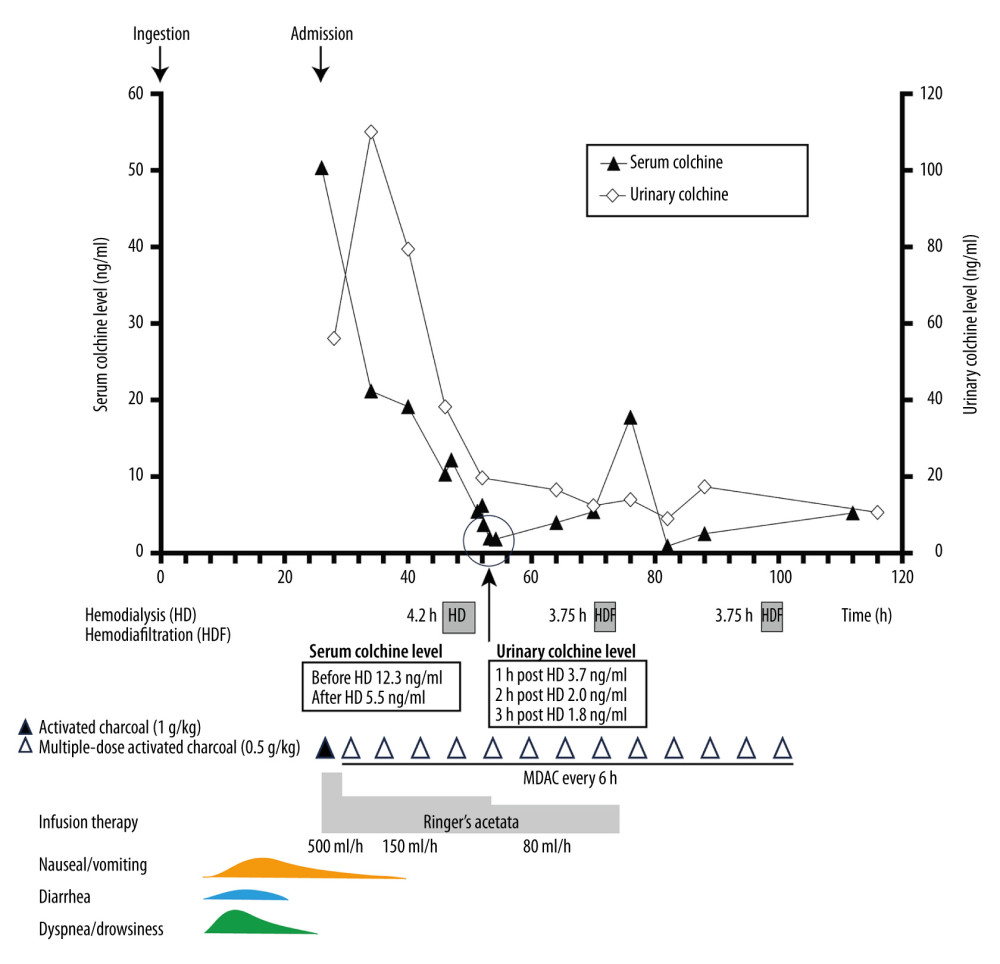
CASE REPORT: A man in his 20s presented with nausea, vomiting, diarrhea, drowsiness, and dyspnea. He ingested 2 dried Colchicum autumnale bulbs (estimated colchicine: 6.24-15.6 mg) with an energy drink. Activated charcoal with laxative was administered at 27 hours post ingestion (h-PI), followed by multiple-dose activated charcoal (MDAC) every 6 hours (13 doses, 27-101 h-PI), high-volume fluid infusion (Ringer’s acetate), and blood purification (hemodialysis [HD] at 48-52 h-PI; hemodiafiltration [HDF] at 71-75 and 98-102 h-PI). Serum colchicine was measured at 16 time points and urine at 11 time points. Serum colchicine at 27 h-PI was 50.3 ng/mL, and was temporally associated with a rapid decrease to 21.2 ng/mL at 35 h-PI, coinciding with initiation of infusion and activated charcoal. A secondary rise to 17.74 ng/mL occurred at 77 h-PI, approximately 2.4 hours after HDF1 completion. Urine colchicine was 110.0 ng/mL at 29 h-PI, then gradually decreased. Serum colchicine changed modestly (12.16 to 5.46 ng/mL) during HD.
CONCLUSIONS: In this case of severe colchicine poisoning, serial serum and urine concentration monitoring provided a time-resolved profile across concurrent interventions. The observed temporal associations support hypothesis generation regarding the potential roles of renal elimination and MDAC-mediated interruption of enterohepatic recirculation in colchicine clearance, while recognizing that concurrent therapies preclude attribution of effects to any single intervention.
Keywords: Charcoal, Colchicine
Introduction
Colchicine is a lipophilic alkaloid used to treat gout flares. Its therapeutic margin is narrow, and peak plasma concentrations (Cmax) are reported to be 6.50 ± 1.03 ng/mL at 1.07 ± 0.55 hours (Tmax) after oral administration [1]. Toxicity occurs easily without the ingestion of markedly high doses.
Case Report
A man in his 20s was transferred to the emergency department (ED) with nausea, vomiting, diarrhea, drowsiness, and dyspnea. He had previously been treated for autism spectrum disorder and attention-deficit hyperactivity disorder but was taking no home medications at the time of admission. This was the patient’s first episode of intentional overdose; no prior history of self-poisoning was documented. On arrival at the ED, he reported ingesting 2 dried
The serum colchicine level (Figure 1) was 50.3 ng/mL at 27 h-PI (on arrival). It decreased to 21.2 ng/mL at 35 h-PI, temporally associated with initiation of fluid infusion and activated charcoal, and further to 12.16 ng/mL immediately before HD (48.0 h-PI; circuit line). After HD, the level was 5.46 ng/mL (52.2 h-PI; circuit line). Before HDF1, the level was 3.98 ng/mL (65.0 h-PI; peripheral vein). The next available sample, which was obtained approximately 2.4 hours after HDF1 completion, was 17.74 ng/mL (77.0 h-PI), representing a secondary rise. Before HDF2, the level was 2.54 ng/mL (89.0 h-PI); the subsequent sample at 113.0 h-PI was 5.24 ng/mL. No samples were drawn immediately before or after the HDF sessions via the extracorporeal circuit, limiting interpretation of HDF-specific effects. The urine colchicine level was 110.0 ng/mL at 29 h-PI (timed specimen), then gradually decreased through 89 h-PI. A spot urine at 117 h-PI confirmed further decline. Urine creatinine levels fell markedly between 41 and 53 h-PI (429.7 to 43.5 mg/dL), likely reflecting dilution from high-volume infusion. Urinary excretion rates could not be calculated without concurrent urine volumes. Renal function remained preserved throughout (creatinine 0.53–0.65 mg/dL; estimated glomerular filtration rate [eGFR] > 130 mL/min/1.73 m2).
The patient was discharged on the fifth day of hospitalization after a psychiatric consultation, and was followed up as an outpatient. No obvious abnormalities were observed after hospitalization.
The CARE guidelines were followed in the preparation of this case report.
Discussion
LIMITATIONS:
This case has several important limitations. First, multiple interventions (MDAC, high-volume fluid therapy, HD, and HDF) were administered concurrently, precluding attribution of observed concentration changes to any single therapy. Second, renal clearance cannot be quantified from the available data, as urine volume was not measured at individual colchicine sampling intervals, and urine creatinine was available at only 2 time points. Urine colchicine concentrations were neither corrected for urine volume nor normalized by urine creatinine; therefore, these data reflect concentration only and should not be interpreted as reflecting excretion rate or renal clearance. Third, no serum samples were drawn immediately before HDF1/HDF2 start or immediately after HDF end from the circuit line; post-HDF values likely reflect a mixed signal of HDF effects and post-session redistribution. Fourth, plant identification relied solely on the patient’s verbal report, and the dose estimate was based on published colchicine content ranges without weighing the actual material. Fifth, no comprehensive multi-drug toxicology screen was performed (only alcohol screening, which was below the detection limit), and the energy drink’s composition and quantity were unknown; these factors cannot be excluded as potential contributors to the observed kinetics. Sixth, colchicine concentrations were measured using a commercially available LC-MS/MS screening platform (Shimadzu Corp); a case-specific analytical validation was not performed, and the assay was not independently validated for the purposes of this report. These limitations should be considered when interpreting the findings, which are presented as hypothesis-generating observations rather than definitive conclusions.
Conclusions
The most common symptoms of colchicine poisoning are gastrointestinal, including nausea, vomiting, diarrhea, and dehydration. In this case, serial monitoring of serum and urine colchicine over a detailed treatment timeline revealed temporal associations between fluid therapy, MDAC administration, and concentration changes. These findings support the hypothesis that promotion of renal excretion through fluid therapy and inhibition of colchicine reabsorption from the enterohepatic circulation using MDAC may contribute to colchicine elimination, while acknowledging that concurrent interventions and the absence of quantitative clearance data preclude definitive conclusions.
References
1. Ferron GM, Rochdi M, Jusko WJ, Scherrmann JM, Oral absorption characteristics and pharmacokinetics of colchicine in healthy volunteers after single and multiple doses: J Clin Pharmacol, 1996; 36; 874-83
2. Angelidis C, Kotsialou Z, Kossyvakis C, Colchicine pharmacokinetics and mechanism of action: Curr Pharm Des, 2018; 24; 659-63
3. Erden A, Karagoz H, Gümüscü HH, Colchicine intoxication: A report of two suicide cases: Ther Clin Risk Manag, 2013; 9; 505-9
4. Finkelstein Y, Aks SE, Hutson JR, Colchicine poisoning: The dark side of an ancient drug: Clin Toxicol (Phila), 2010; 48; 407-14
5. Labib S, Boujraf S, Berdai A, Harandou M, Fatal colchicine intoxication: Saudi J Anaesth, 2014; 8; 394-95
6. Eddleston M, Fabresse N, Thompson A, Anti-colchicine Fab fragments prevent lethal colchicine toxicity in a porcine model: A pharmacokinetic and clinical study: Clin Toxicol (Phila), 2018; 56; 773-81
7. Çankaya N, Bulduk İ, Çolak AM: Saudi J Biol Sci, 2019; 26; 345-51
8. Putterman C, Ben-Chetrit E, Caraco Y, Levy M, Colchicine intoxication: Clinical pharmacology, risk factors, features, and management: Semin Arthritis Rheum, 1991; 21; 143-55
9. Deng H, Xiang P, Zhang S, Delayed elimination in humans after ingestion of colchicine: Two fatal cases of colchicine poisoning: J Forensic Sci, 2023; 68; 1425-30
10. Grossenbacher F, Giodarno Orsini G, Cazaubon Y, Myocardial infarction in relation to colchicine poisoning: A precipitating cause of refractory toxic cardiogenic shock: Therapie, 2021; 76; 51-53
11. Schreiber L, Morovič M, Špacayová K, Halko R, Colchicine extract suicidal lethal poisoning confirmation using high-resolution accurate mass spectrometry: A case study: J Forensic Sci, 2019; 64; 1274-80
12. Zawahir S, Gawarammana I, Dargan PI: Clin Toxicol (Phila), 2017; 55; 914-18
13. Niel E, Scherrmann JM, Colchicine today: Jt Bone Spine, 2006; 73; 6728
14. Pérez Marín M, Prod’Hom S, de Villiers SF, Case report: Colchicine toxicokinetic analysis in a poisoned child requiring extracorporeal life support: Front Pediatr, 2021; 9; 658347
15. Lu X, Liu Y, Wang C, Pathogenic characteristics and treatment in 43 cases of acute colchicine poisoning: Toxicol Res (Camb), 2021; 10; 885-92
16. Ramezani M, Mostafazadeh B, Rahimi M, Colchicine poisoning treated with hemoperfusion and hemodialysis: A case report: Clin Case Rep, 2022; 10; e6419
17. Mullins ME, Pinnick RV, Terhes JM, Life-threatening diphenhydramine overdose treated with charcoal hemoperfusion and hemodialysis: Ann Emerg Med, 1999; 33; 104-7
18. Ben-Chetrit E, Scherrmann JM, Zylber-Katz E, Levy M, Colchicine disposition in patients with familial Mediterranean fever with renal impairment: J Rheumatol, 1994; 21; 710-13
Tables
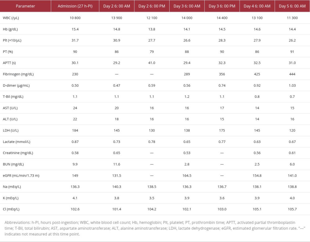 Table 1. Laboratory data on admission and serial values during hospitalization (h-PI = hours post ingestion).
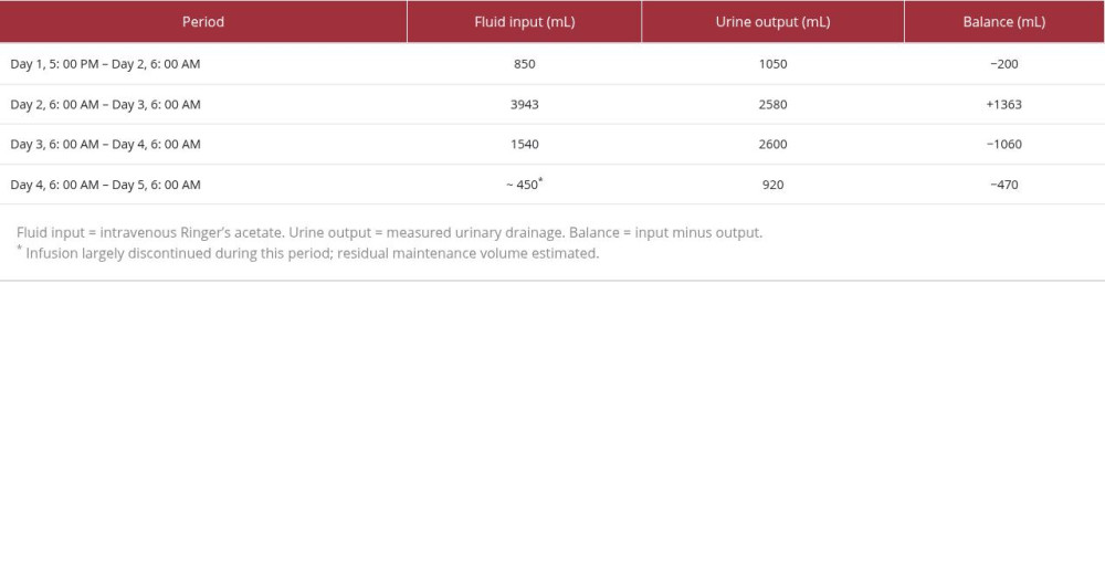 Table 2. Fluid input/output balance by period.
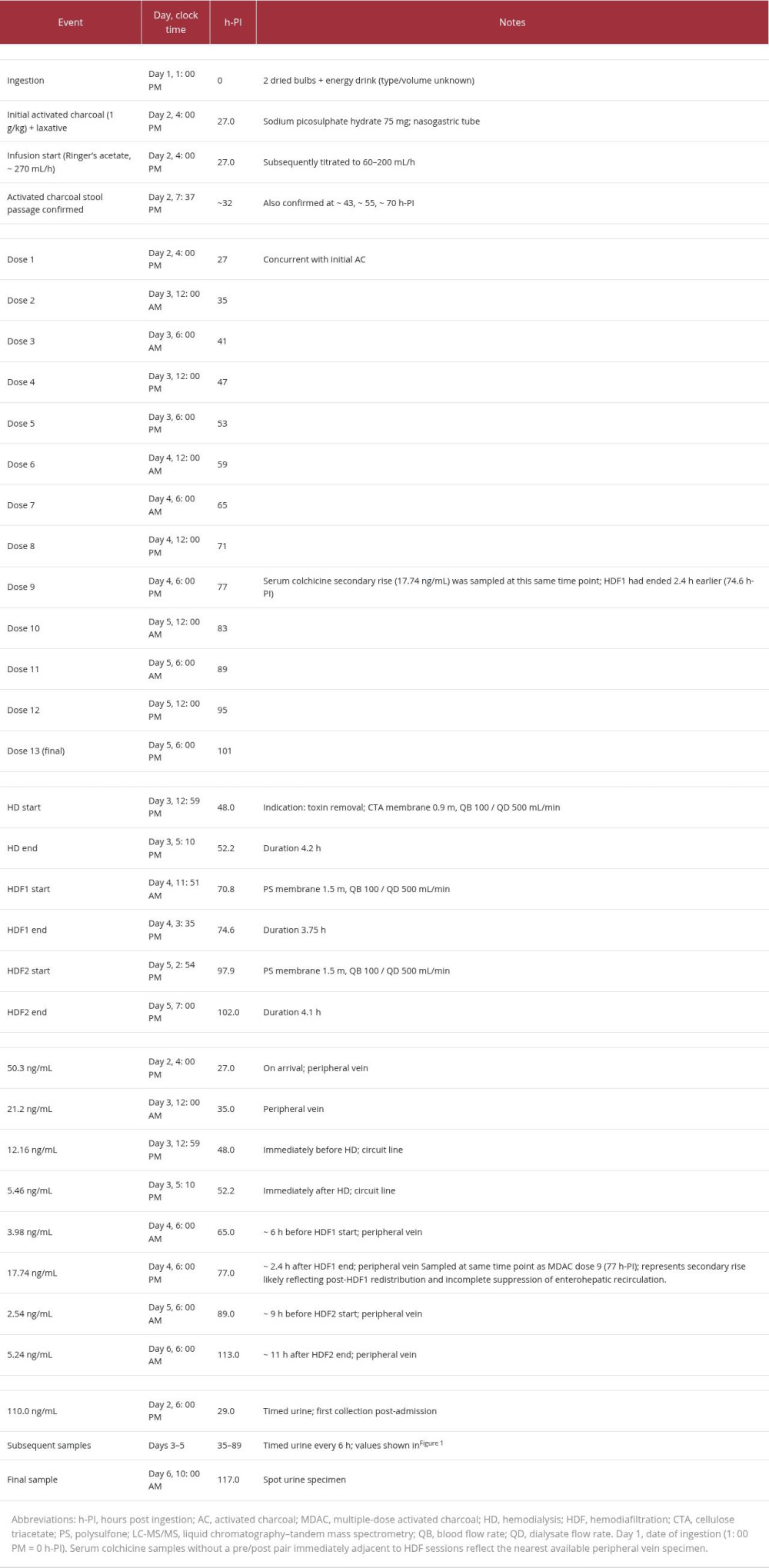 Table 3. Intervention and monitoring timeline (h-PI = hours post ingestion).Table 1. Laboratory data on admission and serial values during hospitalization (h-PI = hours post ingestion).Table 2. Fluid input/output balance by period.Table 3. Intervention and monitoring timeline (h-PI = hours post ingestion).
 In Press
Case report 
Am J Case Rep In Press; DOI: 10.12659/AJCR.953543
Case report
Am J Case Rep In Press; DOI: 10.12659/AJCR.954216
Case report 
Am J Case Rep In Press; DOI: 10.12659/AJCR.954932
Case report 
Am J Case Rep In Press; DOI: 10.12659/AJCR.954146
Most Viewed Current Articles
07 Dec 2021 : Case report
28,712,695
DOI :10.12659/AJCR.934347
Am J Case Rep 2021; 22:e934347
06 Dec 2021 : Case report  180,721
DOI :10.12659/AJCR.934406
Am J Case Rep 2021; 22:e934406
21 Jun 2024 : Case report
124,419
DOI :10.12659/AJCR.944371
Am J Case Rep 2024; 25:e944371
08 May 2026 : Case report
95,462
DOI :10.12659/AJCR.952158
Am J Case Rep 2026; 27:e952158