


31 August 2026: Articles 
RadiationAssociated Undifferentiated Pleomorphic Sarcoma of the Neck 2 Decades After External Beam Radiation for Medullary Thyroid Carcinoma: A Case Report and Literature Review
Unusual clinical course, Challenging differential diagnosis, Rare disease
Tanya Odisho ABDEF 1,2, Sedighe Esmaeilzade ABDEF 3, Alexander Laskaris BEF 2, Sungmi Jung ABDEF 4, James Man Git Tsui DEF 1,3, Sinziana Dumitra ABDEF 5,2, Elliot Mitmaker ABDEF 1,2*
DOI: 10.12659/AJCR.953580
Am J Case Rep 2026; 27:e953580


Abstract
BACKGROUND: Radiation therapy is critical for treating many solid tumors but increases the risk of secondary malignancies due to DNA damage. Radiation-associated sarcomas (RAS) occur in 0.03% to 0.8% of irradiated patients, typically 5 to 20 years after treatment. Undifferentiated pleomorphic sarcoma (UPS) is a common RAS subtype with poor 5-year survival (12%-14%). Risk rises with doses ≥50 Gy and is especially concerning in previously irradiated head and neck regions, where surgery is challenging. Germline mutations in DNA repair genes (eg, ATM) further increase susceptibility.
CASE REPORT: A 54-year-old man treated in 2003 for multifocal medullary thyroid carcinoma (thyroidectomy, bilateral neck dissection, adjuvant 59.4 Gy radiation) presented 22 years later with a painless neck mass. Imaging showed a 1.9-cm enhancing lesion within the prior radiation field. Excision revealed a 4-cm, FNCLCC grade 3 intramuscular UPS meeting the modified Cahan criteria for RAS. Immunohistochemistry excluded recurrent thyroid carcinoma. Margins were positive, but re-excision and re-irradiation were not feasible. The sarcoma tumor board recommended close surveillance and germline testing. This case involved high-grade UPS arising 2 decades after neck irradiation. Compared with sporadic UPS, RAS-associated UPS has worse disease-specific survival (~52% vs 76%) and higher local recurrence (~55% vs 24%), emphasizing the importance and difficulty of achieving negative margins in previously irradiated fields.
CONCLUSIONS: As cancer survival improves, lifelong vigilance for RAS remains essential. New masses in irradiated areas require prompt evaluation. Multidisciplinary management and consideration of genetic testing are critical, as treatment options are often limited by prior therapy.
Keywords: Radiation Effects, Sarcoma, Thyroid (USP)
Introduction
An uncommon thyroid malignancy, medullary thyroid carcinoma (MTC) originates from parafollicular C cells responsible for calcitonin production [1]. Primary treatment typically involves total thyroidectomy accompanied by central and often bilateral cervical lymph node dissection [2], although complete resection may be unachievable in advanced disease. Aggressive surgical procedures in the cervical region carry significant morbidity, including recurrent laryngeal nerve injury with vocal cord dysfunction, dysphagia, parathyroid dysfunction, and injury to adjacent neurovascular structures [1]. When residual disease or a high risk of locoregional recurrence is present, selective use of adjuvant external beam radiation therapy (EBRT) may be considered, as supported by the American Thyroid Association [1]. In such settings, EBRT has been shown to improve locoregional disease control, with long-term follow-up studies demonstrating substantially higher 10-year regional control rates of 86% with EBRT compared to 52% with surgery alone [3].
However, despite its benefits, EBRT is associated with late toxicities, including secondary malignancies with latency periods of 7 to 16 years [4–8]. Thyroid cancer survivors face an estimated 7% risk of second primary malignancy [9], which appears to increase with higher cumulative radiation exposure (relative risk 1.03) [4]. RAS develops in 0.03% to 0.8% of irradiated patients and carries worse overall and disease-specific survival than sporadic sarcomas, especially in the head and neck region where surgical management is complex [2,4,6,7,10]. Reported 5-year survival rates in this setting are 24% to 38% [8,11–14].
According to the modified Cahan criteria, RAS refers to a histologically distinct sarcoma that develops within a previously irradiated field following an adequate latency interval [15–17]. Molecularly, ionizing radiation induces genomic instability and DNA damage that can contribute to malignant transformation, frequently involving mutations in RB1, ATM and TP53 [18,19]. Undifferentiated pleomorphic sarcoma (UPS) is the most frequent RAS subtype and is characterized by marked cellular pleomorphism, absence of lineage-specific differentiation, and frequent TP53 mutations, distinguishing it from sporadic sarcomas [2,20,21]. Diagnosis is made by exclusion following histologic and immunohistochemical evaluation [20,21].
We describe a case of UPS arising in the cervical region nearly 2 decades after EBRT administered for MTC in a 54-year-old man. This case highlights the need for lifelong surveillance following radiation therapy, illustrates the diagnostic and therapeutic challenges encountered in previously irradiated tissues, and raises consideration of potential underlying genetic susceptibility to radiation-related malignancies.
Case Report
A 54-year-old man with a history of MTC diagnosed in 2002 presented in early 2025 with a progressively enlarging right-sided neck mass (Figure 1). His initial management had consisted of total thyroidectomy with bilateral neck dissection. This was followed by adjuvant external beam radiation therapy (EBRT), which was delivered and completed in April 2003 using a 3D conformal radiation therapy (3D-CRT) technique, with a total dose of 59.4 Gy in 33 fractions to the thyroid bed and involved cervical lymph node regions. Pathology confirmed multifocal MTC with nodal metastases. Surgical margins were negative, and the patient remained under biannual surveillance for 22 years with persistently elevated but stable calcitonin levels.
In 2025, rising calcitonin levels (> 110 ng/L) and a right submandibular mass raised a concern for recurrent MTC. CT imaging revealed a 1.9 × 1.1 × 1.5 cm well-demarcated hypodense lesion inferior to the right submandibular space, inseparable from the thyrohyoid muscle with mild extracapsular extension (Figure 1C). No additional disease was identified. Following multidisciplinary discussion, excisional biopsy was pursued in the context of a suspected metastatic MTC lymph node.
Intraoperatively, the mass was located between the thyroid cartilage and hyoid bone, densely adherent to the thyrohyoid muscle within a previously operated and irradiated field. Circumferential resection including muscle was performed without evidence of invasion into adjacent cartilage or bone. Postoperative CT demonstrated surgical scarring and fibrosis without a definite residual mass, although interpretation was limited by distorted anatomy (Figure 1D).
Histopathology revealed a 4.0-cm, high-grade intramuscular UPS without lymphoid tissue (Figure 2A). Immunohistochemistry was positive for vimentin and focally positive for SMA and desmin, and negative for epithelial and neuroendocrine markers, excluding recurrent MTC (Figure 2B, 2C). The tumor was FNCLCC grade 3/3 with < 5% necrosis and positive margins.
Postoperatively, serum calcitonin levels remained persistently elevated but stable, measuring 108 ng/L 1 month after resection and 110 ng/L at the most recent follow-up in May 2026. Importantly, there has been no progressive upward trend over time. Despite this biochemical abnormality, serial imaging and histopathologic evaluation have shown no evidence of recurrent or residual medullary thyroid carcinoma. Immunohistochemical studies of the resected lesion were negative for neuroendocrine differentiation, further excluding metastatic or recurrent MTC. The etiology of the persistently elevated calcitonin remains unclear. In the absence of radiologic or pathologic correlation, this is considered an unexplained persistent biochemical elevation without structural disease correlate. Continued surveillance has therefore been recommended, as occult microscopic disease cannot be entirely excluded.
The case was reviewed by the multidisciplinary tumor board, where re-irradiation was deemed contraindicated due to prior radiation dose exposure. Surgical re-excision was also considered excessively morbid given the tumor’s proximity to the trachea. The agreed-upon management plan was close surveillance with serial imaging and referral for genetic evaluation. Given the patient’s significant family history of malignancy and parental consanguinity, there was concern for an underlying hereditary cancer predisposition syndrome; therefore, a comprehensive germline evaluation was performed. The patient underwent a multigene hereditary cancer predisposition panel (“Sarcoma Panel with flex testing for medullary thyroid carcinoma (MTC) and hereditary breast and ovarian cancer (HBOC) predisposition syndrome”) which analyzed 63 cancer susceptibility genes, including but not limited to
Surveillance MRI of the neck performed in September 2025 demonstrated no evidence of locoregional recurrence at the site of the previously resected right neck mass and no suspicious cervical lymphadenopathy. However, a CT scan of the chest obtained during the same period revealed bilateral pulmonary nodules concerning for metastatic disease.
The patient was subsequently referred to Medical Oncology and systemic therapy options, including immunotherapy and clinical trial enrollment, were reviewed in multidisciplinary discussion. Given the diagnosis of metastatic RAS-UPS, the patient was initiated on palliative doxorubicin administered every 3 weeks. Follow-up PET imaging in April 2026 demonstrated recurrent disease within the right neck, as well as a mixed metabolic response of the bilateral pulmonary metastases to treatment. In light of these findings, the treatment regimen was subsequently switched to liposomal doxorubin (Caelyx), which the patient has tolerated well to date. He remains clinically stable and is being closely monitored.
Discussion
Radiation therapy is widely employed as an adjunctive modality in the management of solid tumors in both curative and palliative settings and is a key component of multidisciplinary cancer care. Despite its therapeutic benefits, ionizing radiation has been associated with delayed toxicities, including the development of secondary malignancies driven by radiation-induced DNA damage and disruption of cell-cycle control [22–24]. With increasing numbers of long-term cancer survivors, the occurrence of radiation-associated secondary cancers has become more frequently recognized.
The most common radiation-associated malignancies include squamous cell carcinoma and sarcoma [13,25–27]. Among radiation-induced sarcomas, undifferentiated pleomorphic sarcoma (UPS) has been frequently reported and is often associated with an aggressive clinical course and less favorable outcomes compared with sporadic counterparts [2]. Our case fulfills established RAS diagnostic criteria, including prior radiation exposure, prolonged latency, development within the radiation field, and distinct histology. Latency periods typically range from 8 to 10 years but can be longer [24,28–30].
Although EBRT is not routinely indicated for all MTC cases [1,31], our patient received adjuvant radiation for multifocal disease with nodal involvement. The risk of RAS has been shown to increase with higher cumulative radiation doses, particularly those exceeding approximately 50 Gy [29,32]. Consistent with this observation, the sarcoma in our patient developed more than 2 decades after exposure to a total dose of 59.4 Gy. Similar cases have been described following high-dose head and neck radiation [33,34]. Notably, our patient remained disease-free for more than 2 decades prior to RAS onset.
Clinically, RAS commonly presents as a painless enlarging mass within an irradiated field. In our case, the diagnostic evaluation was initially influenced by elevated serum calcitonin levels, which raised concern for recurrence of medullary thyroid carcinoma. Radiologic imaging demonstrated a well-circumscribed lesion with local invasion, and definitive diagnosis relied on histopathologic examination and immunohistochemical profiling. The tumor had features consistent with a high-grade UPS and showed expression of mesenchymal markers, including vimentin, smooth muscle actin, and desmin. In contrast, markers typically associated with neuroendocrine differentiation, such as calcitonin, chromogranin, cytokeratins, and epithelial membrane antigen, were absent, effectively excluding recurrent MTC (Figure 2).
Management of RAS remains challenging. Complete surgical excision with histologically negative margins is the most important determinant of long-term survival in patients with RAS [6,19], whereas systemic therapy offers limited benefit beyond palliative anthracycline-based regimens. However, achieving clear margins is often difficult in previously irradiated and surgically altered anatomical fields. In the present case, the initial resection yielded positive margins, and re-excision was not pursued due to the anticipated functional and structural morbidity. Additional radiation was contraindicated in the setting of prior cumulative dose constraints. The patient was therefore initially managed with close clinical and radiologic surveillance. He subsequently developed locoregional recurrence and pulmonary metastases, prompting initiation of systemic therapy.
Although the patient had a notable family history of malignancy, germline testing was not performed at the time of initial diagnosis of medullary thyroid carcinoma, as routine clinical practice at that time primarily focused on targeted RET mutation analysis rather than expanded multigene panel testing in the absence of a defined hereditary syndrome. Genetic evaluation was subsequently pursued after the diagnosis of RAS, when suspicion for an underlying DNA repair defect or hereditary cancer predisposition syndrome was increased. Inherited variants in genes responsible for DNA damage repair, particularly ATM, are linked to heightened radiosensitivity and an elevated risk of secondary malignancies [4,35]; therefore, our patient underwent genetic evaluation to assess for an inherited predisposition and to guide future surveillance strategies for himself and family members. While no germline mutations were detected, the breadth of his family history suggests that an underlying hereditary predisposition cannot be entirely excluded, particularly in the context of radiation-associated tumorigenesis, which has been linked to defects in DNA damage repair pathways.
Conclusions
This case demonstrates the importance of long-term surveillance in cancer survivors treated with radiation therapy, particularly in the head and neck region. It emphasizes the need for heightened clinical suspicion when new lesions arise within irradiated fields and illustrates the diagnostic and therapeutic challenges posed by radiation-associated sarcomas. Because of their aggressive nature and unfavorable outcomes, particularly when complete resection is not possible, early recognition and coordinated multidisciplinary management are crucial. Reporting such cases contributes to improved awareness of these uncommon but serious late effects of radiation treatment.
Figures
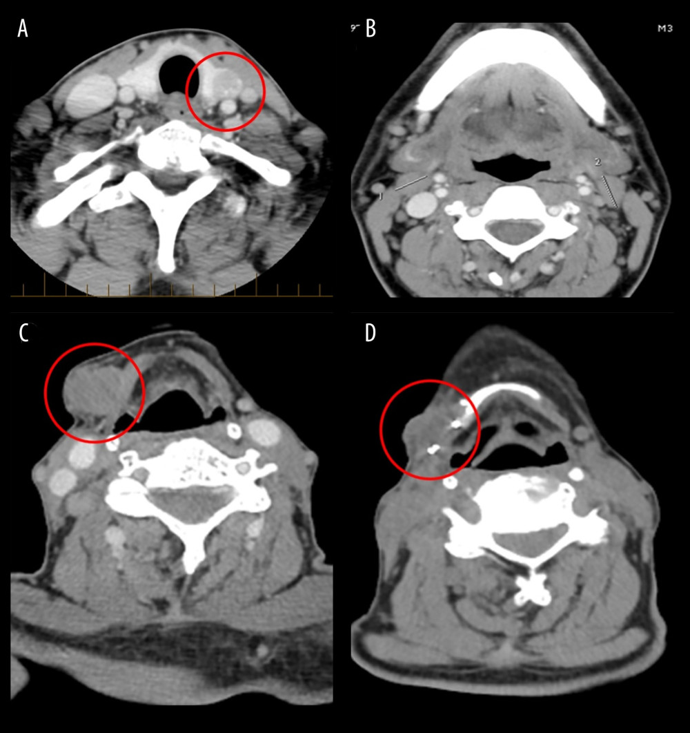 Figure 1. CT imaging showing the progression of neck soft tissue masses. In 2002, a 12-mm nodule was identified in the left thyroid lobe (A), along with multiple bilateral nodes suggestive of metastases, the largest in the jugulodigastric and right omohyoid regions measuring 16 mm (B). Images from 2025 after remission reveal a well-defined 1.9 × 1.7 × 1.5 cm lesion inferior to the right submandibular space, suspected enlarged lymph node with extracapsular extension (C). Postsurgical imaging shows resection of a right cervical sarcomatous mass between the right hyoid and thyroid cartilages (D).
 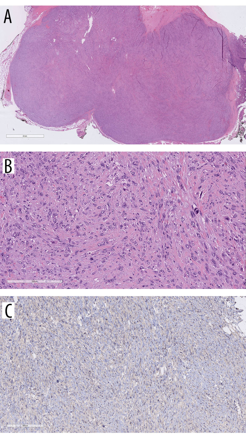 Figure 2. Histopathology of the resected mass. A 4.0-cm relatively well-circumscribed tumor was excised (A). The tumor displays a pattern less arrangement of atypical spindled cells with no identifiable line of differentiation (B). Immunohistochemical stains confirm no evidence of thyroid cell differentiation and no reactivity for calcitonin depicted (C).
 References
1. Wells SA, Asa SL, Dralle H, Revised American Thyroid Association guidelines for the management of medullary thyroid carcinoma: Thyroid, 2015; 25(6); 567-610
2. Dineen SP, Roland CL, Feig R, Radiation-associated undifferentiated pleomorphic sarcoma is associated with worse clinical outcomes than sporadic lesions: Ann Surg Oncol, 2015; 22(12); 3913-20
3. Brierley J, Tsang R, Simpson WJ, Medullary thyroid cancer: Analyses of survival and prognostic factors and the role of radiation therapy in local control: Thyroid, 1996; 6(4); 305-10
4. Berrington de Gonzalez A, Curtis RE, Kry SF, Proportion of second cancers attributable to radiotherapy treatment in adults: A cohort study in the US SEER cancer registries: Lancet Oncol, 2011; 12(4); 353-60
5. Riad S, Biau D, Holt GE, The clinical and functional outcome for patients with radiation-induced soft tissue sarcoma: Cancer, 2012; 118(10); 2682-92
6. Gladdy RA, Qin LX, Moraco N, Do radiation-associated soft tissue sarcomas have the same prognosis as sporadic soft tissue sarcomas?: J Clin Oncol, 2010; 28(12); 2064-69
7. Cha C, Antonescu CR, Quan ML, Long-term results with resection of radiation-induced soft tissue sarcomas: Ann Surg, 2004; 239(6); 903-10
8. Yeang MS, Tay K, Ong WS, Outcomes and prognostic factors of post-irradiation and de novo sarcomas of the head and neck: A histologically matched case-control study: Ann Surg Oncol, 2013; 20(9); 3066-75
9. Brown AP, Chen J, Hitchcock YJ, The risk of second primary malignancies up to three decades after the treatment of differentiated thyroid cancer: J Clin Endocrinol Metab, 2008; 93(2); 504-15
10. Mito JK, Mitra D, Doyle LA, Radiation-associated sarcomas: An update on clinical, histologic, and molecular features: Surg Pathol Clin, 2019; 12(1); 139-48
11. Chan JY, Wong ST, Lau GI, Wei WI, Postradiation sarcoma after radiotherapy for nasopharyngeal carcinoma: Laryngoscope, 2012; 122(12); 2695-99
12. Xi M, Liu MZ, Wang H, Radiation-induced sarcoma in patients with nasopharyngeal carcinoma: A single-institution study: Cancer, 2010; 116(23); 5479-86
13. Lou J, Jiang L, Dai X, Radiation-induced sarcoma of the head and neck following radiotherapy for nasopharyngeal carcinoma: A single institutional experience and literature review: Front Oncol, 2020; 10; 526360
14. Debnam JM, Guha Thakurta N, Mahfouz YMM, Radiation-associated head and neck sarcomas: Spectrum of imaging findings: Oral Oncol, 2012; 48; 155-61
15. Cahan WG, Woodard HQ, Sarcoma arising in irradiated bone; Report of 11 cases: Cancer, 1948; 1(1); 3-29
16. Murray EM, Werner D, Greeff EA, Taylor DA, Postradiation sarcomas: 20 cases and a literature review: Int J Radiat Oncol Biol Phys, 1999; 45(4); 951-61
17. Singh GK, Yadav V, Singh P, Bhowmik KT, Radiation-induced malignancies making radiotherapy a “two-edged sword”: A review of literature: World J Oncol, 2017; 8(1); 1-6
18. Hall EJ, Wuu CS, Radiation-induced second cancers: The impact of 3D-CRT and IMRT: Int J Radiat Oncol Biol Phys, 2003; 56(1); 83-88
19. Torres MA, Ballo MT, Butler CE, Management of locally recurrent soft-tissue sarcoma after prior surgery and radiation therapy: Int J Radiat Oncol Biol Phys, 2007; 67(4); 1124-29
20. Lesluyes T, Baud J, Pérot G, Genomic and transcriptomic comparison of post-radiation versus sporadic sarcomas: Mod Pathol, 2019; 32(12); 1786-94
21. Hong DC, Yang J, Sun C, Genomic profiling of radiation-induced sarcomas reveals the immunologic characteristics and its response to immune checkpoint blockade: Clin Cancer Res, 2023; 29(15); 2869-84
22. Berrington de González A, Curtis RE, Gilbert E, Second solid cancers after radiation therapy: A systematic review of the epidemiologic studies of the radiation dose-response relationship: Radiat Res, 2011; 178(3); AVO-AVR
23. Thariat J, Hannoun-Levi JM, Myint AS, Past, present, and future of radiotherapy for the benefit of patients: Nat Rev Clin Oncol, 2013; 10(1); 52-60
24. Khanna L, Prasad SR, Yedururi S, Second malignancies after radiation therapy: update on pathogenesis and cross-sectional imaging findings: Radiographics, 2021; 41(3); 876-94
25. Giannini L, Incandela F, Fiore M, Radiation-induced sarcoma of the head and neck: A review of the literature: Front Oncol, 2018; 8; 449
26. Vakil J, Cuartas-Mesa MC, Jian Hua L, When radiation therapy becomes a foe: A rare case of radiation-induced angiosarcoma of head and neck: Cureus, 2023; 15(2); e35122
27. Ferreira IV, Cattan MES, Chone CT, Radiation-induced osteosarcoma in the head and neck region: Case report and literature review: Oral Oncol, 2025; 162; 107216
28. Amendola BE, Amendola MA, McClatchey KD, Miller CH, Radiation-associated sarcoma: A review of 23 patients with postradiation sarcoma over a 50-year period: Am J Clin Oncol, 1989; 12(5); 411-15
29. Patel SG, See AC, Williamson PA, Radiation induced sarcoma of the head and neck: Head Neck, 1999; 21(4); 346-54
30. Smith JB, Cass LM, Simpson MC, Radiation-associated sarcoma of the head and neck: Incidence, latency, and survival: Laryngoscope, 2022; 132(5); 1034-41
31. Terezakis SA, Lee KS, Ghossein RA, Role of adjuvant radiation therapy in medullary thyroid carcinoma: Int J Radiat Oncol Biol Phys, 2011; 80(3); 735-40
32. Sheppard DG, Libshitz HI, Post-radiation sarcomas: A review of the clinical and imaging features in 63 cases: Clin Radiol, 2001; 56(1); 22-29
33. Takumi Y, Obata K, Kondo A, Treating radiation-induced sarcoma of the head and neck: A case report: Otolaryngol Case Rep, 2021; 19; 100283
34. Sharma D, Das R, Ahmed S, Radiation-induced undifferentiated pleomorphic sarcoma in thyroid: A rare occurrence: Indian J Otolaryngol Head Neck Surg, 2025; 77(1); 453-57
35. Sherborne AL, Davidson PR, Yu K, Mutational analysis of ATM, BRCA1, BRCA2 and TP53 genes in radiation-induced sarcomas: Radiother Oncol, 2015; 117(3); 466-71
Figures
Figure 1. CT imaging showing the progression of neck soft tissue masses. In 2002, a 12-mm nodule was identified in the left thyroid lobe (A), along with multiple bilateral nodes suggestive of metastases, the largest in the jugulodigastric and right omohyoid regions measuring 16 mm (B). Images from 2025 after remission reveal a well-defined 1.9 × 1.7 × 1.5 cm lesion inferior to the right submandibular space, suspected enlarged lymph node with extracapsular extension (C). Postsurgical imaging shows resection of a right cervical sarcomatous mass between the right hyoid and thyroid cartilages (D).Figure 2. Histopathology of the resected mass. A 4.0-cm relatively well-circumscribed tumor was excised (A). The tumor displays a pattern less arrangement of atypical spindled cells with no identifiable line of differentiation (B). Immunohistochemical stains confirm no evidence of thyroid cell differentiation and no reactivity for calcitonin depicted (C). In Press
Case report 
Am J Case Rep In Press; DOI: 10.12659/AJCR.953543
Case report 
Am J Case Rep In Press; DOI: 10.12659/AJCR.954216
Case report 
Am J Case Rep In Press; DOI: 10.12659/AJCR.954932
Case report 
Am J Case Rep In Press; DOI: 10.12659/AJCR.954146
Most Viewed Current Articles
07 Dec 2021 : Case report
28,712,695
DOI :10.12659/AJCR.934347
Am J Case Rep 2021; 22:e934347
06 Dec 2021 : Case report  180,721
DOI :10.12659/AJCR.934406
Am J Case Rep 2021; 22:e934406
21 Jun 2024 : Case report
124,419
DOI :10.12659/AJCR.944371
Am J Case Rep 2024; 25:e944371
08 May 2026 : Case report
95,462
DOI :10.12659/AJCR.952158
Am J Case Rep 2026; 27:e952158