


23 September 2026: Articles 
Refractory Chylous Ascites After Robot-Assisted Radical Prostatectomy With Sequential Anatomically Distinct Lymphatic Leak Sites Identified by Repeat Lymphangiography
Unusual clinical course, Unusual or unexpected effect of treatment, Educational Purpose (only if useful for a systematic review or synthesis)
Takayuki Owaki ABCDEFG 1,2*, Yasuhito Funahashi ABE 1,3, Kosuke Tochigi B 1,4, Ryota Hyodo BDF 5, Wataru Nakane B 1,6, Masaya Matsushima B 5,7, Shusuke Akamatsu E 1
DOI: 10.12659/AJCR.954777
Am J Case Rep 2026; 27:e954777


Abstract
BACKGROUND: Chylous ascites is a rare, potentially life-threatening complication of pelvic surgery, characterized by progressive malnutrition, immune dysfunction, and massive fluid accumulation. When the condition is refractory, its management represents a complex challenge requiring sequential escalation across multiple therapeutic modalities.
CASE REPORT: A 72-year-old man developed refractory chylous ascites after robot-assisted radical prostatectomy with extended pelvic lymph node dissection for high-grade prostate cancer (Gleason score 5+4=9). Despite conservative measures, consisting of low-fat diet and continuous subcutaneous octreotide infusion, the ascites persisted. Repeated large-volume paracentesis with cell-free concentrated ascites reinfusion therapy (CART) and total parenteral nutrition (TPN) were required to manage worsening malnutrition. Lymphangiography and N-butyl cyanoacrylate (NBCA) embolization provided no sustained relief. Subsequent surgical lymphatic ligation identified active leakage alongside floating solidified NBCA in the internal iliac region, confirming the prior embolization site. Although the leak was temporarily controlled by ligation, chylous drainage recurred on postoperative day 2. Repeat lymphangiography revealed a new, anatomically distinct leak in the external iliac region, suggesting dynamic redistribution of lymphatic flow through a collateral pathway. Repeat NBCA embolization of this secondary site achieved definitive resolution. Supported by TPN and CART, the patient was discharged on postoperative day 117 with improved nutritional status. He had no recurrence over a 5-year follow-up, with clinical and laboratory assessment every 3 months.
CONCLUSIONS: This case suggests that lymphatic leak sites may shift during treatment. Repeat lymphangiography may be considered when chylous drainage recurs after apparently successful intervention. Multidisciplinary management, including TPN and CART, was integral to sustaining this patient through a prolonged and complex treatment course.
Keywords: Ascites, Embolization, Therapeutic, Prostatectomy
Introduction
Lymphocele and minor lymphorrhea following extended pelvic lymph node dissection (ePLND) are well-recognized surgical complications, with a reported incidence of up to 22% to 33% after robot-assisted radical prostatectomy (RARP) with ePLND [1]. However, chylous ascites—the accumulation of triglyceride-rich chylous fluid in the peritoneal cavity due to the disruption of intestinal lymphatics—is a much rarer and more severe manifestation. Unlike simple lymphoceles, chylous ascites leads to profound nutritional consequences: the loss of protein, fat, fat-soluble vitamins, and lymphocytes leads to progressive malnutrition, immunosuppression, and significant morbidity. In severe or prolonged cases, it can be life-threatening [2].
From an intensive care perspective, the management of refractory chylous ascites represents a complex challenge involving nutritional optimization, fluid and electrolyte management, prevention of infectious complications, and coordination of multiple therapeutic modalities including dietary modification, pharmacological treatment, interventional radiology, and surgery [3,4]. No standardized management protocol currently exists, and treatment is largely guided by case series and expert opinion [5,6].
Reports of chylous ascites following RARP with ePLND remain limited, and cases requiring sequential escalation across conservative therapy, interventional radiology, and surgical management are particularly uncommon [7,8]. We present a case in which sequential multidisciplinary management—including conservative measures, concentrated ascites reinfusion therapy (CART), total parenteral nutrition (TPN), lymphatic embolization, surgical ligation, and repeat embolization—was required to achieve resolution. During treatment, a second, anatomically distinct lymphatic leak in the external iliac region was identified by repeat lymphangiography after successful control of the primary internal iliac leak, suggesting possible redistribution of lymphatic flow through collateral pathways. This case highlights the potential value of repeat lymphangiography when chylous drainage recurs after apparently successful intervention.
Case Report
A 72-year-old man was referred to our hospital for an elevated prostate-specific antigen of 13.7 ng/mL. Prostate needle biopsy revealed adenocarcinoma staged as cT2bN0M0 with a Gleason score of 5 + 4 = 9 (Grade Group 5). The patient underwent RARP with bilateral ePLND. Dissected nodal stations included common iliac, external iliac, internal iliac, and obturator nodes. The total operative time was 3 hours 43 minutes, with estimated blood loss of 150 mL. Final pathology confirmed pT2c, with a Gleason score of 5 + 4 = 9, negative surgical margins, and no nodal involvement (0 of 28 nodes; pN0).
The patient was mobilized and began oral intake on postoperative day (POD) 1. From POD 2, drain output increased progressively and became milky in appearance. Biochemical analysis of the drain fluid confirmed chylous content (triglyceride concentration markedly elevated: 274 mg/dL). A low-fat diet was initiated, and, with no signs of systemic compromise at that time, the drain was removed on POD 7 and the patient was discharged with expectant management on POD 8.
The patient remained ambulatory with preserved activities of daily living for approximately 2 months postoperatively, with abdominal circumference measured at 84 cm at 1 month. At 3 months, abdominal circumference had increased to 91 cm, with progressive distension and early satiety impairing oral intake, necessitating readmission for intensive management.
On readmission, laboratory investigations revealed hypoalbuminemia (albumin 2.9 g/dL) and mild leukopenia. Conservative management was initiated with a low-fat diet and continuous subcutaneous octreotide infusion (300 μg/day), but no clinical improvement was observed.
On POD 87, lymphangiography was performed. Given ongoing fluid accumulation, repeated large-volume paracentesis (approximately 3000 mL per session) was performed on multiple occasions, with cell-free CART to mitigate protein and oncotic losses. However, the ascites reaccumulated within 2 to 3 days of each session, indicating a high-output leak with ongoing nutritional depletion (Figure 1A).
On POD 94, lymphatic embolization was performed under computed tomography guidance. The fistulous tract and proximal lymphatic stump near the leak site were punctured, and an N-butyl cyanoacrylate (NBCA)-Lipiodol mixture (1: 1 ratio, 1.5 mL total) was injected to occlude the leaking lymphatics. However, no sustained clinical improvement was achieved.
On POD 102, the patient underwent surgical lymphatic ligation via a midline lower abdominal incision. Intraoperatively, approximately 6800 mL of chylous ascites was evacuated. In the internal iliac region, solidified NBCA was identified floating within the operative field, directly confirming the previously embolized site. Active chyle leakage was identified medial to the lymph node dissection bed in the internal iliac region. Mass ligation of the tissue medial to the internal iliac artery successfully arrested the leak intraoperatively. Fibrin sealant was applied and a drain was placed.
Postoperatively, drain output on day 1 was minimal and serous. However, on day 2, output abruptly increased again, exceeding 1000 mL/day. Albumin continued to decline, reaching a nadir of 1.8 g/dL. Given progressive hypoalbuminemia and the inability to meet nutritional requirements enterally, TPN was initiated to provide adequate protein and caloric support while minimizing chyle production. TPN was continued as the primary nutritional route while further intervention was planned.
On POD 108, repeat lymphangiography revealed a new leak site in the external iliac region, anatomically distinct from the original internal iliac leak. This finding suggested a dynamic redistribution of lymphatic flow, with a collateral pathway becoming the dominant route of lymphatic egress following ligation of the primary site. An inguinal lymph node superior to the groin crease was punctured under ultrasound guidance, and an NBCA-Lipiodol mixture (1: 3 ratio, 3.5 mL total) was injected to embolize the lymphatics distal to the new leak (Figure 1B).
Following this second embolization, drain output decreased progressively. The drain was clamped, and no further ascites reaccumulation was observed on serial imaging or clinical examination. The drain was removed 5 days after clamping. The patient was discharged on POD 117 with improved nutritional parameters (albumin 2.3 g/dL at discharge). The patient was followed every 3 months for 5 years with clinical assessment, laboratory evaluation including serum albumin, and abdominal circumference measurement. No recurrence of chylous ascites, lower extremity edema, or abdominal distension was observed throughout the follow-up period (Figure 2).
Discussion
CONSERVATIVE AND NUTRITIONAL MANAGEMENT:
Initial management aims to reduce chyle production: a low-fat or medium-chain triglyceride–based diet limits chylomicron synthesis and thoracic duct flow, and octreotide further reduces splanchnic and gastrointestinal lymphatic secretion to promote spontaneous closure of leaks [3,6,10]. These measures failed in this patient, likely reflecting extensive lymphatic disruption with persistent retrograde flow. TPN was therefore used to eliminate enteral fat absorption and rest the bowel, while CART recovered protein from drained ascitic fluid to offset the ongoing oncotic losses [10,11]. Together, TPN and CART sustained the patient’s nutritional status through repeated interventions until definitive resolution.
LYMPHANGIOGRAPHY AND LYMPHATIC EMBOLIZATION:
Lymphangiography with Lipiodol serves a dual diagnostic and therapeutic role, inducing a local sclerosing reaction that can obliterate the leak, while NBCA embolization offers a minimally invasive option with reported success rates of approximately 60% to 70% for thoracic duct interventions and is generally attempted before surgery, given its lower morbidity [12–15]. In this case, embolization on POD 94 did not achieve sustained resolution, although intraoperative identification of floating solidified NBCA later confirmed the treated site as the primary leak.
SURGICAL LIGATION:
In this case, the primary leak occurred in the internal iliac region, an anatomically complex area where complete ligation of all lymphatic channels during ePLND is technically difficult; meticulous dissection remains the main means of reducing this risk, although the complication cannot be entirely eliminated. Surgical ligation is indicated when conservative and interventional measures fail to control chylous leakage [2,3]. Identification of the precise leak site is the central challenge [2]. Active leakage was confirmed medial to the internal iliac artery, and mass ligation successfully arrested the intraoperative leak. However, postoperative drainage recurred on day 2 after ligation—a pattern that has been described in cases in which multiple or collateral lymphatic channels contribute to the leak and are not simultaneously controlled [16]. The recurrence prompted repeat imaging and ultimately revealed a distinct secondary leak site, emphasizing that surgical success at one anatomical location does not preclude emergence of flow through alternative lymphatic pathways [17,18].
DYNAMIC SHIFT IN LYMPHATIC LEAK SITE: A CLINICALLY INSTRUCTIVE FINDING:
The most notable feature of this case is the anatomical shift in lymphatic leakage—from the internal iliac to the external iliac region—following surgical ligation. The intraoperative exclusion of recurrence at the primary site (confirmed by the presence of solidified NBCA) and the identification of a new leak by repeat lymphangiography suggest that lymphatic flow may have been redistributed through collateral pathways after closure of the primary leak, which represents the most plausible mechanism for this sequential change. Nevertheless, the possibility that the external iliac leak was pre-existing but initially occult—masked by the dominant flow through the primary internal iliac site—cannot be entirely ruled out.
Anatomically, the plausibility of this redistribution is supported by direct fluoroscopic evidence. Following pelvic lymphadenectomy, intranodal lymphangiography has documented retrograde chylous flow from the cisterna chyli descending through the lumbar lymphatics to leak from disrupted common iliac lymphatic vessels into the peritoneal cavity [9]. In this case, occlusion of the primary internal iliac pathway by surgical ligation likely elevated lymphatic pressure sufficiently to redirect chylous flow through such pre-existing collateral networks, ultimately decompressing at the external iliac region as the path of least resistance.
Dynamic alteration of lymphatic drainage following intervention is well recognized in the context of thoracic duct embolization and surgical ligation. Persistent collateral channels have been shown to account for treatment failure even after apparently successful occlusion of the dominant leak [17,18]. However, sequential shift of the leakage site to a distinct anatomical location—confirmed by repeat lymphangiography and corroborated by intraoperative findings— has not been well documented in the post-RARP/ePLND setting, and this case contributes detailed documentation with direct implications for re-imaging strategy. These findings suggest that repeat lymphangiography may be considered when drainage recurs after apparently successful intervention, as the leak site may shift during treatment. In this patient, repeat lymphangiography guided curative repeat embolization and avoided a potentially unnecessary second surgical procedure (Figure 3).
Conclusions
We report a case of refractory chylous ascites following RARP with ePLND that required sequential multidisciplinary management—including dietary modification, octreotide, repeated paracentesis, CART, TPN, lymphangiography, lymphatic embolization, surgical ligation, and repeat embolization—before achieving definitive resolution. A key finding was the dynamic shift in lymphatic leak site from the internal to the external iliac region following ligation. Nutritional support including TPN and CART was integral to sustaining the patient through a prolonged treatment course. This case underscores the importance of a flexible, stepwise multidisciplinary approach in managing this rare but serious complication [5]. This case suggests that recurrent chylous drainage after apparently successful intervention may reflect a change in the dominant lymphatic leak site. Repeat lymphangiography may be considered in cases of recurrent postoperative chylous ascites when recurrence follows apparently successful intervention.
Figures
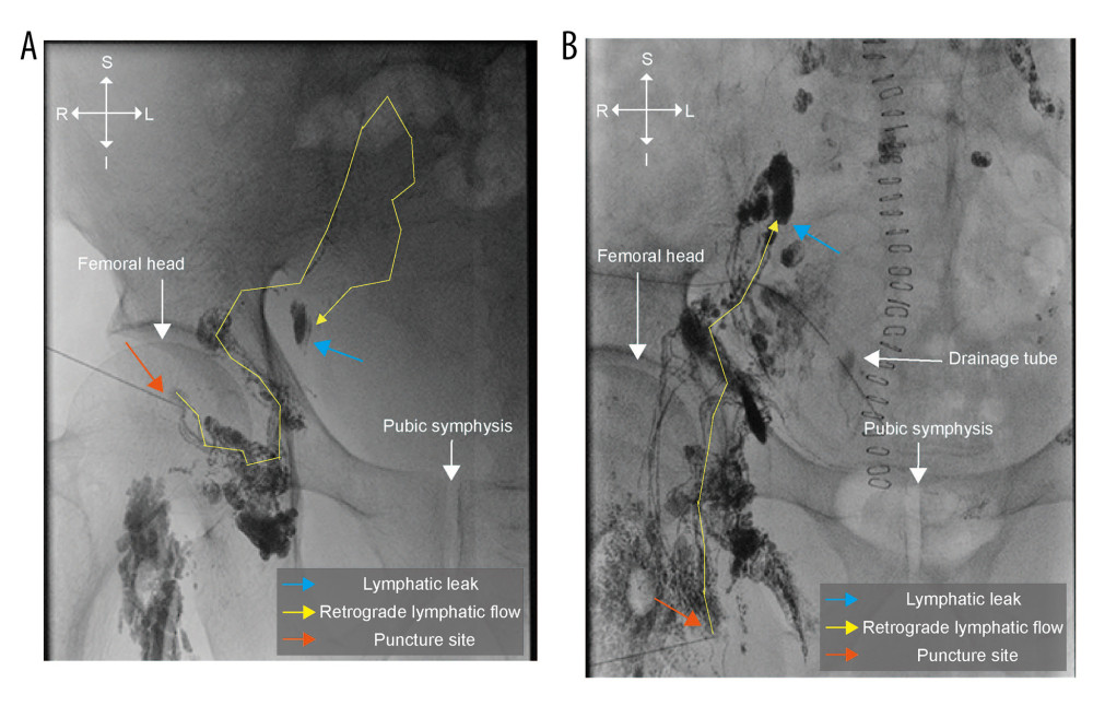 Figure 1. Lymphangiography images(A) Initial lymphangiography performed on postoperative day (POD) 87. The cyan arrow indicates the internal iliac lymphatic leakage site. The yellow line and arrow demonstrate retrograde lymphatic flow through the pelvic lymphatic channels. The orange arrow indicates the puncture site. (B) Repeat lymphangiography performed on POD 108. The cyan arrow indicates a new lymphatic leak in the external iliac region, anatomically distinct from the original internal iliac leak site. The yellow line and arrow demonstrate retrograde lymphatic flow through the collateral pathway. The orange arrow indicates the puncture site. These findings suggest redistribution of lymphatic flow through collateral pathways following ligation of the primary leak.
 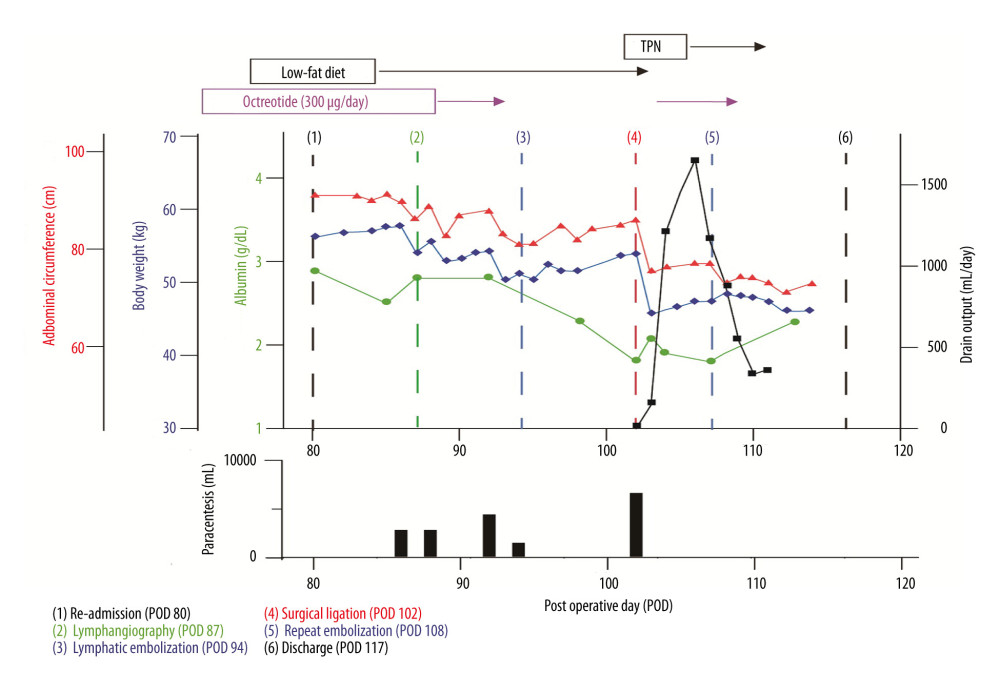 Figure 2. Clinical course during the second admissionAlbumin (green), body weight (blue), and abdominal circumference (red) are plotted against postoperative day (POD) on the left axes. Drain output (black) is shown on the right axis. Upper bars indicate the duration of low-fat diet, octreotide, and total parenteral nutrition (TPN). Lower panel shows paracentesis volumes with cell-free concentrated ascites reinfusion therapy (CART). Numbered dashed vertical lines indicate key clinical events: (1) re-admission (POD 80); (2) lymphangiography (POD 87); (3) lymphatic embolization (POD 94); (4) lymphatic ligation (POD 102); (5) repeat embolization (POD 108); (6) discharge (POD 117).
 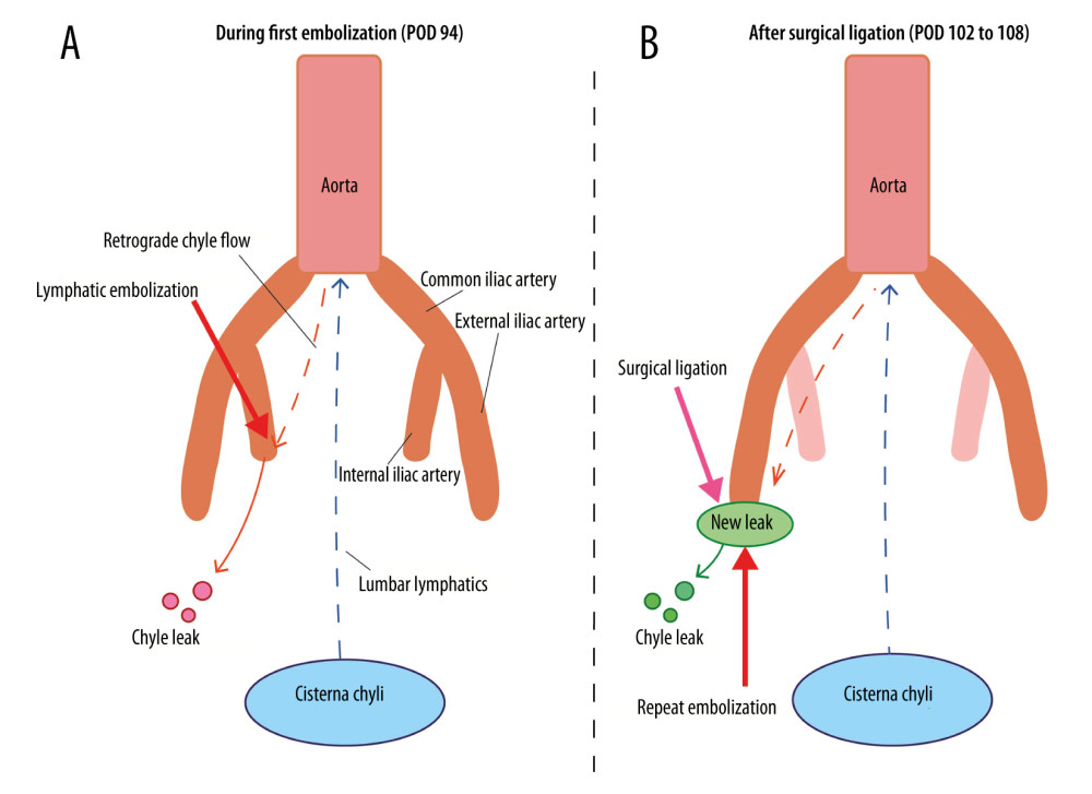 Figure 3. Schematic diagram of the hypothesized mechanism of dynamic redistribution of lymphatic leakage(A) During the first lymphatic embolization on postoperative day (POD) 94, retrograde chylous flow through the lumbar lymphatics may have contributed to persistent leakage from the internal iliac region (red droplets), despite ongoing embolization. (B) After surgical ligation of the internal iliac leak (POD 102), lymphatic flow may have been redirected through a collateral pathway (orange dashed arrow), resulting in a new leak in the external iliac region (green ellipse and droplets). Repeat embolization of this secondary leak site achieved definitive resolution. These schematics represent the authors’ proposed explanation for the observed sequential change in leak site and do not depict a definitively proven anatomical sequence.
 References
1. Gloger S, Ubrig B, Boy A, Bilateral peritoneal flaps reduce incidence and complications of lymphoceles after robotic radical prostatectomy with pelvic lymph node dissection-results of the prospective randomized multicenter trial ProLy: J Urol, 2022; 208(2); 333-40
2. Bhardwaj R, Vaziri H, Gautam A, Chylous ascites: A review of pathogenesis, diagnosis and treatment: J Clin Transl Hepatol, 2018; 6(1); 105-13
3. Lizaola B, Bonder A, Trivedi HD, Review article: The diagnostic approach and current management of chylous ascites: Aliment Pharmacol Ther, 2017; 46(9); 816-24
4. Ashida H, Kisaki S, Enoki K, Ojiri H, Interventional radiology in management of postoperative chylous ascites: Interv Radiol (Higashimatsuyama), 2025; 10; e20230039
5. Rose KM, Huelster HL, Roberts EC, Contemporary management of chylous ascites after retroperitoneal surgery: Development of an evidence-based treatment algorithm: J Urol, 2022; 208(1); 53-61
6. DiBattista JV, Odenwald MA, Te H, Successful treatment of refractory chylous ascites with octreotide in a patient with decompensated cirrhosis: ACG Case Rep J, 2024; 11(4); e01322
7. Han J, Aron M, Refractory lymphatic ascites after radical prostatectomy: Urology, 2020; 145; 9-10
8. Lv S, Wang Q, Zhao W, A review of the postoperative lymphatic leakage: Oncotarget, 2017; 8(40); 69062-75
9. Oka T, Ishimaru H, Hirao M, A case of chylous ascites caused by reflux from the cisterna chyli to the disrupted common iliac lymphatic vessel: Fluoroscopic records during intranodal lymphangiography: BJR Case Rep, 2024; 10(1); uaae004
10. Duletzke NT, Kiraly LN, Martindale RG, Chylothorax and chylous ascites: Overview, management, and nutrition: Nutr Clin Pract, 2023; 38(3); 557-63
11. Hanafusa N, Isoai A, Ishihara T, Safety and efficacy of cell-free and concentrated ascites reinfusion therapy (CART) in refractory ascites: Post-marketing surveillance results: PLoS One, 2017; 12(5); e0177303
12. Kawasaki R, Sugimoto K, Fujii M, Therapeutic effectiveness of diagnostic lymphangiography for refractory postoperative chylothorax and chylous ascites: Correlation with radiologic findings and preceding medical treatment: Am J Roentgenol, 2013; 201(3); 659-66
13. Litchinko A, Monnard E, Tappero C, Egger B: Front Radiol, 2025; 5; 1537744
14. Kim SW, Hur S, Kim SY, The efficacy of lymph node embolization using N-butyl cyanoacrylate compared to ethanol sclerotherapy in the management of symptomatic lymphorrhea after pelvic surgery: J Vasc Interv Radiol, 2019; 30(2); 195-202e1
15. Kayama E, Nishimoto K, Kaneko G, Embolization using N-butyl cyanoacrylate for postoperative lymphatic leakage: A case report: Clin Genitourin Cancer, 2018; 16(2); e355-e71
16. Jun H, Hur S, Interventional radiology treatment for postoperative chylothorax: Korean J Thorac Cardiovasc Surg, 2020; 53(4); 200-4
17. Wang S, Jiang W, Post-esophagectomy chylothorax refractory to mass ligation of thoracic duct above diaphragm: A case report: J Cardiothorac Surg, 2022; 17(1); 259
18. Takahashi T, Kisu I, Kiyokawa S, Successful treatment of chylous leakage with delayed presentation after endometrial cancer surgery using dietary therapy, octreotide, and computed tomography-guided lymphangiography: A case report and literature review: J Obstet Gynaecol Res, 2025; 51(9); e70066
Figures
Figure 1. Lymphangiography images(A) Initial lymphangiography performed on postoperative day (POD) 87. The cyan arrow indicates the internal iliac lymphatic leakage site. The yellow line and arrow demonstrate retrograde lymphatic flow through the pelvic lymphatic channels. The orange arrow indicates the puncture site. (B) Repeat lymphangiography performed on POD 108. The cyan arrow indicates a new lymphatic leak in the external iliac region, anatomically distinct from the original internal iliac leak site. The yellow line and arrow demonstrate retrograde lymphatic flow through the collateral pathway. The orange arrow indicates the puncture site. These findings suggest redistribution of lymphatic flow through collateral pathways following ligation of the primary leak.Figure 2. Clinical course during the second admissionAlbumin (green), body weight (blue), and abdominal circumference (red) are plotted against postoperative day (POD) on the left axes. Drain output (black) is shown on the right axis. Upper bars indicate the duration of low-fat diet, octreotide, and total parenteral nutrition (TPN). Lower panel shows paracentesis volumes with cell-free concentrated ascites reinfusion therapy (CART). Numbered dashed vertical lines indicate key clinical events: (1) re-admission (POD 80); (2) lymphangiography (POD 87); (3) lymphatic embolization (POD 94); (4) lymphatic ligation (POD 102); (5) repeat embolization (POD 108); (6) discharge (POD 117).Figure 3. Schematic diagram of the hypothesized mechanism of dynamic redistribution of lymphatic leakage(A) During the first lymphatic embolization on postoperative day (POD) 94, retrograde chylous flow through the lumbar lymphatics may have contributed to persistent leakage from the internal iliac region (red droplets), despite ongoing embolization. (B) After surgical ligation of the internal iliac leak (POD 102), lymphatic flow may have been redirected through a collateral pathway (orange dashed arrow), resulting in a new leak in the external iliac region (green ellipse and droplets). Repeat embolization of this secondary leak site achieved definitive resolution. These schematics represent the authors’ proposed explanation for the observed sequential change in leak site and do not depict a definitively proven anatomical sequence. In Press
Case report 
Am J Case Rep In Press; DOI: 10.12659/AJCR.953543
Case report
Am J Case Rep In Press; DOI: 10.12659/AJCR.954216
Case report 
Am J Case Rep In Press; DOI: 10.12659/AJCR.954932
Case report 
Am J Case Rep In Press; DOI: 10.12659/AJCR.954146
Most Viewed Current Articles
07 Dec 2021 : Case report
28,712,695
DOI :10.12659/AJCR.934347
Am J Case Rep 2021; 22:e934347
06 Dec 2021 : Case report  180,721
DOI :10.12659/AJCR.934406
Am J Case Rep 2021; 22:e934406
21 Jun 2024 : Case report
124,419
DOI :10.12659/AJCR.944371
Am J Case Rep 2024; 25:e944371
08 May 2026 : Case report
95,462
DOI :10.12659/AJCR.952158
Am J Case Rep 2026; 27:e952158